Review
. 2022 Mar 21;11(1):19.
doi: 10.1186/s40035-022-00293-2.

Propagation of tau and α-synuclein in the brain: therapeutic potential of the glymphatic system

Affiliations
Review

Propagation of tau and α-synuclein in the brain: therapeutic potential of the glymphatic system

Douglas M Lopes et al. Transl Neurodegener. .

Abstract

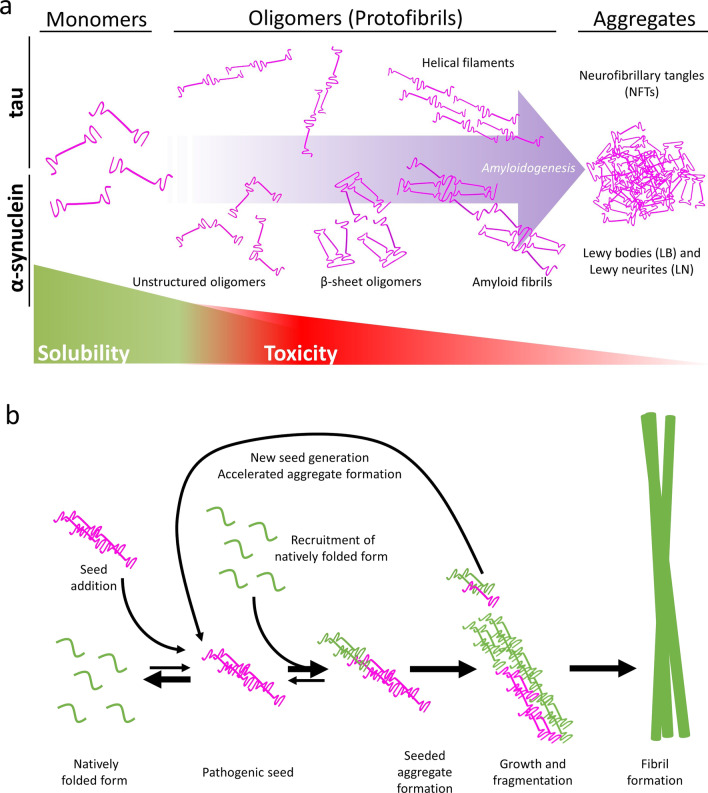
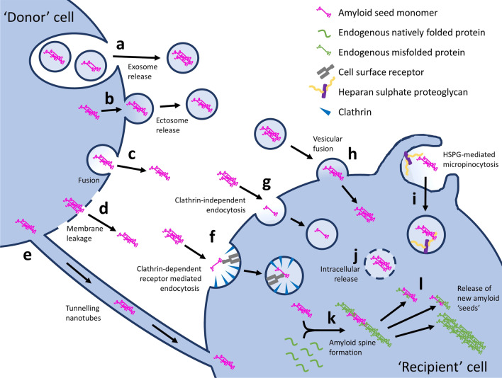
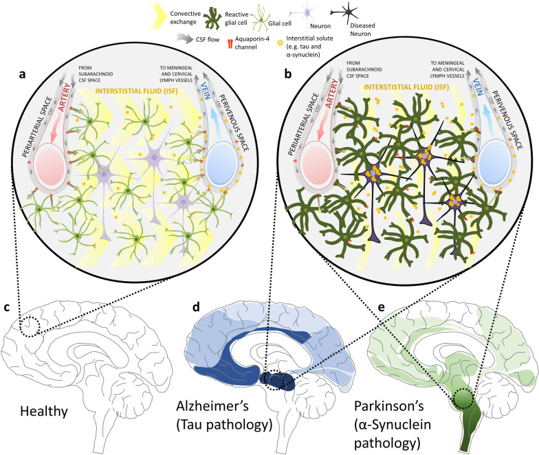
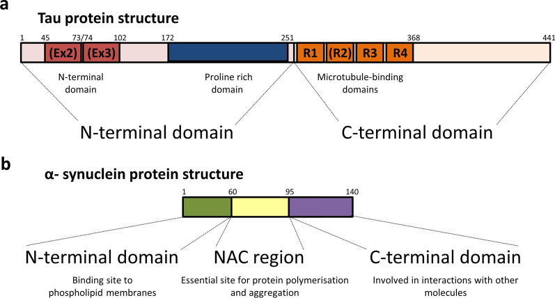
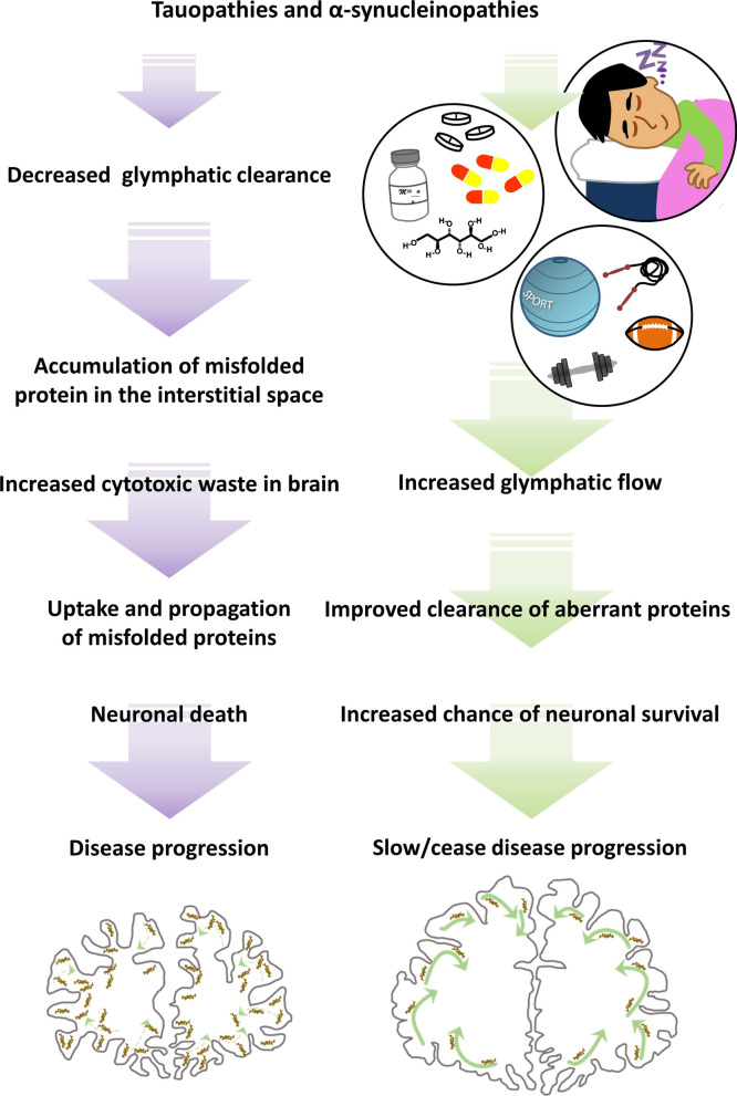
Many neurodegenerative diseases, including Alzheimer's disease and Parkinson's disease, are characterised by the accumulation of misfolded protein deposits in the brain, leading to a progressive destabilisation of the neuronal network and neuronal death. Among the proteins that can abnormally accumulate are tau and α-synuclein, which can propagate in a prion-like manner and which upon aggregation, represent the most common intracellular proteinaceous lesions associated with neurodegeneration. For years it was thought that these intracellular proteins and their accumulation had no immediate relationship with extracellular homeostasis pathways such as the glymphatic clearance system; however, mounting evidence has now suggested that this is not the case. The involvement of the glymphatic system in neurodegenerative disease is yet to be fully defined; however, it is becoming increasingly clear that this pathway contributes to parenchymal solute clearance. Importantly, recent data show that proteins prone to intracellular accumulation are subject to glymphatic clearance, suggesting that this system plays a key role in many neurological disorders. In this review, we provide a background on the biology of tau and α-synuclein and discuss the latest findings on the cell-to-cell propagation mechanisms of these proteins. Importantly, we discuss recent data demonstrating that manipulation of the glymphatic system may have the potential to alleviate and reduce pathogenic accumulation of propagation-prone intracellular cytotoxic proteins. Furthermore, we will allude to the latest potential therapeutic opportunities targeting the glymphatic system that might have an impact as disease modifiers in neurodegenerative diseases.

Keywords: Aquaporin-4; Clearance; Glymphatic; Propagation; Synucleinopathy; Tauopathy.


Conflict of interest statement

The authors declare that they have no competing interests.

Figures

Fig. 1
Fig. 1
The formation of cytotoxic species of tau and α-synuclein leading to formation of insoluble filaments. a Monomeric forms of these proteins, which are notably soluble, are capable of recruiting and binding to similar structures, giving rise to oligomeric configurations and more complex structures—helical filaments in the case of tau and amyloid fibrils in α-synuclein protein. This process leads to the formation of insoluble aggregates (tau neurofibrillary tangles and α-synuclein Lewy bodies and neurites) which can accumulate intracellularly and lead to cell death. b As indicated by the arrows, the formation of a pathological seed is an energetically unfavourable event, and as such is rare. Once a seed has formed however, monomers of the protein in its natively folded form can change shape and join the initially formed seed in creation of a seeded aggregate. Fragmentation of this aggregate generates new seed forms, accelerating the formation of further aggregates. Recruitment of further monomers/oligomers results in its growth and formation of fibrils (Adapted from Goedert [97])
Fig. 2
Fig. 2
Cell-to-cell spread of prion-like protein species. Prions are proposed to be spread from ‘donor’ to ‘recipient’ cells by numerous mechanisms. ‘Naked’ prions can be released into the extracellular space through either exosomal release or leakage through damaged cell membranes (c and d, respectively), where they can, in turn, become internalised by mechanisms such as direct pinocytosis, a process thought to occur via both clathrin-dependent receptor mediated endocytosis (f) and clathrin-independent endocytosis (g), resulting in intracellular release (j). Heparan sulphate proteoglycans (HSPGs), transmembrane and lipid-anchored cell surface receptors that interact with a variety of ligands, have also been shown to mediate internalisation of prions, via HSPG-mediated micropinocytosis (i): an endocytic process characterized by binding of the prion ligand to surface-bound HSPG, actin-driven membrane ruffling, internalization of extracellular fluids, and formation of large intracellular vacuoles. Prions can also be released within exosomes (a) or ectosomes (b), which can be internalised through vesicular fusion (h). Tunnelling nanotubes, F-actin containing membranous bridges which connect the cytoplasm of remote cells with one another, can also facilitate cell-to-cell exchange (e). Ultimately, once inside the new host cell, prion-like amyloid-prone proteins are thought to initiate amyloid assembly, through recruitment of endogenous natively folded protein monomers or oligomers leading to formation of an initial segment of an amyloid spine (k). This spine can then break, inducing further intracellular propagation of amyloid formation and release of new amyloid ‘seeds’ (l), which can then be released and propagate to new unaffected cells by the aforementioned cell-to-cell translocation pathways
Fig. 3
Fig. 3
The glymphatic system in neurodegenerative disease. a An overall view of the glymphatic system and its main components in health (c): the CSF that flows alongside the arteries (from the subarachnoid CSF spaces) moves across the brain parenchyma via convective exchange (yellow chevrons). The movement is driven by pressure gradients and the convective transport is facilitated by a network of glial projections (green cells) and aquaporin-4 channels expressed on their endfeet (red structures). As the CSF passes through the brain parenchyma, it carries the interstitial solutes present in that space towards the perivenous space, from where it gets cleared out of the brain towards meningeal and cervical lymph vessels. b Failure of the glymphatic system in neurodegenerative disease (e.g. d and e): aquaporin-4 becomes depolarised from astrocytic endfeet, leading to reduced convective exchange of paravascular CSF with interstitial fluid. This leads to reduced parenchymal clearance of proteins such as tau (d depicted in blue in the advanced Braak stage Alzheimer’s scenario) and α-synuclein (e depicted in green in the advanced Braak stage Parkinson’s), facilitating their cell-to-cell propagation throughout the brain
Fig. 4
Fig. 4
Schematic structure of tau and α-synuclein proteins. a Tau protein contains three main domains: an N-terminal domain (red/pink), a proline-rich domain (blue), and a microtubule-binding domain (orange). Tau isoforms can range between 352 and 441 amino acids in length; containing zero, one, or two N-terminal repeats (encoded by exons 2 and 3—shown in dark red), and three or four C-terminal microtubule-binding domains (presence or absence of R2, encoded by exon 10)—which define the 3R and 4R tau species. b In contrast, α-synuclein is a relatively short protein (140 amino acids in length), but can also be divided into three regions: an N-terminal domain (green), a non-amyloid-component (NAC) region (yellow), and a C-terminal domain (purple), with each of their correspondent functions shown
Fig. 5
Fig. 5
The glymphatic system in tauopathies and α-synucleinopathies. During the development of these neurodegenerative diseases (purple arrows, left), it is believed that the glymphatic function and cytotoxic protein clearance become impaired, leading to the accumulation of aberrant proteins, further seeding and propagation, and destabilisation of the neuronal network, ultimately leading to disease progression. Recent studies suggest that manipulation of the glymphatic system (green arrows, right panel)—e.g. good sleep hygiene, leading a healthy lifestyle or the administration of osmotic drugs—may have the potential to increase glymphatic function and alleviate and reduce pathogenic accumulation of intracellular cytotoxic proteins. These plausible therapeutics have the potential to increase neuronal survival and therefore delay or even prevent the progression of disease. Protein accumulation (depicted by brown aggregates) as a result of the extent of glymphatic function in the two scenarios (appropriately weighted green arrows) is represented schematically in coronal diagrams of a degenerated (left) and healthy (right) human brain cross-section in the lower section of the figure

Similar articles

Cited by

References

    1. GBD 2016 Dementia Collaborators Global, regional, and national burden of Alzheimer's disease and other dementias, 1990–2016: a systematic analysis for the Global Burden of Disease Study 2016. Lancet Neurol. 2019;18(1):88–106. - PMC - PubMed
    1. Hou Y, Dan X, Babbar M, Wei Y, Hasselbalch SG, Croteau DL, et al. Ageing as a risk factor for neurodegenerative disease. Nat Rev Neurol. 2019;15(10):565–581. - PubMed
    1. Wyss-Coray T. Ageing, neurodegeneration and brain rejuvenation. Nature. 2016;539(7628):180–186. - PMC - PubMed
    1. Palop JJ, Mucke L. Amyloid-beta-induced neuronal dysfunction in Alzheimer's disease: from synapses toward neural networks. Nat Neurosci. 2010;13(7):812–818. - PMC - PubMed
    1. Herms J, Dorostkar MM. Dendritic spine pathology in neurodegenerative diseases. Annu Rev Pathol. 2016;11:221–250. - PubMed

Publication types

MeSH terms

Substances