. 2022 Jun:104:102570.
doi: 10.1016/j.ceca.2022.102570. Epub 2022 Mar 11.

Probing single molecule mechanical interactions of syntaxin 1A with native synaptobrevin 2 residing on a secretory vesicle

Affiliations

Probing single molecule mechanical interactions of syntaxin 1A with native synaptobrevin 2 residing on a secretory vesicle

Wei Liu et al. Cell Calcium. 2022 Jun.

Abstract

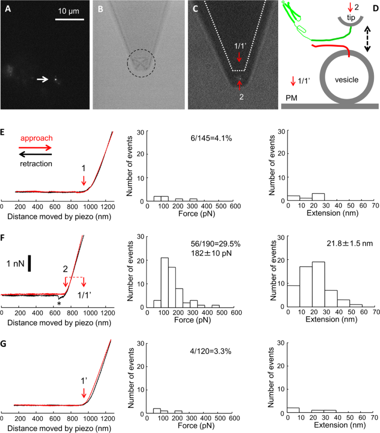
Interactive mechanical forces between pairs of individual SNARE proteins synaptobrevin 2 (Sb2) and syntaxin 1A (Sx1A) may be sufficient to mediate vesicle docking. This notion, based on force spectroscopy single molecule measurements probing recombinant Sx1A an Sb2 in silico, questioned a predominant view of docking via the ternary SNARE complex formation, which includes an assembly of the intermediate cis binary complex between Sx1A and SNAP25 on the plasma membrane to engage Sb2 on the vesicle. However, whether a trans binary Sx1A-Sb2 complex alone could mediate vesicle docking in a cellular environment remains unclear. To address this issue, we used atomic force microscopy (AFM) in the force spectroscopy mode combined with fluorescence imaging. Using AFM tips functionalized with the full Sx1A cytosolic domain, we probed native Sb2 studding the membrane of secretory vesicles docked at the plasma membrane patches, referred to as "inside-out lawns", identified based on fluorescence stains and prepared from primary culture of lactotrophs. We recorded single molecule Sx1A-Sb2 mechanical interactions and obtained measurements of force (∼183 pN) and extension (∼21.6 nm) necessary to take apart Sx1A-Sb2 binding interactions formed at tip-vesicle contact. Measured interactive force between a single pair of Sx1A-Sb2 molecules is sufficient to hold a single secretory vesicle docked at the plasma membrane within distances up to that of the measured extension. This finding further advances a notion that native vesicle docking can be mediated by a single trans binary Sx1A-Sb2 complex in the absence of SNAP25.

Keywords: Atomic force microscopy; Docking; Exocytosis; Force spectroscopy; Membrane lawns; Nanomanipulation; SNARE proteins; Single molecule measurements; Vesicle docking.


Conflict of interest statement

Conflicts of interest/Competing interests. The authors declare no conflicts and competing financial interests.

Conflicts of interest

All authors declare no conflicts of interest.

Figures

Figure 1.
Figure 1.
Preparations of inside-out lactotoph lawns and functionalized AFM tips. A-C) Fluorescently/FM1–43FX labeled vesicles attached to the cytosolic leaflet of the lactotroph plasma membrane lawns colocalize highly with the immunolabeled synaptobrevin 2 (Sb2), a vesicle membrane SNARE, and with prolactin (PRL), a vesicle cargo protein. A, Top row) Confocal images of immunolabeled Sb2 (green) and FM1–43FX (red). Colocalized pixels (white) are displayed in the Mask image (right). A, bottom row) Confocal images of immunolabeled PRL (green) and FM1–43FX (red). Colocalized pixels (white) are displayed in the Mask image (right). Scale bar, 5 μm. B) Differential interference contrast (DIC) image of a lactotroph lawn. Note that many vesicles, granule-like structures, are visible as puncta darker than their surroundings. Scale bar, 5 μm. C) Graph showing fluorescence colocalization (mean ± s.e.m.) of FM1–43FX with either immunolabeled Sb2 or PRL. Numbers at the bottom of bars indicate the number of lawns analyzed. D) Recombinant syntaxin 1A (Sx1A) cytosolic domain is attached to the nickel coated AFM cantilever/tip through a six histidine residues tag (H6) at its C terminus. Bright field images (BF) of non-functionalized (left) and Sx1A-H6-functionalized (right) cantilevers that were subjected to indirect immunochemistry (TRITC). Arrow points to a light reflection off the integral pyramidal tip at the bottom of the cantilever. Cantilevers incubated with Sx1A-H6 (+) were successfully functionalized as indicated by the positive immunoreactivity when compared to the control cantilevers where Sx1A-H6 (−) was omitted from the incubation solution. Scale bar, 50 μm
Figure 2.
Figure 2.
Mechanical single-molecule probing of interactions between recombinant syntaxin 1A cytosolic tail (Sx1A) and a native vesicle SNARE synaptobrevin 2 (Sb2) on secretory vesicles docked to the lactotroph plasma membrane lawns. A) A fluorescence image of a lawn with a prominent fluorescent punctum (indicated by the arrow) representing a docked secretory vesicle loaded with FM1–43. Scale bar, 10 μm (for A-C). B) A low light bright field illumination image of the corresponding field of view in A shows a Sx1A functionalized AFM cantilever with an integral pyramidal tip (dashed circle), located ~1 μm above the lawn. C) Combined fluorescence and bright field illumination image of the same lawn/vesicle and the cantilever, the later positioned so that the tip is directly above the vesicle (Position 2). Dotted profile indicates the original location of the cantilever in A, whereby its tip was ~6 μm away on the ordinate from the vesicle above the inner leaflet of the plasma membrane (Position 1). In temporal experimental sequence, this tip first probed the plasma membrane, then the vesicle, and finally returned to the original position, which we referred to as position 1’, to re-probe the initial plasmalemmal site. The red arrows indicate the positions of the AFM tip apex in the particular positions. D) Schematic of the experimental approach. Recombinant cytosolic domain of Sx1A (Sx1A-H6; green) is attached to the nickel coated cantilever tip through histidine residue tags (H6) at its C terminus leaving the domain free to interact with the native vesicle SNARE synaptobrevin 2 (Sb2, red). These two proteins are brought to near proximity (approach; dashed arrow pointing down) by the means of piezoelectric element and then taken apart (retraction, dashed arrow pointing up). The red arrows and numbers indicate positions as described in C. PM, plasma membrane. E-G) Force-distance (extension) curves (left graphs) using a Sx1A-H6 functionalized tip and probing either the plasma membrane (E and G) or native Sb2 on the vesicle. The approach part of curves is shown in red, while retraction in black traces. Diameter of a secretory vesicle was estimated based on its height (in this example ~205 nm) determined from the difference (F, red dashed horizontal line) in distances moved by z-piezo to contact the plasma membrane at position 1 (E; red vertical arrow) and the secretory vesicle at position 2 (F; red vertical arrow). Re-probing of the same plasma membrane site (position 1’) at the end of experiment (G) confirms the distance moved by z-piezo to contact the plasma membrane (red arrow). Retraction part of a force-distance curve in F shows the Sx1A-Sb2 intermolecular “bond” to increasingly extents as the tip moves vertically further away from the vesicle, which leads to increased application of the force on the intermolecular bond till it ruptures (asterisk). This is the measure of the force (ordinate) necessary to rupture single Sx1A-Sb2 pair binding interactions. The extension induced can be calculated from the z-axis distance moved by the piezo (abscissa) from the point of contact to the point of rupturing the intermolecular interaction. Distributions of the forces and extensions at rupture for Sx1A-Sb2 single intermolecular bonds are displayed in F (mid and right graphs, respectively). In E and G, data likely represent interaction between Sx1A and the plasma membrane associated SNARE SNAP25. Probability of interactions (percentage) between Sx1A tip and SNAREs is given within mid graphs as a ratio (percentage) between the number of successful events and the total number of force-distance curves performed. Force and extension measurements are provided as mean ± s.e.m. in F (mid and right graphs, respectively); such numbers are not given in E and G due to paucity of successful interactions. Scale bar (for all force-curves in E-G), 1 nN. Data in E-G originate from measurements obtained from four vesicles and the paired locations at plasma membranes. Retraction velocity is 0.6 μm/s, corresponding to force loading rate of ~7.8 nN/s. Drawing is not to scale.
Figure 3.
Figure 3.
Specificity of mechanical single-molecule interactions between recombinant Sx1A cytosolic tail and native Sb2. A) In control force spectroscopy (double arrow) experiments, recombinant cytosolic domain of Sx1A (green) attached to AFM tip forms parsimonious interactions (3.6%) with protein(s), likely SNAP25, at the plasma membrane (PM) (top), while it readily interacts (26.3%; mid and bottom graphs) with Sb2 on vesicles (n=4). Graphs represent distributions of forces (mid) and extensions (bottom) recorded from interactions between Sx1A-functionalized tips and vesicles. B) The pre-formed binary Sx1A-SNAP25B complex binds to Sb2 to form the ternary SNARE complex and thus prevents interactions between Sx1A on the AFM tip and complexed Sb2. Re-probing of the same vesicle and plasma membrane site, respectively, shows reduced interactions (6.4%) between Sx1A-functionalized tip and Sb2 in the presence of the binary Sx1A-SNAP25B complex (mid and bottom graphs), while interactions of the same tip with the plasma membrane are unaffected (4.0%; top drawing). Probability of interactions (percentage) between Sx1A-funcionalized tip and plasma membrane or native Sb2 is given as a ratio between the number of successful events and the total number of force-distance curves performed. Force and extension measurements in A are provided as mean ± s.e.m. In B, such numbers are not given due to paucity of successful interactions. Data originate from measurements obtained from four vesicles and their paired locations at plasma membrane. Retraction velocity is 0.6 μm/s, corresponding to force loading rate of ~7.8 nN/s. Drawings are not to scale.

Similar articles

Cited by

References

    1. Parpura V, Baker BJ, Jeras M, Zorec R, Regulated exocytosis in astrocytic signal integration, Neurochem Int, 57 (2010) 451–459. - PMC - PubMed
    1. Sollner T, Whiteheart SW, Brunner M, Erdjument-Bromage H, Geromanos S, Tempst P, Rothman JE, SNAP receptors implicated in vesicle targeting and fusion, Nature, 362 (1993) 318–324. - PubMed
    1. Jahn R, Scheller RH, SNAREs--engines for membrane fusion, Nat Rev Mol Cell Biol, 7 (2006) 631–643. - PubMed
    1. Sutton RB, Fasshauer D, Jahn R, Brunger AT, Crystal structure of a SNARE complex involved in synaptic exocytosis at 2.4 A resolution, Nature, 395 (1998) 347–353. - PubMed
    1. McNally JM, Woodbury DJ, Lemos JR, Syntaxin 1A Drives Fusion of Large Dense-Core Neurosecretory Granules Into a Planar Lipid Bilayer, Cell Biochem Biophys, 41 (2004) 11–24. - PubMed

Publication types

MeSH terms