. 2022;14(1):151-171.
doi: 10.1016/j.jcmgh.2022.03.003. Epub 2022 Mar 18.

Interleukin 1β Blockade Reduces Intestinal Inflammation in a Murine Model of Tumor Necrosis Factor-Independent Ulcerative Colitis

Affiliations

Interleukin 1β Blockade Reduces Intestinal Inflammation in a Murine Model of Tumor Necrosis Factor-Independent Ulcerative Colitis

Marina Liso et al. Cell Mol Gastroenterol Hepatol. 2022.

Abstract

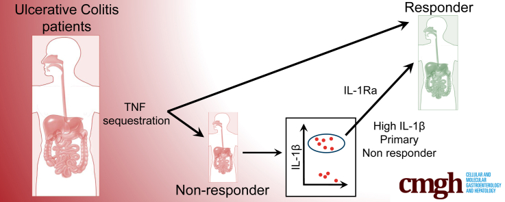
Background & aims: Inflammatory bowel diseases are multifactorial diseases commonly treated with either immunomodulatory drugs or anti-tumor necrosis factor (TNF). Currently, failure to respond to anti-TNF therapy (assessed no earlier than 8-12 weeks after starting treatment) occurs in 20%-40% of patients enrolled in clinical trials and in 10%-20% in clinical practice. Murine models of inflammatory bowel disease provide important tools to better understand disease mechanism(s). In this context and among the numerous models available, Winnie-TNF-knockout (KO) mice recently were reported to show characteristics of ulcerative colitis (UC) that are independent of TNF, and with increased interleukin (IL)1β production.

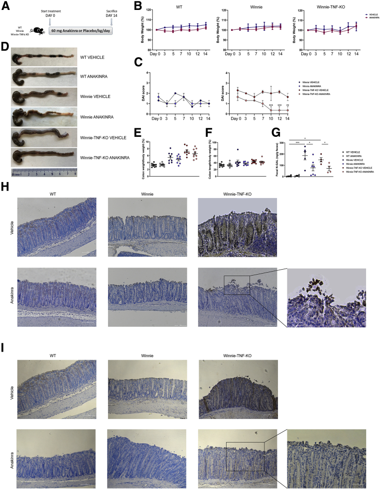
Methods: Herein, the efficacy of recombinant IL1-receptor antagonist (anakinra) administration was evaluated in Winnie-TNF-KO mice, used as a UC model of primary anti-TNF nonresponders.

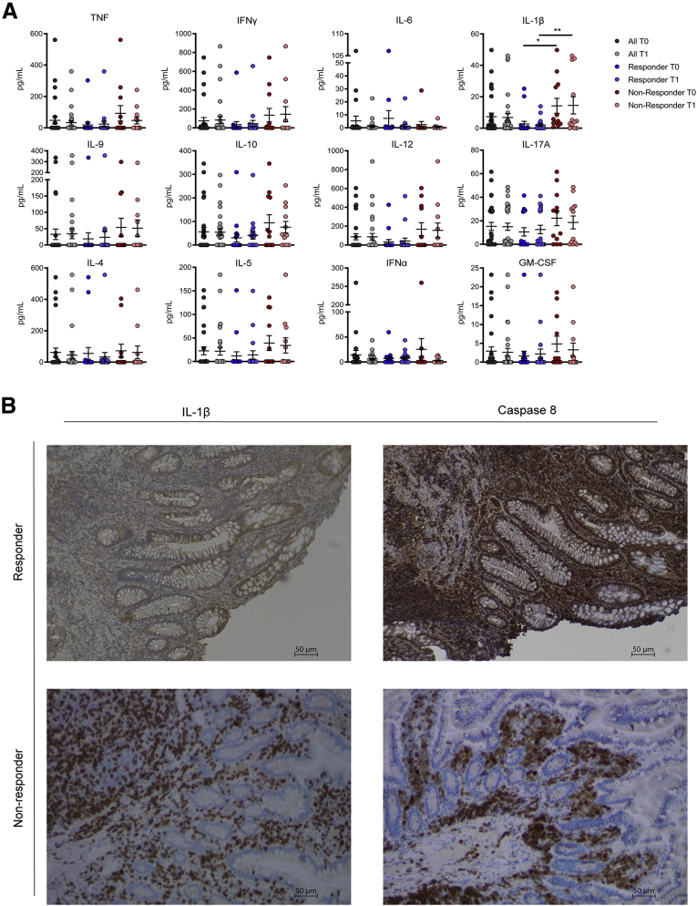
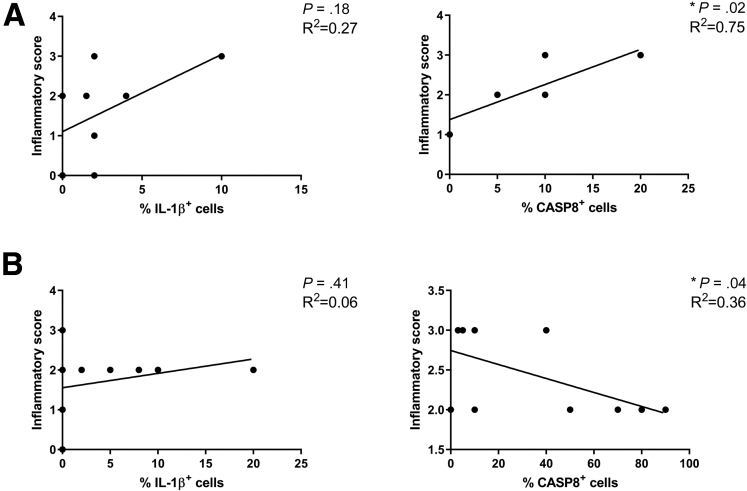
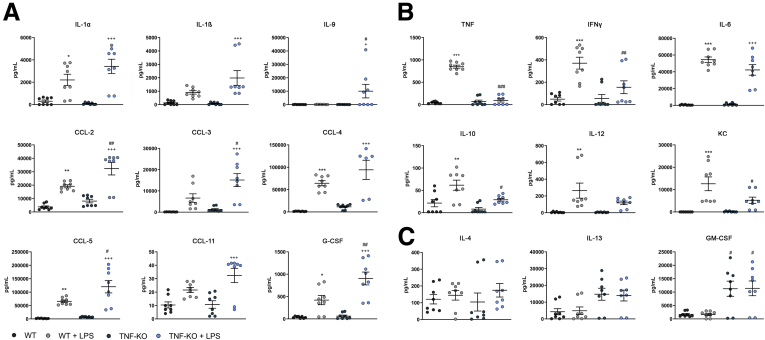
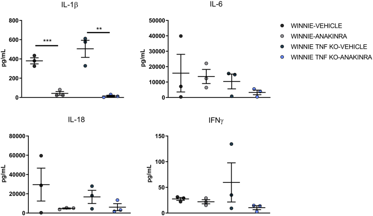
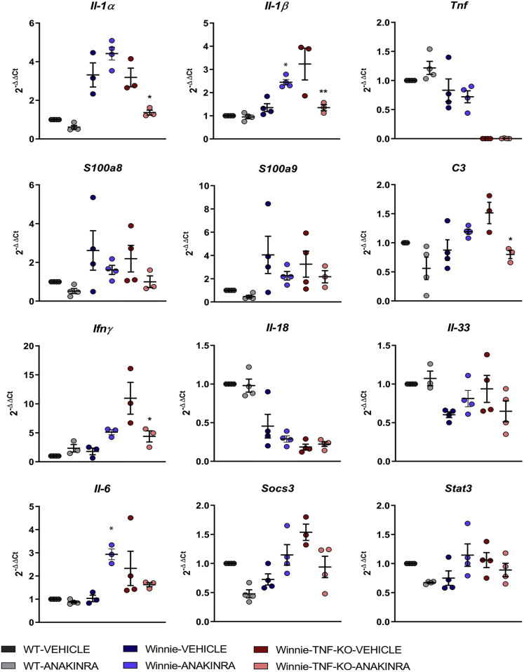
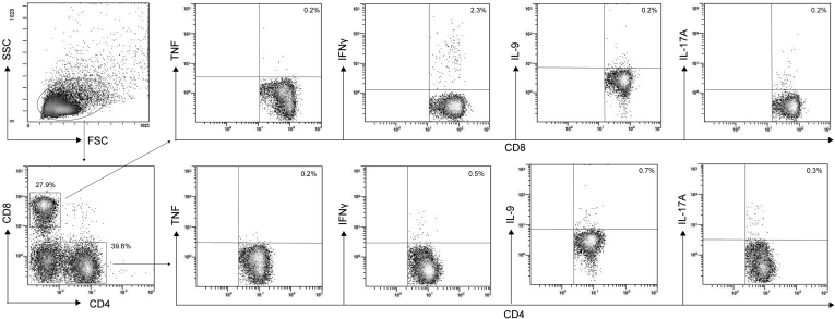
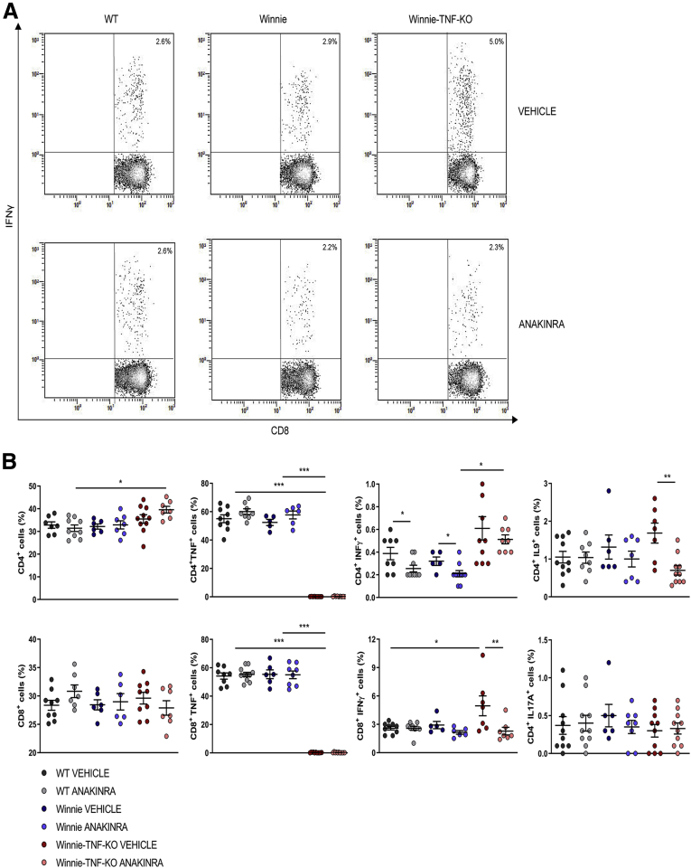
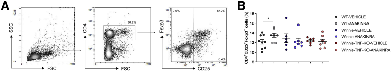
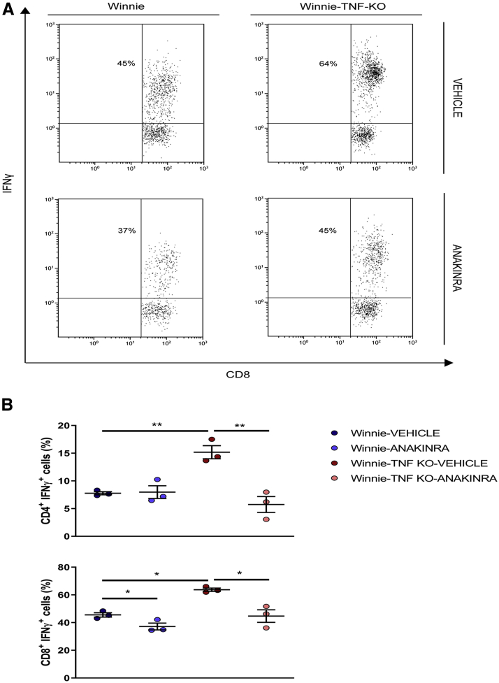
Results: We analyzed gut mucosal biopsy specimens and circulating cytokine profiles of a cohort of 30 UC patients; approximately 75% of primary nonresponders were characterized by abundant IL1β in both the serum and local intestinal tissues. In Winnie-TNF-KO mice, administration of anakinra efficiently reduced the histologic score of the distal colon, which represents the most common site of inflammation in Winnie mice. Furthermore, among lamina propria and mesenteric lymph node-derived T cells, interferon γ-expressing CD8+ T cells were reduced significantly after anakinra administration.

Conclusions: Our study provides new insight and alternative approaches to treat UC patients, and points to anti-IL1 strategies (ie, anakinra) that may be a more effective therapeutic option for primary nonresponders to anti-TNF therapy.

Keywords: Cytokines; TNF; Ulcerative Colitis.


Figures

None
Graphical abstract
Figure 1
Figure 1
(A) Assessment of cytokine levels in sera of patients before and after IFX infusion. All data sets from each time point (T0 and T1) are grouped and shown as "All" (in grey, N=30), the responder group is shown in blue (N = 18), and the nonresponder group is shown in red (N = 12). T0 refers to before IFX infusion; T1 refers to 12 weeks after the first IFX infusion. (B) IHC analysis for IL1β and caspase 8 in formalin-fixed, paraffin-embedded tissues obtained from responder and nonresponder patients at T0. Magnification, 10×. ∗P < .05, ∗∗P < .01.
Figure 2
Figure 2
Linear regression between inflammatory score and the percentage of IL1β or caspase 8 (CASP8)-positive cells in the (A) nonresponder (N = 8) and (B) responder (N = 12) groups at T0.
Figure 3
Figure 3
Murine BMDC cytokine (CK) and chemokine (CC) profiles after LPS exposure determined by multiplex assay. BMDCs from WT and TNF-KO mice were exposed to LPS for 24 hours (N = 8 for each group). Dark grey and blue circles represent the CK or CC level from WT and TNF-KO BMDCs, respectively; light grey and light blue circles show the CK and CC levels after LPS exposure. (A) CK and CC were increased in the TNF-KO BMDCs. (B) CK and CC were decreased in the TNF-KO BMDCs. (C) There was no effect in CK and CC secretion mediated by LPS stimulation. ∗/+/#P < .05, ∗∗/++/##P < .01, and ∗∗∗/+++/P < .001. ∗WT versus WT+LPS; +TNF-KO versus TNF-KO +LPS; #WT+LPS versus TNF-KO +LPS. G-CSF, granulocyte colony-stimulating factor; KC, keratinocyte-derived chemokine.
Figure 4
Figure 4
Macroscopic features and measurements of colonic parameters at the end of treatment with anakinra. (A) Schematic representation of the experimental design. WT, Winnie and Winnie-TNF-KO mice were treated with IP injection of anakinra or vehicle for 14 days (N = 9 for each group). (B) Effect of anakinra or vehicle on mice body weight from day 0 until the end of treatment. (C) DAI score recorded for Winnie and Winnie-TNF-KO mice treated with anakinra or vehicle. (D) Representative images of whole colons for each experimental group. (E and F) Measurement of colon weight/body weight and colon length/body weight indices (%), respectively. (G) Detection of N-GAL level in feces of WT, Winnie, and Winnie-TNF-KO mice treated with anakinra or vehicle. (H and I) IHC analysis for IL1β and caspase 8, respectively, in formalin-fixed, paraffin-embedded tissues obtained from WT, Winnie, and Winnie-TNF-KO mice treated with anakinra or vehicle. (10× and magnified 20× on the right). ∗P < .05, ∗∗P < .01, and ∗∗∗P < .001.
Figure 5
Figure 5
Cytokine profile of colonic tissue culture from Winnie and Winnie-TNF-KO mice treated with vehicle or anakinra (N = 3 for each group). ∗∗P < .005, ∗∗∗P < .001.
Figure 6
Figure 6
Relative expression of Il1α, Il1β, Tnf, S100a8, S100a9, C3, Ifnγ, Il18, Il33, Il6, Socs3, and Stat3 in WT (N = 3), Winnie (N = 3), and Winnie-TNF-KO (N = 4) mice treated with vehicle or anakinra.P < .05 (anakinra vs vehicle).
Figure 7
Figure 7
Gene expression analysis of the 88 genes expressed in the distal colon of WT (N = 3), Winnie (N = 3), and Winnie-TNF-KO (N = 4) mice treated with vehicle or anakinra. ∗p < 0.05 (Anakinra vs vehicle)
Figure 8
Figure 8
Gating strategy for intracellular staining. Representative density plot analysis of intracellular staining of TNF, IFNγ, IL9, and IL17A from CD4+ and CD8+ cells from MLN of Winnie-TNF-KO mice treated with anakinra (N = 9 for each group). SSC, side scatter.
Figure 9
Figure 9
(A) Representative density plot analysis of intracellular staining of IFNγ from CD8+T cells in MLN of WT, Winnie, and Winnie-TNF-KO mice, treated with vehicle or anakinra. (B) Frequencies of CD4+ and CD8+ T cells and intracellular staining of TNF, IFNγ, IL9, and IL17A from CD4+ and CD8+ T cells, in the MLNs of WT, Winnie, and Winnie-TNF-KO mice treated with anakinra or vehicle. ∗P < .05, ∗∗P < .01, and ∗∗∗P < .001.
Figure 10
Figure 10
Regulatory T cell (Treg) staining of CD4+cells isolated from MLNs. (A) Representative density plot of Treg gating strategy. (B) Frequencies of CD4+CD25+Foxp3+ cells in MLNs of WT, Winnie, and Winnie-TNF-KO mice treated with anakinra or vehicle (N = 9 for each group). ∗P < .05. FSC, forward scatter; SSC, side scatter.
Figure 11
Figure 11
(A) Representative density plot analysis of intracellular staining of IFNγ from CD8+T cells in LP of Winnie and Winnie-TNF-KO mice, treated with vehicle or anakinra. (B) Frequencies of IFNγ+ from CD4+ and CD8+ T cells in the LP of Winnie and Winnie-TNF-KO mice treated with anakinra or vehicle. ∗P < .05, ∗∗P < .01.

References

    1. Murch S.H., Lamkin V.A., Savage M.O., Walker-Smith J.A., MacDonald T.T. Serum concentrations of tumour necrosis factor alpha in childhood chronic inflammatory bowel disease. Gut. 1991;32:913. - PMC - PubMed
    1. Tsukada Y., Nakamura T., Iimura M., Iizuka B.E., Hayashi N. Cytokine profile in colonic mucosa of ulcerative colitis correlates with disease activity and response to granulocytapheresis. Am J Gastroenterol. 2002;97:2820–2828. - PubMed
    1. Braegger C.P., Nicholls S., Murch S.H., Stephens S., MacDonald T.T. Tumour necrosis factor alpha in stool as a marker of intestinal inflammation. Lancet. 1992;339:89–91. - PubMed
    1. Sherman M., Tsynman D.N., Kim A., Arora J., Pietras T., Messing S., St Hilaire L., Yoon S., Decross A., Shah A., Saubermann L. Sustained improvement in health-related quality of life measures in patients with inflammatory bowel disease receiving prolonged anti-tumor necrosis factor therapy. J Dig Dis. 2014;15:174–179. - PubMed
    1. Pisani L.F., Moriggi M., Gelfi C., Vecchi M., Pastorelli L. Proteomic insights on the metabolism in inflammatory bowel disease. World J Gastroenterol. 2020;26:696–705. - PMC - PubMed

Publication types

MeSH terms

Substances