. 2022 May 17;98(20):e2046-e2059.
doi: 10.1212/WNL.0000000000200660. Epub 2022 Mar 21.

Spectrum of Phenotypic, Genetic, and Functional Characteristics in Patients With Epilepsy With KCNC2 Pathogenic Variants

Affiliations

Spectrum of Phenotypic, Genetic, and Functional Characteristics in Patients With Epilepsy With KCNC2 Pathogenic Variants

Niklas Schwarz et al. Neurology. .

Abstract

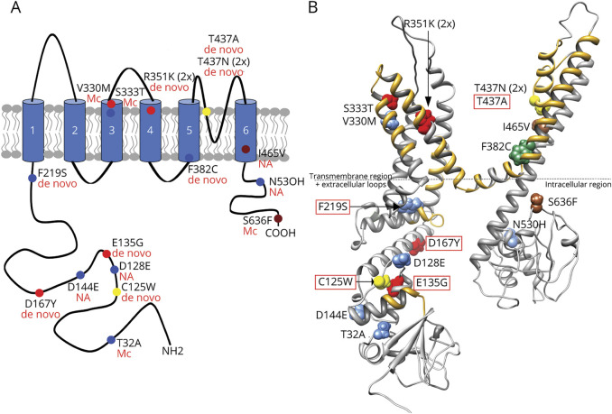
Background and objectives: KCNC2 encodes Kv3.2, a member of the Shaw-related (Kv3) voltage-gated potassium channel subfamily, which is important for sustained high-frequency firing and optimized energy efficiency of action potentials in the brain. The objective of this study was to analyze the clinical phenotype, genetic background, and biophysical function of disease-associated Kv3.2 variants.

Methods: Individuals with KCNC2 variants detected by exome sequencing were selected for clinical, further genetic, and functional analysis. Cases were referred through clinical and research collaborations. Selected de novo variants were examined electrophysiologically in Xenopus laevis oocytes.

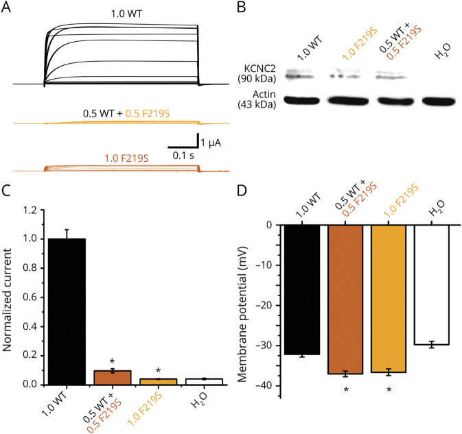
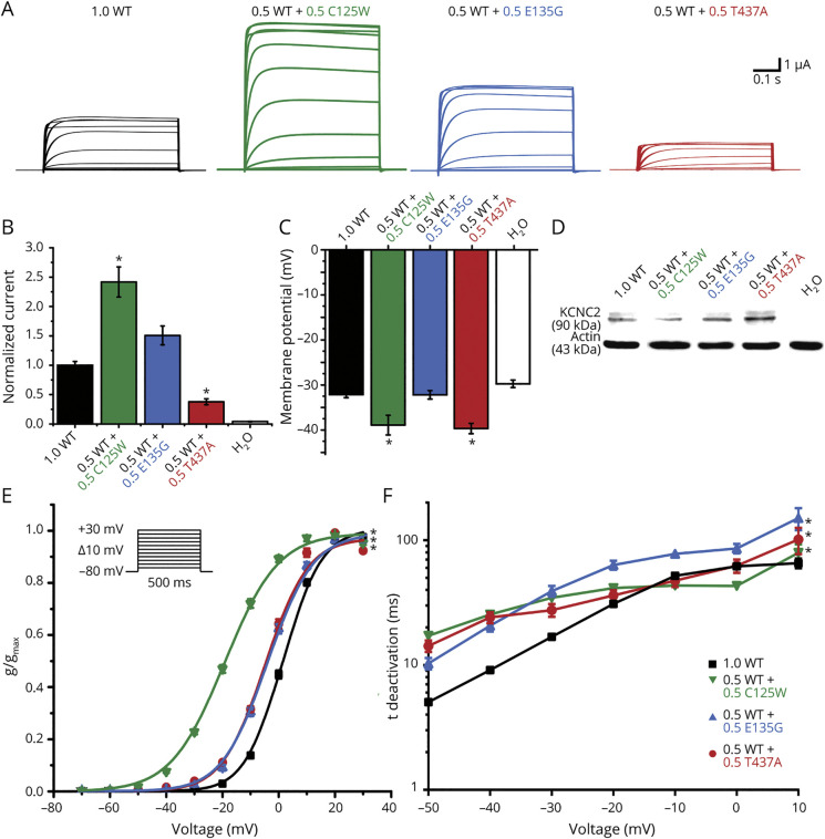
Results: We identified novel KCNC2 variants in 18 patients with various forms of epilepsy, including genetic generalized epilepsy (GGE), developmental and epileptic encephalopathy (DEE) including early-onset absence epilepsy, focal epilepsy, and myoclonic-atonic epilepsy. Of the 18 variants, 10 were de novo and 8 were classified as modifying variants. Eight drug-responsive patients became seizure-free using valproic acid as monotherapy or in combination, including severe DEE cases. Functional analysis of 4 variants demonstrated gain of function in 3 severely affected DEE cases and loss of function in 1 case with a milder phenotype (GGE) as the underlying pathomechanisms.

Discussion: These findings implicate KCNC2 as a novel causative gene for epilepsy and emphasize the critical role of KV3.2 in the regulation of brain excitability.


Figures

Figure 1
Figure 1. Structure of KV3.2 Encoded by KCNC2
A) Schematic structure of the KV3.2 subunit. The subunit consists of 6 transmembrane segments (1–6) with long C- and N-terminal regions. The N-terminal plays a crucial role for the tetramerization of the channel. The 4th transmembrane segment works as the voltage sensor and the extracellular loop between the 5th and the 6th transmembrane segment forms the selectivity filter for K+ ions. Variants are color-coded according to the phenotype of the patient: red = developmental and epileptic encephalopathy, yellow = early-onset absence epilepsy, blue = genetic generalized epilepsy, green = myoclonic-atonic epilepsy, and brown = focal epilepsy. (B) The 3D structure of KV3.2 predicted by RaptorX with KCNC2 variants and phenotypes included. The golden areas within the structure are highly conserved regions characterized by paralog conservation (Paraz score) and depletion of population variants (missense tolerance ratio [MTR] score). Extracellular loops are shown above the dotted line; the intracellular N- and C-terminal regions are shown below the line. The splice variant and E608K are not shown within the structure. E608K is only expressed in transcript number NM_139136 and not on NM_139137. which was used to create the structure. Red-rimmed variants were selected for functional analysis either measured here or previously described by us.
Figure 2
Figure 2. Electrophysiologic Analysis of the p.F219S KCNC2 Variant
Functional analysis for the F219S variant compared with wild-type (WT). The figures show that the milder phenotype F219S-GGE has a dramatically dominant negative effect in a sense of a loss of function. (A) Representative traces of KV3.2 currents in Xenopus laevis oocytes expressing WT, F219S, or a 1:1 mixture of both in response to the voltage steps from −70 mV to +30 mV. (B) Immunoblot analysis for lysates of X laevis oocytes injected with cRNA for KV3.2 WT, F219S, equal amounts of WT + F219S, or water. All channels showed a band at about 90 kDa. (C) Mean current amplitudes of analyzed oocytes injected with WT (n = 101), F219S (n = 39), equal amounts of WT + F219S (n = 29), or water (n = 44). (D) Resting membrane potentials of oocytes injected with WT (n = 101), F219S (n = 39), equal amounts of WT + F219S (n = 29), or water (n = 44). Shown are means ± SEM. Statistically significant differences between WT channels and the tested groups were verified by analysis of variance on ranks (indicated by asterisks).
Figure 3
Figure 3. Electrophysiologic Analysis of Selected KCNC2 Variants
Functional analysis of the variants C125W, E135G, and T437A compared with wild-type (WT). The more severe phenotypes C125W-EOAE (increased current amplitude/activation at more hyperpolarized potentials/slow deactivation), E135G-DEE (normal current/activation at more hyperpolarized potentials/slow deactivation), and T437A-EOAE (reduced current amplitude/activation at more hyperpolarized potentials/slow deactivation) demonstrate gain of function. (A) Representative traces of KV3.2 currents recorded in Xenopus laevis oocytes expressing wild-type (WT) or the different variants (C125W, E135G, T437A) in response to voltage steps from −70 mV to +30 mV (with an increment of 10 mV). (B) Mean current amplitudes of oocytes injected with WT (n = 101) and equal amounts of WT + C125W (n = 40), WT + E135G (n = 31), WT + T437A (n = 41), or water (1.0, n = 44). (C) Resting membrane potentials of oocytes injected with WT (n = 101) and equal amounts of WT + C125W (n = 40), WT + E135G (n = 31), WT + F219S (n = 29), WT + T437A (n = 41), or water (n = 44). Shown are means ± SEM. Statistically significant differences between WT channels and the tested groups were verified by analysis of variance on ranks (indicated by asterisks). (D) Immunoblot analysis for lysates of X laevis oocytes injected with cRNA for KV3.2 WT and equal amounts of WT + C125W, WT + E135G, WT + T437A, or water. All channels showed a band at about 90 kDa. E. Mean voltage-dependent activation of KV3.2 channel for WT (n = 101), equal amounts of WT + C125W (n = 40), WT + E135G (n = 31), and WT + T437A (n = 42). Lines illustrate Boltzmann function fit to the data points. All activation curves showed a significant shift to more hyperpolarized potentials in comparison with WT channel alone. All data are shown as means ± SEM. (F) Mean voltage-dependent deactivation time constant of KV3.2 channel WT (n = 72), WT + C125W (n = 40), WT + E135G (n = 12), and WT + T437A (n = 20). All deactivation curves showed a significantly slower deactivation in comparison with channels only containing WT subunit. All data are shown as means ± SEM.

References

    1. World Health Organization. WHO Epilepsy: A Public Health Imperative. World Health Organization; 2019.
    1. GBD 2016 Neurology Collaborators. Global, regional, and national burden of neurological disorders, 1990-2016: a systematic analysis for the Global Burden of Disease Study 2016. Lancet Neurol. 2019;18(5):459-480. - PMC - PubMed
    1. Weber YG, Biskup S, Helbig KL, Von Spiczak S, Lerche H. The role of genetic testing in epilepsy diagnosis and management. Expert Rev Mol Diagn. 2017;17(8):739-750. - PubMed
    1. Butler A, Wei AG, Baker K, Salkoff L. A family of putative potassium channel genes in Drosophila. Science. 1989;243(4893):943-947. - PubMed
    1. Rudy B, McBain CJ. Kv3 channels: voltage-gated K+ channels designed for high-frequency repetitive firing. Trends Neurosci. 2001;24(9):517-526. - PubMed

Publication types

Substances