. 2022 Mar 21;14(6):2607-2627.
doi: 10.18632/aging.203958. Epub 2022 Mar 21.

2, 3, 5, 4'-tetrahydroxystilbene-2-O-beta-D-glucoside protects against neuronal cell death and traumatic brain injury-induced pathophysiology

Affiliations

2, 3, 5, 4'-tetrahydroxystilbene-2-O-beta-D-glucoside protects against neuronal cell death and traumatic brain injury-induced pathophysiology

Yu-Hsin Chen et al. Aging (Albany NY). .

Abstract

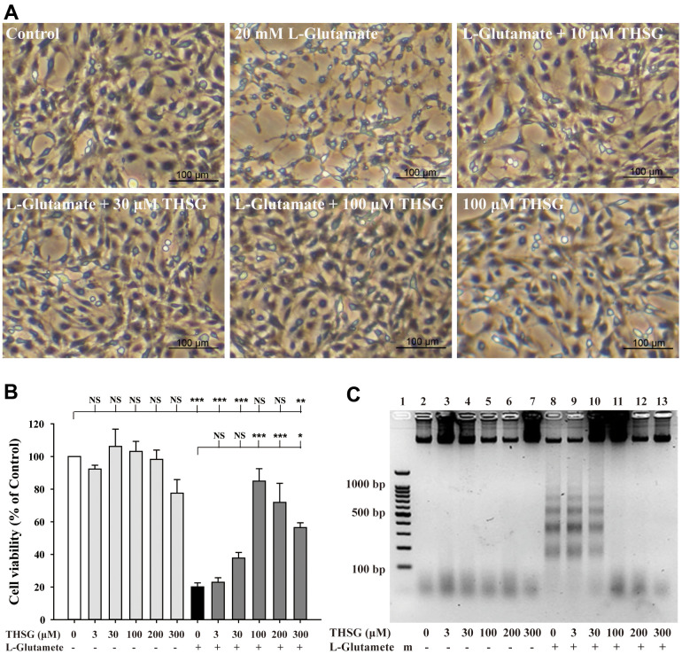
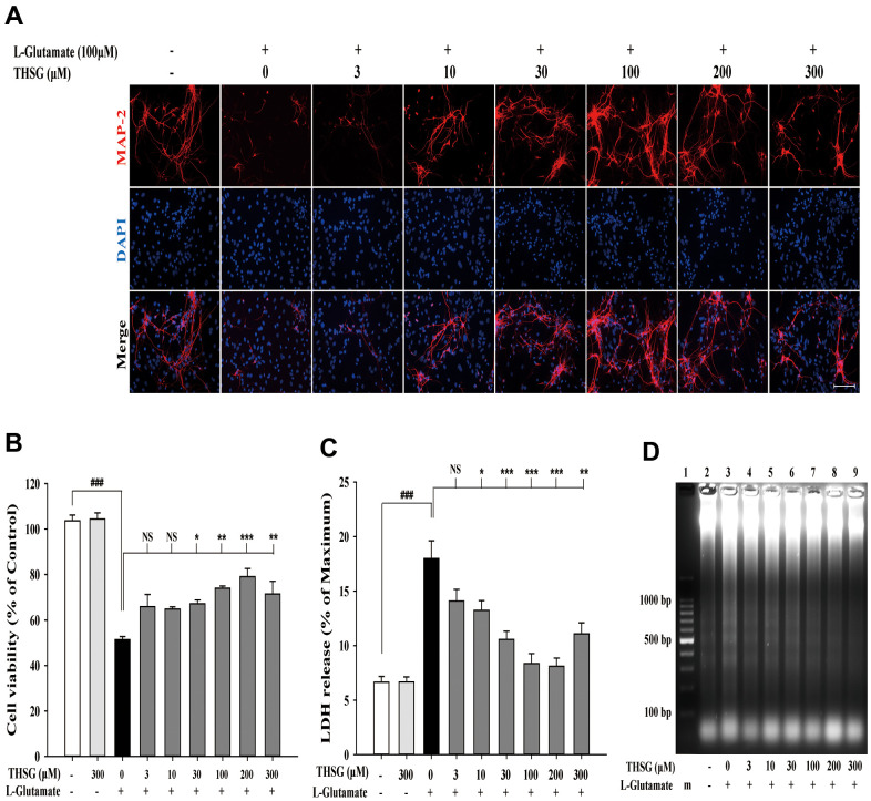
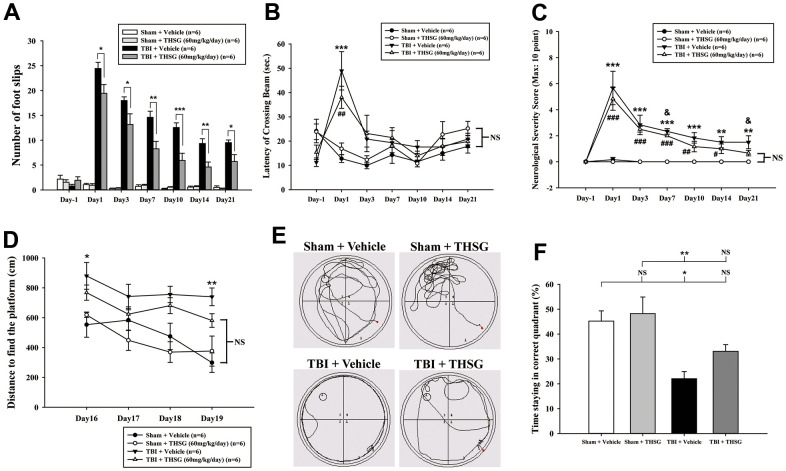
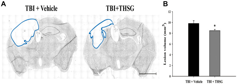
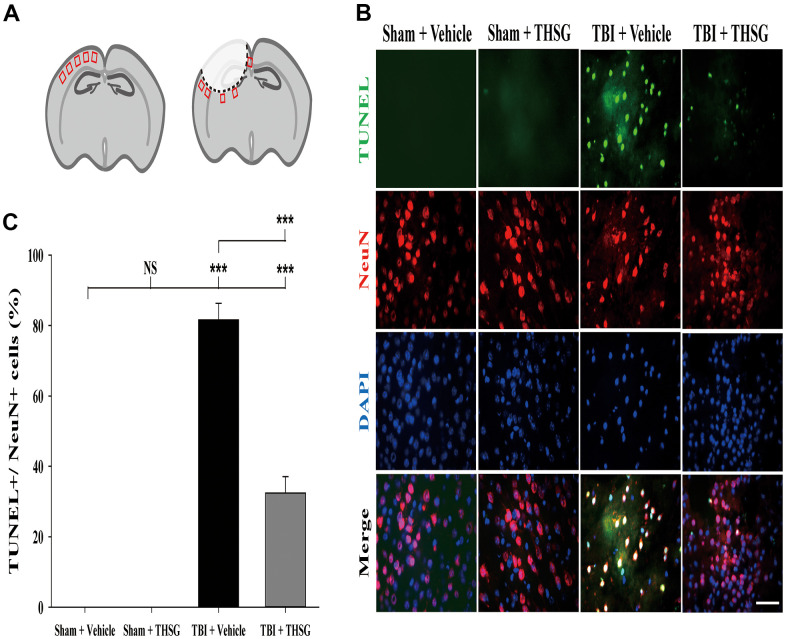
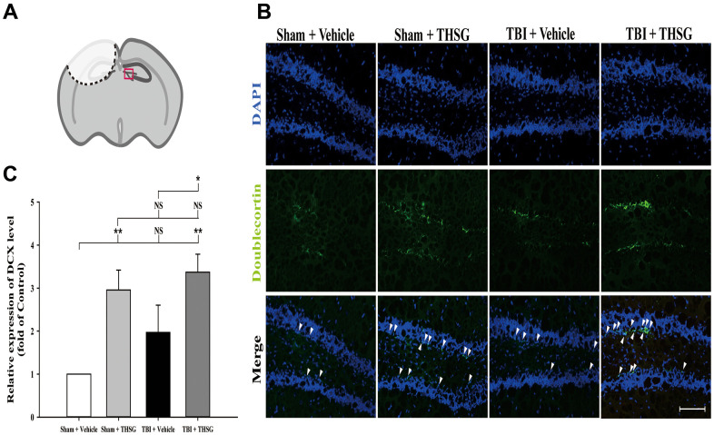
Traumatic brain injury (TBI) is a global health issue that affects at least 10 million people per year. Neuronal cell death and brain injury after TBI, including apoptosis, inflammation, and excitotoxicity, have led to detrimental effects in TBI. 2, 3, 5, 4'-tetrahydroxystilbene-2-O-beta-D-glucoside (THSG), a water-soluble compound extracted from the Chinese herb Polygonum multiflorum, has been shown to exert various biological functions. However, the effects of THSG on TBI is still poorly understood. THSG reduced L-glutamate-induced DNA fragmentation and protected glial and neuronal cell death after L-glutamate stimulation. Our results also showed that TBI caused significant behavioral deficits in the performance of beam walking, mNSS, and Morris water maze tasks in a mouse model. Importantly, daily administration of THSG (60 mg/kg/day) after TBI for 21 days attenuated the injury severity score, promoted motor coordination, and improved cognitive performance post-TBI. Moreover, administration of THSG also dramatically decreased the brain lesion volume. THSG reduced TBI-induced neuronal apoptosis in the brain cortex 24 h after TBI. Furthermore, THSG increased the number of immature neurons in the subgranular zone (SGZ) of the dentate gyrus (DG) of the hippocampus. Our results demonstrate that THSG exerts neuroprotective effects on glutamate-induced excitotoxicity and glial and neuronal cell death. The present study also demonstrated that THSG effectively protects against TBI-associated motor and cognitive impairment, at least in part, by inhibiting TBI-induced apoptosis and promoting neurogenesis.

Keywords: Chinese herb; apoptosis; cognitive dysfunction; excitotoxicity; traumatic brain injury.


Conflict of interest statement

CONFLICTS OF INTEREST: The authors declare that they have no conflicts of interest.

Figures

Figure 1
Figure 1
Effects of THSG on glutamate-induced DNA fragmentation and excitotoxicity. C6 neural glioma cells were treated with various concentrations of THSG (10, 30, or 100 μM) for 2 h and then treated with L-glutamate (20 mM) for 24 h. C6 glioma cell morphology images are shown in (A). Cells treated with 20 mM L-glutamate for 24 h caused drastic cell death, as demonstrated by the poor and shrunken cell morphology. Scale bar = 100 μm. Cell viability measured by the MTT assay is shown in (B) (n = 4). Note: THSG significantly rescued glioma neural cells from glutamate neurotoxicity, and the best effective dose of THSG was 100 μM, which completely prevented glutamate-induced cell death. NS = no significant difference; ** P > 0.01; *** P < 0.001 between the groups. Statistical analysis was performed using ANOVA for repeated measures followed by Tukey’s test of least significant difference. NS = no significantly difference; *, P > 0.05; **, P < 0.01; ***, P < 0.001. (C) Gel electrophoresis showing the effects of THSG at a range of concentrations (3-300 μM) on L-glutamate-induced DNA fragmentation in glioma neural cells.
Figure 2
Figure 2
THSG ameliorates glutamate-induced neuronal morphological change and excitotoxicity in primary cortical neurons. (A) Immunofluorescence images of treatment with or without various concentrations of THSG for 2 h followed with 100 μM L-glutamate for another 24 h; Staining performed with the primary antibody anti-MAP-2 (specific marker of neuronal dendrites, red) and a nuclear-specific dye DAPI (blue). Cells treated with L-glutamate for 24 h causes dendritic shrinkage; application of THSG 2 h prior to the treatment with glutamate improves the glutamate-induced neuron shrinkage. Merged images show the labeling co-localization. Scale bar = 100 μm. Cells were pre-treated for 2 h with THSG prior to treatment with L-glutamate for another 24 h; cell viability of primary cortical culture neurons was evaluated by MTT assay (B) and by lactate dehydrogenase (LDH) assay (C) (n = 4). Statistical analysis was carried out using ANOVA for repeated measures, followed by Tukey’s test of least significant difference. NS = no significant difference; *, P > 0.05; **, P < 0.01; ***, P < 0.001. (D) Gel electrophoresis showing the effects of THSG at a range of concentrations (3–300 μM) on L-glutamate-induced DNA fragmentation in primary cortical culture neurons.
Figure 3
Figure 3
Administration of THSG improves neurological outcomes and cognitive functions following post-TBI. (A) Evaluation of motor coordination by beam-walking test in THSG treatments after TBI. Data represented as the mean ± SEM (n = 6 per group). *, P < 0.05; **, P < 0.01 and ***, P < 0.001 TBI + Vehicle vs. TBI + THSG group. (B) Latency of beam crossing in beam-walking task. Data represented as the mean ± SEM (n = 6 per group). NS = no significantly difference between groups; ***, P < 0.001 TBI + Vehicle vs. Sham + Vehicle group; ##, P < 0.01 TBI + THSG vs. Sham + Vehicle group. (C) Neurological function measured by mNSS. Data represented as the mean ± SEM (n = 6 each group). NS = no significantly difference between TBI + THSG and Sham + Vehicle group; **, P < 0.01 and ***, P < 0.001 TBI + Vehicle vs. Sham + Vehicle group. #, P < 0.05; ##, P < 0.01 and ###, P < 0.001 TBI + THSG vs. Sham + Vehicle group. &, P < 0.05 TBI + Vehicle vs. TBI + THSG group. (D) Cognitive performance measured by the Morris water maze test. Data represented as the mean ± SEM (n = 6 per group). NS = no significantly difference between TBI + THSG and Sham + Vehicle group; *, P < 0.05 and **, P < 0.01 vs. Sham + Vehicle group. (E) Representative images showed the swimming path of the maze task without platform at day 19 following THSG treatments. The circle in the specific quadrant outlines the original position of the hidden platform. Once in the probe trial, mice were released at the opposite site (red spot) for 60 seconds. (F) Spatial memory evaluated by probe test of Morris water maze. Data represented as the mean ± SEM (n = 6 per group). NS = no significantly difference, *, P < 0.01 vs. Sham + Vehicle group. **, P < 0.01 vs. TBI + Vehicle group.
Figure 4
Figure 4
THSG treatment reduces cortical lesion volume post TBI. (A) Microphotograph representing the brain section of TBI and TBI with THSG treatment at 21 days post TBI. Lines indicate the areas of the lesion was measured (blue). Scale bar = 2 mm. (B) Quantification of the lesion size from sections. Data represent the mean ± SEM. *, P < 0.05 compare with TBI + Vehicle group (n = 6 for each group).
Figure 5
Figure 5
Administration of THSG decreased TBI-induced neural apoptosis in the brain cortex post TBI. (A) Schematic illustration of regions of interest (ROIs) in cerebral cortex with sham (left) or TBI (right). The sampling areas are shown in red squares. (B) Representative double staining immunofluorescence with TUNEL assay (green) and NeuN (a maker for neurons, red) and DAPI (blue) counterstain in brain cortex from the Sham + vehicle, Sham + THSG, TBI + vehicle, and TBI + THSG group. Labeling co-localization is shown as yellow in the merged images. Scale bar = 50 μm. (C) Quantification of the co-localization of the TUNEL positive and NeuN, as well as the counterstaining with DAPI in cortical brain tissues from the Sham + vehicle, TBI + vehicle, and TBI + THSG groups. Data are expressed as the mean ± SEM (n = 6 per group). ***, P < 0.001 between groups.
Figure 6
Figure 6
Administration of THSG increases the number of hippocampal immature neurons in the subgranular zone of dentate gyrus post TBI. (A) Illustrations of the region of interest with red square in the brain section sample. (B) Representative immunofluorescence staining by anti-doublecortin (DCX, a marker for immature neurons, green) and counterstained with DAPI (blue), 21 days after THSG treatments in the subgranular zone of dentate gyrus. Labeling co-localization is shown with white arrows in the merged images. Scale bar = 100 μm. Mice were divided into three groups: Sham + Vehicle, Sham + THSG, TBI + vehicle, and TBI + THSG groups. (C) Quantitative results of the expression of DCX shown as the fold of the Sham + Vehicle group. Data represented as the mean ± SEM (n=6 for each group). NS = no significantly difference; *, P < 0.05; **, P < 0.01 between groups.

Similar articles

Cited by

References

    1. Dewan MC, Rattani A, Gupta S, Baticulon RE, Hung YC, Punchak M, Agrawal A, Adeleye AO, Shrime MG, Rubiano AM, Rosenfeld JV, Park KB. Estimating the global incidence of traumatic brain injury. J Neurosurg. 2018; 130: 1080–97. 10.3171/2017.10.JNS17352 - DOI - PubMed
    1. Corps KN, Roth TL, McGavern DB. Inflammation and neuroprotection in traumatic brain injury. JAMA Neurol. 2015; 72:355–62. 10.1001/jamaneurol.2014.3558 - DOI - PMC - PubMed
    1. Kokkinou M, Kyprianou TC, Kyriakides E, Constantinidou F. A population study on the epidemiology and outcome of brain injuries in intensive care. NeuroRehabilitation. 2020; 47:143–52. 10.3233/NRE-203111 - DOI - PubMed
    1. Smith JA, Park S, Krause JS, Banik NL. Oxidative stress, DNA damage, and the telomeric complex as therapeutic targets in acute neurodegeneration. Neurochem Int. 2013; 62:764–75. 10.1016/j.neuint.2013.02.013 - DOI - PMC - PubMed
    1. Davis CK, Vemuganti R. DNA damage and repair following traumatic brain injury. Neurobiol Dis. 2021; 147:105143. 10.1016/j.nbd.2020.105143 - DOI - PubMed

Publication types