MDCT Findings in Gastrointestinal Perforations and the Predictive Value according to the Site of Perforation
- PMID: 35314633
- PMCID: PMC8938822
- DOI: 10.3390/tomography8020056
MDCT Findings in Gastrointestinal Perforations and the Predictive Value according to the Site of Perforation
Abstract
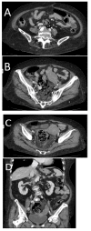
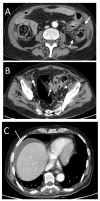
Background: Gastrointestinal perforations are a frequent cause of acute abdominal symptomatology for patients in the emergency department. The aim of this study was to investigate the findings of multidetector-row computed tomography of gastrointestinal perforations and analyze the impact of any imaging signs on the presurgical identification of the perforation site. Methods: We retrospectively reviewed emergency MDCT findings of 93 patients submitted to surgery for gastrointestinal perforation at two different institutions. Two radiologists separately reviewed the emergency MDCT examinations performed on each patient, before and after knowing the surgical diagnosis of the perforation site. A list of findings was considered. Positive predictive values were estimated for each finding with respect to each perforation site, and correspondence analysis (CA) was used to investigate the relationship between the findings and each of the perforation types. Results: We did not find inframesocolic free air in sigmoid colorectal perforations, and in rare cases, only supramesocolic free fluid in gastroduodenal perforations was found. A high PPV of perivisceral fat stranding due to colonic perforation and general distension of upstream loops and collapse of downstream loops were evident in most patients. Conclusions: Our data could offer additional information on the perforation site in the case of doubtful findings to support surgeons, especially in planning a laparoscopic approach.
Keywords: MDCT; acute abdomen; emergency; intestinal perforations; pneumoperitoneum; surgery.
Conflict of interest statement
The authors declare no conflict of interest.
Figures






Similar articles
-
Comparison between the site of multislice CT signs of gastrointestinal perforation and the site of perforation detected at surgery in forty perforated patients.Radiol Med. 2004 Sep;108(3):208-17. Radiol Med. 2004. PMID: 15343135 English, Italian.
-
[Use of multidetector computed tomography for locating the site of gastrointestinal tract perforations].Cir Esp. 2013 May;91(5):316-23. doi: 10.1016/j.ciresp.2012.06.004. Epub 2012 Oct 1. Cir Esp. 2013. PMID: 23036254 Spanish.
-
Accuracy of MDCT in predicting site of gastrointestinal tract perforation.AJR Am J Roentgenol. 2006 Nov;187(5):1179-83. doi: 10.2214/AJR.05.1179. AJR Am J Roentgenol. 2006. PMID: 17056902
-
Gastrointestinal tract perforation: MDCT findings according to the perforation sites.Korean J Radiol. 2009 Jan-Feb;10(1):63-70. doi: 10.3348/kjr.2009.10.1.63. Korean J Radiol. 2009. PMID: 19182505 Free PMC article. Review.
-
Role of Multidetector Computed Tomography in the Diagnosis of Colorectal Perforations.Semin Ultrasound CT MR. 2016 Feb;37(1):49-53. doi: 10.1053/j.sult.2015.10.007. Epub 2015 Oct 17. Semin Ultrasound CT MR. 2016. PMID: 26827738 Review.
Cited by
-
Endoscopic management of duodenal perforation caused by foreign bodies in adults: A retrospective study.United European Gastroenterol J. 2024 Apr;12(3):390-398. doi: 10.1002/ueg2.12505. Epub 2023 Dec 30. United European Gastroenterol J. 2024. PMID: 38159037 Free PMC article.
-
Gastroduodenal Perforation in Cancer Patients: Association with Chemotherapy and Prognosis.Med Sci (Basel). 2023 Mar 28;11(2):26. doi: 10.3390/medsci11020026. Med Sci (Basel). 2023. PMID: 37092495 Free PMC article.
-
Preoperative CT diagnosis of perforated Meckel's diverticulitis in a young patient: a case report.J Basic Clin Physiol Pharmacol. 2023 Nov 29;35(1-2):93-97. doi: 10.1515/jbcpp-2023-0237. eCollection 2024 Jan 1. J Basic Clin Physiol Pharmacol. 2023. PMID: 38015811
-
Applications of Hydrogels in Emergency Therapy.Gels. 2025 Mar 23;11(4):234. doi: 10.3390/gels11040234. Gels. 2025. PMID: 40277670 Free PMC article. Review.
-
Emergency Laparoscopic Removal of an Ingested Fishbone Perforating the Stomach After Endoscopy: A Case Report.Cureus. 2025 Mar 23;17(3):e81060. doi: 10.7759/cureus.81060. eCollection 2025 Mar. Cureus. 2025. PMID: 40271311 Free PMC article.
References
-
- Taourel P., Baert A.L., Reiser M.F., Hricak H., Knauth M. Medical Radiology Diagnostic Imaging, CT of the Acute Abdomen, Bowel Perforations. Springer; Berlin/Heidelberg, Germany: 2011. pp. 309–327.
-
- Simonetti I., Puglia M., Tarotto L., Palumbo F., Esposito F., Sciuto A., Ragozzino A. When traditions become dangerous: Intestinal perforation from unusual foreign body—Case report and short literature review. Eur. J. Radiol. Open. 2019;6:152–155. doi: 10.1016/j.ejro.2019.04.002. - DOI - PMC - PubMed
MeSH terms
LinkOut - more resources
Full Text Sources

