. 2022 Mar 15;8(2):798-814.
doi: 10.3390/tomography8020066.

The "Black Pattern", a Simplified Ultrasound Approach to Non-Traumatic Abdominal Emergencies

Affiliations

The "Black Pattern", a Simplified Ultrasound Approach to Non-Traumatic Abdominal Emergencies

Stefania Tamburrini et al. Tomography. .

Abstract

Background: A key issue in abdominal US is the assessment of fluid, which is usually anechoic, thus appearing "black". Our approach focuses on searching for fluid in non-traumatic patients, providing a new, simplified method for point-of-care US (POCUS).

Objective: Fluid assessment is based on a three-step analysis that we can thus summarize. 1. Look for black where it should not be. This means searching for effusions or collections. 2. Check if black is too much. This means evaluating anatomical landmarks where fluid should normally be present but may be abnormally abundant. 3. Look for black that is not clearly black. This means evaluating fluid aspects, whether wholly anechoic or not (suggesting heterogeneous or corpusculated fluid).

Discussion: Using this simple method focused on US fluid presence and appearance should help clinicians to make a timely diagnosis. Although our simplified, systematic algorithm of POCUS may identify abnormalities; this usually entails a second-level imaging. An accurate knowledge of the physio-pathological and anatomical ultrasound bases remains essential in applying this algorithm.

Conclusion: The black pattern approach in non -traumatic emergencies may be applied to a broad spectrum of abnormalities. It may represent a valuable aid for emergency physicians, especially if inexperienced, involved in a variety of non-traumatic scenarios. It may also be a simple and effective teaching aid for US beginners.

Keywords: abdomen; abdominal ultrasound; emergency; emergency ultrasound; ultrasound.


Conflict of interest statement

The authors declare no conflict of interest.

Figures
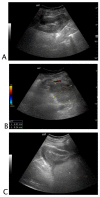
Figure 1
Figure 1
Black where it should not be. A 26-year-old female patient with abdominal tenderness and fainting. At ultrasound, an inhomogeneous subcapsular fluid collection (white arrow) was detected all around the right kidney (A). On the lower pole of the kidney, fluid collection was detected (B). On the upper pole of the right kidney, a hyperechoic mass (*) was seen, referable to angiomyolipoma (C). A suspected ultrasound diagnosis of hemo-retroperitoneum for spontaneous angiomyolipoma bleeding was formulated. The patient underwent CT angiography that confirmed active bleeding of the right kidney’s upper pole angiomyolipoma. The patient underwent endovascular arterial embolization. Final diagnosis: spontaneous bleeding of renal angiomyolipoma.
Figure 2
Figure 2
Black where it should not be. A 72-year-old male patient with abdominal pain and malaise after right nephrectomy. The right renal lodge was occupied by loculated fluid collection with fluid/fluid levels and clot (A). Perihepatic free fluid with inhomogeneous clot was detected (B). A suspected ultrasound diagnosis of post-surgical bleeding was formulated. The patient underwent CT angiography that confirmed the bleeding, and arterial endovascular embolization. Final diagnosis: post-nephrectomy bleeding.
Figure 3
Figure 3
Black where it should not be. A 62-year-old male patient with left lower quadrant abdominal pain. Sigmoid colon appeared stratified with muscular prevalence (white arrow) and perivisceral fat was markedly hypoechogenic (*). Adjacent to the bowel wall, a loculated inhomogeneous fluid collection was detected. A suspected diagnosis of complicated diverticulitis was formulated. Final diagnosis: Hinchey stage II diverticulitis (pelvic abscess > 4 cm).
Figure 4
Figure 4
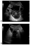
Black where it should not be. A 78-year-old male patient with abdominal pain who underwent robotic prostatectomy 20 days before. (A) Free fluid characterized by thick septa and inhomogeneous echogenicity was seen in all abdominal quadrants. (B) The bladder was empty and catheterized. Small hyperechogenic air bubbles were detected on the outer border of the bladder. A suspected diagnosis of post-surgical bladder perforation was formulated. The patient underwent CT with retrograde cystography without intravenous contrast for acute renal impairment, which confirmed the ultrasound diagnosis of bladder perforation. The patient underwent surgery. Final diagnosis: post-surgical bladder perforation.
Figure 5
Figure 5
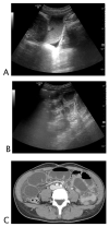
Black is too much, and it is where it should not be. A 68-year-old woman with abdominal distension, pain, and vomit. At ultrasound, (A) hypokinetic dilated small bowel loops were visualized on the right flank with thin parietal walls and poor evidence of valvulae, and free fluid was detected between bowel loops. On the left flank, (B) fluid between undilated bowel loops with regular representation of valvulae were detected. A suspected diagnosis of decompensated mechanical small bowel obstruction with fulcrum on the right flank was formulated. The patient underwent abdomino–pelvic CT; (C) pelvis that confirmed mechanical obstruction determined by adhesions on the right flank. Final diagnosis: decompensated small bowel obstruction.
Figure 6
Figure 6
Black where it should not be. A 75-year-old man with recent history of low back pain treated with intramuscular anti-inflammatory therapy. He presented at the emergency department with pain in the left thigh on administration site. The ultrasound showed a corpuscle collection with the typical “swirl sign” during probe compression due to the movement of pus within the fluid collection. Final diagnosis: pyogenic liver abscess.
Figure 7
Figure 7
Black where it should not be. A 60-year-old male patient who underwent laparotomy gastrectomy with fever and abdominal pain. At ultrasound, a pluriloculated fluid collection was detected in the abdominal wall with a fistulous connection in the peritoneal cavity. No free fluid in the abdomen was detected. A suspected diagnosis of infected collection along the laparotomy suture was formulated. Final diagnosis: abscess along the laparotomy suture with peritoneal fistulous connection.
Figure 8
Figure 8
Black where it should not be. A 35-year-old man with a significant history of intravenous drug abuse presented with fever and local pain in the right forearm. The arm was warm and with tight skin. The ultrasound showed a necrotizing fasciitis with subcutaneous thickening, air, and fascial fluid. He underwent decompressive fasciotomy in association with large spectrum antibiotic therapy.
Figure 9
Figure 9
Black that is not clearly black. A 71-year-old female patient complaining of drug-resistant abdominal pain for the last three days. No history of malignancies or liver disease. At ultrasound, the portal vein was dilated and occupied by inhomogeneous echogenic material (A). The color Doppler evaluation confirmed the absence of flow in the portal vein (B). Hypoechogenic areas were detected in the spleen (C). A suspected diagnosis of portal vein thrombosis with associated spleen infarction was formulated. Final diagnosis: portal vein thrombosis in unknown thrombophilic disorder.
Figure 10
Figure 10
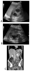
Black is too much, black is not clearly black, and black is where it should not be. A 47-year-old asthenic female patient with unintentional severe weight loss and left flank pain. At ultrasound, the left kidney was hardly recognizable; dilated upper calices with thick parietal walls and inhomogeneous urine (A) on the lower pole staghorn calculi were detected (B). Perinephric and subcapsular fluid was visualized. A suspected ultrasound diagnosis of complicated pyonephrosis was formulated and CT was performed in order to stage the pathology. (C) Final CT diagnosis: stage III xanthogranulomatous pyelonephritis with iliopsoas and lumbar muscle invasion.

Similar articles

Cited by

  • Left side jejunal diverticulitis: US and CT imaging findings.
    Comune R, Liguori C, Guida F, Cozzi D, Ferrari R, Giardina C, Iacobellis F, Galluzzo M, Tonerini M, Tamburrini S. Comune R, et al. Radiol Case Rep. 2024 Apr 20;19(7):2785-2790. doi: 10.1016/j.radcr.2024.04.003. eCollection 2024 Jul. Radiol Case Rep. 2024. PMID: 38680749 Free PMC article.
  • CT prognostic signs of postoperative complications in emergency surgery for acute obstructive colonic cancer.
    Pezzullo F, Comune R, D'Avino R, Mandato Y, Liguori C, Lassandro G, Tamburro F, Galluzzo M, Scaglione M, Tamburrini S. Pezzullo F, et al. Radiol Med. 2024 Apr;129(4):525-535. doi: 10.1007/s11547-024-01778-y. Epub 2024 Mar 21. Radiol Med. 2024. PMID: 38512630
  • CTA Imaging of Peripheral Arterial Injuries.
    Tamburrini S, Lassandro G, Tiralongo F, Iacobellis F, Ronza FM, Liguori C, Comune R, Pezzullo F, Galluzzo M, Masala S, Granata V, Basile A, Scaglione M. Tamburrini S, et al. Diagnostics (Basel). 2024 Jun 26;14(13):1356. doi: 10.3390/diagnostics14131356. Diagnostics (Basel). 2024. PMID: 39001246 Free PMC article. Review.
  • Multimodality Imaging Features of Papillary Renal Cell Carcinoma.
    Comune R, Tiralongo F, Bicci E, Saturnino PP, Ronza FM, Bortolotto C, Granata V, Masala S, Scaglione M, Sica G, Tamburro F, Tamburrini S. Comune R, et al. Diagnostics (Basel). 2025 Apr 1;15(7):906. doi: 10.3390/diagnostics15070906. Diagnostics (Basel). 2025. PMID: 40218256 Free PMC article.
  • Imaging in Non-Traumatic Emergencies.
    Scaglione M, Masala S, Iacobellis F, Tonerini M, Sica G, Liguori C, Saba L, Tamburrini S. Scaglione M, et al. Tomography. 2023 Jun 12;9(3):1133-1136. doi: 10.3390/tomography9030093. Tomography. 2023. PMID: 37368545 Free PMC article.

References

    1. Rudralingam V., Footitt C., Layton B. Ascites matters. Ultrasound. 2017;25:69–79. doi: 10.1177/1742271X16680653. - DOI - PMC - PubMed
    1. Richards J.R., McGahan J.P. Focused Assessment with Sonography in Trauma (FAST) in 2017: What Radiologists Can Learn. Radiology. 2017;283:30–48. doi: 10.1148/radiol.2017160107. - DOI - PubMed
    1. Rosano N., Gallo L., Mercogliano G., Quassone P., Picascia O., Catalano M., Pesce A., Fiorini V., Pelella I., Vespere G., et al. Ultrasound of Small Bowel Obstruction: A Pictorial Review. Diagnostics. 2021;11:617. doi: 10.3390/diagnostics11040617. - DOI - PMC - PubMed
    1. Edell S.L., Gefter W.B. Ultrasonic differentiation of types of ascitic fluid. AJR Am. J. Roentgenol. 1979;133:111–114. doi: 10.2214/ajr.133.1.111. - DOI - PubMed
    1. Casillas V.J., Amendola M.A., Gascue A., Pinnar N., Levi J.U., Perez J.M. Imaging of nontraumatic hemorrhagic hepatic lesions. Radiographics. 2000;20:367–378. doi: 10.1148/radiographics.20.2.g00mc10367. - DOI - PubMed

LinkOut - more resources