. 2022 Mar 21;13(1):1518.
doi: 10.1038/s41467-022-29193-9.

Siwi cooperates with Par-1 kinase to resolve the autoinhibitory effect of Papi for Siwi-piRISC biogenesis

Affiliations

Siwi cooperates with Par-1 kinase to resolve the autoinhibitory effect of Papi for Siwi-piRISC biogenesis

Hiromi Yamada et al. Nat Commun. .

Abstract

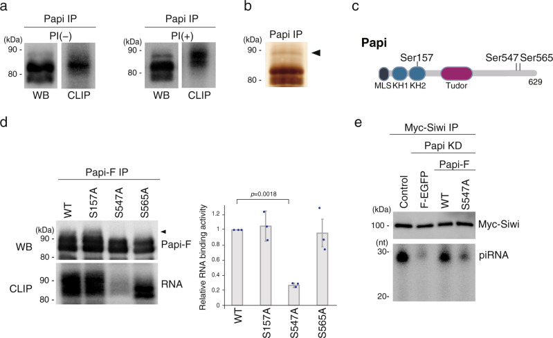
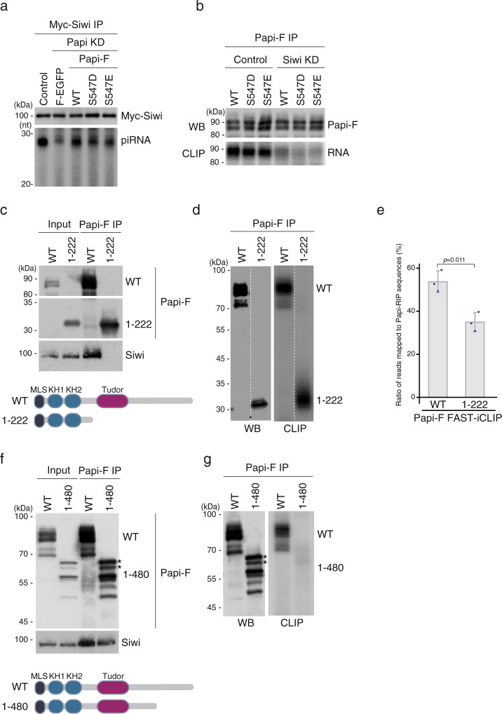
Bombyx Papi acts as a scaffold for Siwi-piRISC biogenesis on the mitochondrial surface. Papi binds first to Siwi via the Tudor domain and subsequently to piRNA precursors loaded onto Siwi via the K-homology (KH) domains. This second action depends on phosphorylation of Papi. However, the underlying mechanism remains unknown. Here, we show that Siwi targets Par-1 kinase to Papi to phosphorylate Ser547 in the auxiliary domain. This modification enhances the ability of Papi to bind Siwi-bound piRNA precursors via the KH domains. The Papi S547A mutant bound to Siwi, but evaded phosphorylation by Par-1, abrogating Siwi-piRISC biogenesis. A Papi mutant that lacked the Tudor and auxiliary domains escaped coordinated regulation by Siwi and Par-1 and bound RNAs autonomously. Another Papi mutant that lacked the auxiliary domain bound Siwi but did not bind piRNA precursors. A sophisticated mechanism by which Siwi cooperates with Par-1 kinase to promote Siwi-piRISC biogenesis was uncovered.


Conflict of interest statement

The authors declare no competing interests.

Figures

Fig. 1
Fig. 1. Phosphorylation of Ser547 of Papi is necessary for Siwi-piRISC biogenesis.
a Western blotting (WB) panels show band patterns of Papi in CLIP in the presence (+) and absence (−) of phosphatase inhibitor (PI). CLIP panels show the Papi-RNA complex labeled with 32P (n = 3). b Silver staining shows Papi immunoisolated from BmN4 cell lysates. The band that was used in the mass spectrometric analysis is indicated with a triangle (n = 3). c Schematic drawing of Papi shows where Ser157, Ser547, and Ser565 are located. MLS mitochondrial localization signal, KH1 and KH2 K-homology domains. d CLIP panel shows that the RNA-binding activity of the S547A mutant was negligible, whereas that of other mutants was comparable to that of WT Papi. The topmost band in WT Papi is indicated with a triangle. Western blotting (WB) shows the band patterns of WT Papi and its mutants in CLIP. The statistical data of the CLIP signals (n = 3) is shown (right). The signal intensities were calculated using ImageJ (National Institutes of Health) Each dot represents the intensity calculated from three independent experiments. P values were calculated by t-test (two-sided). Data are presented as mean values ± SD. e WT Papi but not the S547A mutant restored Siwi-piRISC biogenesis in cells that lacked Papi [Papi knockdown (KD)] (n = 3). The Papi-Flag (Papi-F) used in this experiment was RNAi-resistant. Control, Myc-Siwi was expressed in normal BmN4 cells; Flag-EGFP (F-EGFP) was used as a negative control. piRNAs were visualized by 32P-labeling. The amounts of Myc-Siwi in Input and the knockdown efficiency for Papi are shown in Supplementary Fig. 1e. Source data are provided as a Source Data file.
Fig. 2
Fig. 2. Par-1 kinase is responsible for Papi Ser547 phosphorylation.
a Western blotting shows that WT Papi is concentrated in the mitochondrial fraction (Mito), whereas the Papi ΔMLS mutant, which lacks the mitochondrial localization signal, is in the cytoplasmic fraction (Cyto) (n = 3). Total, total lysates of BmN4 cells. β-Tubulin was used as a marker of the cytoplasmic fraction. The schematic drawing shows the domain structure of WT Papi and the Papi ΔMLS mutant. b Western blotting shows that WT Papi is efficiently phosphorylated in vivo, whereas the Papi ΔMLS mutant is not (n = 3). de-phospho, the immunoprecipitated materials were treated with (+) and without (−) phosphatase prior to western blotting. c Papi-Flag (Papi-F) (upper) was 32P-labeled efficiently upon incubation with the mitochondrial fraction (Mito) of BmN4 cells in the presence of 32P-γ-ATP. Silver-stained Papi-F (lower) used in the assay is shown (n = 3). d Silver staining shows proteins that co-immunoprecipitated with Papi-F after incubation with the mitochondrial fraction of BmN4 cells. IP, proteins co-immunoprecipitated with Papi-F; Elution, proteins bound to Papi-F were eluted with high-salt buffer; h.c., heavy chain of the antibody; l.c., light chain of the antibody (n = 3). e Six kinases identified in the mass spectrometric analysis. PSM, peptide-spectrum match; MW, predicted molecular weight. f The six kinases in (e) were depleted by RNAi (KD) in BmN4 cells, and the levels of Papi-F within the cells were examined by western blotting (n = 3). The RNAi efficiency was examined by RT-qPCR (Supplementary Fig. 2c). β-Tubulin was used as a loading control. g Papi-pSer547 was detected before Par-1 depletion (Control) but not after Par-1 depletion (Par-1 KD) (n = 3). Anti-Papi-pSer547 antibody (Supplementary Fig. 2d, e) was used. h Siwi-piRISC was barely detected in Par-1-depleted cells (Par-1 KD) (n = 3). F-Siwi, Flag-Siwi. piRNAs were visualized by 32P-labeling. Source data are provided as a Source Data file.
Fig. 3
Fig. 3. Siwi targets cytoplasmic Par-1 kinase to mitochondrial Papi for its phosphorylation.
a Western blotting shows that Ser547 of WT Papi is phosphorylated, whereas Ser547 of the Papi ΔMLS mutant is not (n = 3). Anti-Papi (upper) and anti-Papi-pSer547 (lower) antibodies were used. Immunoprecipitation (IP) was performed using anti-Flag antibody. b Western blotting shows that Flag-Par-1 (F-Par-1) is concentrated in cytoplasmic (Cyto) but not in mitochondrial (Mito) fraction (n = 3). β-Tubulin and HSP60 were used as markers of the cytoplasmic fraction and the mitochondrial fraction, respectively. c Flag-Par-1 (F-Par-1) and Siwi co-immunoprecipitated with Papi from normal BmN4 cell lysates (Control) but not from Siwi-lacking lysates (Siwi KD) (n = 3). The cell lysates contained both cytoplasmic and mitochondrial materials. d Papi immunoisolated from normal BmN4 cell lysates (Control) was phosphorylated at Ser547, whereas Papi immunoisolated from Siwi-lacking lysates (Siwi KD) was not (n = 3). Anti-Papi (upper) and anti-Papi-pSer547 (lower) antibodies were used. e Flag-Par-1 (F-Par-1) interacted with Siwi in BmN4 cytoplasmic lysates (n = 3). The lysates did not contain mitochondrial materials. Flag-LacZ (F-LacZ) was used as a negative control. f Immunoprecipitation and western blotting show that the Siwi 9RK mutant binds to Par-1 similarly to WT Siwi (n = 3). g Papi interacting with WT Siwi and the Siwi K611A mutant on the mitochondrial surface was similarly phosphorylated at Ser547 (n = 3). Immunoprecipitation was conducted from the mitochondrial fraction. h Proposed model showing that Siwi binds Par-1 kinase in the cytosol and targets it to mitochondrial Papi. Papi binds symmetrical dimethylarginine-modified Siwi via the Tudor domain. Par-1 then phosphorylates Ser547 of Papi. Papi then binds Siwi-loaded piRNA precursor via the KH domains for Zuc-mediated piRISC maturation (red triangle). The Siwi 9RK mutant cannot bind to Papi and hardly bound to piRNA precursor. Thus, Siwi was loaded with piRNA precursor after its anchoring onto Papi. i Western blotting (upper two) shows that endogenous Siwi bound the KH mutant of Papi as it does to WT Papi (n = 3). Northern blotting (lowest) shows that piRNA precursors and piRNAs were barely found in the Papi KH mutant-Siwi complex (also see Supplementary Fig. 3). Source data are provided as a Source Data file.
Fig. 4
Fig. 4. Auxiliary domain of Papi influences the RNA-binding activity of Papi via the KH domains.
a Papi S547D and S547E variants restored the Siwi-piRISC production in Papi-depleted cells (Papi KD) (n = 3). All Papi-Flag (Papi-F) employed in this experiment was RNAi-resistant. Control, Myc-Siwi was expressed in normal BmN4 cells; Flag-EGFP (F-EGFP) was used as a negative control. piRNAs were visualized by 32P-labeling. The amounts of Myc-Siwi in Input and the knockdown efficiency for Papi are shown in Supplementary Fig. 4a. b CLIP shows that the RNA-binding activity of the S547D and S547E variants was reduced significantly following Siwi depletion (Siwi KD), which is similar to that of the WT (n = 3). Western blotting (WB) shows the band patterns of WT Papi and its mutants in CLIP. c Immunoprecipitation and western blotting show that, unlike WT Papi, the Papi1-222 mutant did not bind Siwi (n = 3). The schematic drawing shows the domain structure of WT Papi and Papi1-222. d CLIP shows that the Papi1-222 mutant binds RNAs (n = 3). Western blotting (WB) shows the band patterns of WT Papi and the mutant in CLIP. e Bar graph shows the mean ratio of reads of Papi-Flag FAST-iCLIP tags that mapped to Papi-RIP sequences (n = 3). Each dot represents the ratio calculated from three independent experiments. P values were calculated by t-test (two-sided). Data are presented as mean values ± SD. f Immunoprecipitation and western blotting show that the Papi1-480 mutant binds Siwi as well as WT Papi (n = 3). Asterisks indicate phosphorylated Papi1-480 (see Supplementary Fig. 4c, d). Schematic drawing shows the domain structure of WT Papi and Papi1-480. g CLIP shows that the Papi1-480 mutant did not bind RNAs (n = 3). Western blotting (WB) shows the band patterns of WT Papi and the Papi1-480 mutant in CLIP. Asterisks, phosphorylated Papi1-480. Source data are provided as a Source Data file.

Similar articles

Cited by

References

    1. Iwasaki YW, Siomi MC, Siomi H. PIWI-interacting RNA: its biogenesis and functions. Annu. Rev. Biochem. 2015;84:405–433. doi: 10.1146/annurev-biochem-060614-034258. - DOI - PubMed
    1. Czech B, Hannon GJ. One loop to rule them all: the ping-pong cycle and piRNA-guided silencing. Trends Biochem. Sci. 2016;41:324–337. doi: 10.1016/j.tibs.2015.12.008. - DOI - PMC - PubMed
    1. Ozata DM, Gainetdinov I, Zoch A, O’Carroll D, Zamore PD. PIWI-interacting RNAs: small RNAs with big functions. Nat. Rev. Genet. 2019;20:89–108. doi: 10.1038/s41576-018-0073-3. - DOI - PubMed
    1. Klattenhoff C, Theurkauf W. Biogenesis and germline functions of piRNAs. Development. 2008;135:3–9. doi: 10.1242/dev.006486. - DOI - PubMed
    1. Kawaoka S, Minami K, Katsuma S, Mita K, Shimada T. Developmentally synchronized expression of two Bombyx mori Piwi subfamily genes, SIWI and BmAGO3 in germ-line cells. Biochem. Biophys. Res. Commun. 2008;367:755–760. doi: 10.1016/j.bbrc.2008.01.013. - DOI - PubMed

Publication types

Substances