. 2022 Mar 21;12(1):4819.
doi: 10.1038/s41598-022-08791-z.

Thioredoxin interacting protein protects mice from fasting induced liver steatosis by activating ER stress and its downstream signaling pathways

Affiliations

Thioredoxin interacting protein protects mice from fasting induced liver steatosis by activating ER stress and its downstream signaling pathways

Hiroyuki Miyahara et al. Sci Rep. .

Erratum in

Abstract

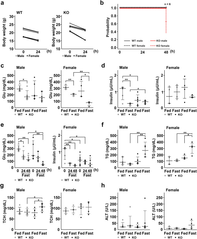
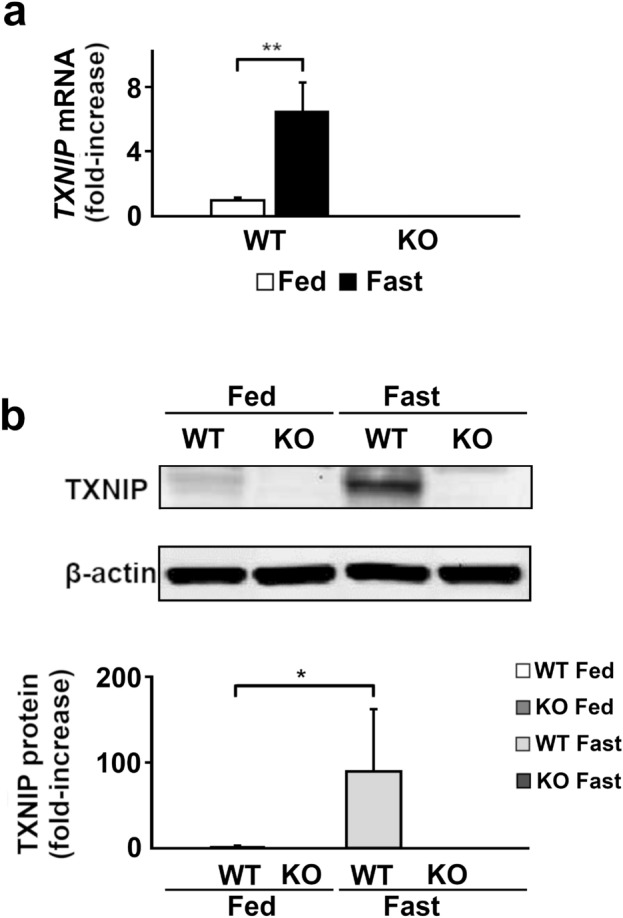
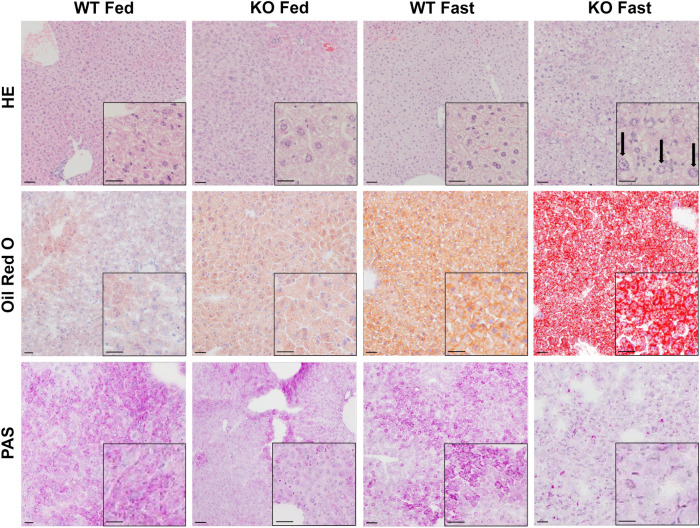
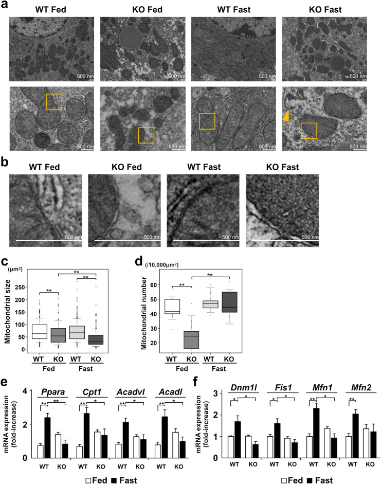
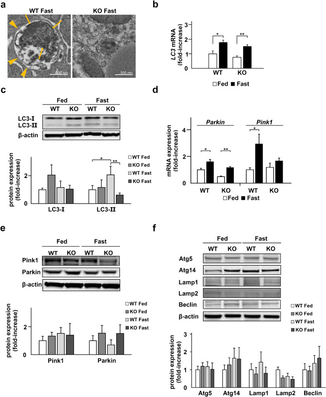
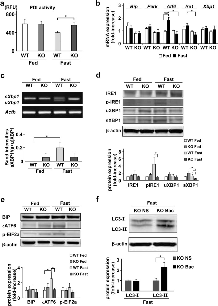
Under normal conditions, fasting results in decreased protein disulfide isomerase (PDI) activity and accumulation of unfolded proteins, leading to the subsequent activation of the unfolded protein response (UPR)/autophagy signaling pathway to eliminate damaged mitochondria. Fasting also induces upregulation of thioredoxin-interacting protein (TXNIP) expression and mice deficient of this protein (TXNIP-KO mice) was shown to develop severe hypoglycemia, hyperlipidemia and liver steatosis (LS). In the present study, we aimed to determine the role of TXNIP in fasting-induced LS by using male TXNIP-KO mice that developed LS without severe hypoglycemia. In TXNIP-KO mice, fasting induced severe microvesicular LS. Examinations by transmission electron microscopy revealed mitochondria with smaller size and deformities and the presence of few autophagosomes. The expression of β-oxidation-associated genes remained at the same level and the level of LC3-II was low. PDI activity level stayed at the original level and the levels of p-IRE1 and X-box binding protein 1 spliced form (sXBP1) were lower. Interestingly, treatment of TXNIP-KO mice with bacitracin, a PDI inhibitor, restored the level of LC3-II after fasting. These results suggest that TXNIP regulates PDI activity and subsequent activation of the UPR/autophagy pathway and plays a protective role in fasting-induced LS.


Conflict of interest statement

The authors declare no competing interests.

Figures

Figure 1
Figure 1
Hyperlipidemia occurs during fasting without changes in Glu levels in TXNIP-KO male mice. (a) Change of body weight was examined and shown by dot plots, revealing that WT and male TXNIP-KO mice lost weight after 24-h fasting and weight loss was more prominent in TXNIP-KO female mice (three mice per group). (b) TXNIP-KO female mice were vulnerable to fasting stress (six mice per group). (ch) Laboratory data were collected from WT and TXNIP-KO mice in fed or fasted state and shown by dot plots; medians and 10–90 percentiles were also shown for each dot plot graph (six mice for male group and four mice for female group). (c,d) Despite Glu levels were statistically lower in female TXNIP-KO mice compared with female WT mice in fasted state, no statistical difference in Glu levels was found between WT and male TXNIP-KO mice in the fasted state. Serum insulin levels also showed no obvious difference between WT and TXNIP-KO mice (both in male and female mice), but serum insulin levels may be high in TXNIP-KO female mice when taking low blood Glu levels into account. (e) Serum Glu and insulin levels of TXNIP-KO male mice in fed state, or after 24-h or 48-h fasting. Although serum Glu level decreased to remarkably lower levels, serum insulin levels were relatively stable in TXNIP-KO mice compared with WT mice, indicating an impairment in the regulation of serum insulin level in response to serum glucose level in TXNIP-KO mice. (f,g) Although serum TG and TCH levels tended to be stable or decrease in WT mice, these values tended to increase in TXNIP-KO mice during fasting, and statistical differences were found in TG and TCH levels between WT and male TXNIP-KO mice and in TG between WT and female TXNIP-KO mice. (h) Serum ALT levels were measured. Although one mouse in WT and TXNIP-KO male mice showed highly increased serum ALT levels, serum ALT levels overall were stable in WT and TXNIP-KO male mice. In female mice, TXNIP-KO female mice tended to show increased serum ALT during fasted state. Reference values (mean ± SD) for male Glu, female Glu, male TG, female TG, male TCH, and female TCH were 306 ± 47 (mg/dL), 302 ± 33 (mg/dL), 104 ± 31 (mg/dL), 104 ± 33 (mg/dL), 91 ± 11 (mg/dL), 79 ± 19 (mg/dL) respectively. *p < 0.05, **p < 0.01. WT wild-type, KO knockout, TXNIP thioredoxin-interacting protein, Glu glucose, TG triglyceride, FFA free fatty acid, TCH total cholesterol, ALT alanine transaminase, SD standard deviation.
Figure 2
Figure 2
The expression of TXNIP is upregulated during fasting. The expression of TXNIP mRNA and protein in fed or fasted states was evaluated by RT-qPCR or western blotting. TXNIP mRNA (a) and protein (b) expression increased significantly in the fasted state in WT mice. TXNIP mRNA and protein expression was completely suppressed in TXNIP-KO mice in both states. *p < 0.05, **p < 0.01 (in each group, six mice were used for RT-qPCR, and ten were used for western blot analysis). TXNIP thioredoxin-interacting protein, WT wild-type, KO knockout mice.
Figure 3
Figure 3
LS occurs during fasting in TXNIP-KO mice. LS during the fasted state in TXNIP-KO mice was verified by H&E, Oil Red O and PAS staining. Prominent hepatic microvesicular steatosis with enlarged nuclei (arrows) was observed in livers of TXNIP-KO mice in the fasted state by H&E staining, and accumulation of lipid droplets was detected by Oil Red O staining (scale bar = 50 μm). PAS staining showed depletion of glycogen in livers of fasted TXNIP-KO mice: depletion of glycogen was found even in the fed state. Representative photographs from each group are shown (six mice per group). H&E hematoxylin–eosin, PAS periodic acid Schiff, TXNIP thioredoxin-interacting protein, WT wild-type mice, KO knockout mice.
Figure 4
Figure 4
Morphology and mitochondrial β-oxidation are altered in TXNIP-KO mice during fasting. The morphology and the expression of β-oxidation-related and mitochondrial fission–fusion related genes, especially in the fasted state, were evaluated by TEM and RT-qPCR, respectively. (a) Top row: Mitochondrial size and number were compared. The size of mitochondria was small and the number of mitochondria increased in TXNIP-KO mice during fasting, although there appeared no obvious difference in the number of mitochondria between the both type of mice. Bottom row: Mitochondrial structures were compared. Although no obvious difference was found in the structure of cristae between WT and TXNIP-KO mice in the fed state, mitochondria were swollen and lacked cristae, and anomalously broken mitochondria (arrow head) were also observed in TXNIP-KO mice in the fasted state (scale bar = 500 nm). (b) The areas indicated by the boxes in figure (a) are enlarged. Compared with WT mice, the irregularity in the outer membrane was found in TXNIP-KO mice during fed state and this membrane irregularities were more evident during the fasted state. (c,d) Ten foci from each group were examined, and the mitochondrial size and number were quantitated. Mitochondrial size was statistically smaller in the liver of TXNIP-KO mice during the fed state, and the difference became more prominent during the fasted state, despite the fact that the number of mitochondria in the fasted state was not different between WT and TXNIP-KO mice. (e) Differences in the expression of β-oxidation-related genes or proteins between WT and TXNIP-KO mice. Overall, β-oxidation-related gene expression was upregulated in WT mice during the fasted state, but this change was not observed in TXNIP-KO mice. Expression of β-oxidation-related genes (Ppara, Cpta, Acadvl, and Acadl) was significantly higher in WT mice than in TXNIP-KO mice during the fasted state, indicating impaired β-oxidation in TXNIP-KO mice during the fasted state. (f) Gene expression of molecules related to mitochondrial fission and fusion (Dnm1l, Fis1, Mfn1, Mfn2) was examined. The expression of these molecules was upregulated in fasted state in WT mice, but this change was not found in TXNIP-KO mice. *p < 0.05. **p < 0.01 (in each group, six mice were used for RT-qPCR). TEM transmission electron microscopy, TXNIP thioredoxin-interacting protein, WT wild-type mice, KO knockout mice, PPARα peroxisome proliferator-activated receptor-α, CPT1 carnitine palmitoyltransferase 1, ACADVL acyl-CoA dehydrogenase very long chain, ACADL acyl-CoA dehydrogenase long chain.
Figure 5
Figure 5
Autophagy is not activated in TXNIP-KO mice during fasting. Deficiency in autophagy was observed in the liver of TXNIP-KO mice. (a) The morphological differences of mitochondria between WT and TXNIP-KO mice were examined by TEM. In the liver of WT mice, autophagy increases during the fasted state, and in the present study, increased number of autophagosomes (arrow heads) containing mitochondria (arrows) were observed. By contrast, autophagosomes were rarely found in the liver of TXNIP-KO mice by TEM analysis. (b,c) The levels of LC3 mRNA and protein were evaluated by RT-qPCR and western blotting, respectively. The expression LC3 mRNA was upregulated during fasting in both WT and TXNIP-KO mice but there was no obvious difference between WT and KO mice. In WT mice the expression of LC3-II, a key molecule in autophagy, was upregulated during the fasted state, but this change was not observed in TXNIP-KO mice. The expression of LC3-II was statistically lower in TXNIP-KO mice during the fasted state. (c,e) The levels of Pink1 and Parkin, key molecules in mitophagy, were examined by RT-qPCR and western blotting, revealing that the expression patterns during the fed and fasted states were similar between WT and KO mice. (f) Other proteins playing essential roles in autophagy, Atg5, Atg14, Lamp1, Lamp2, and Beclin, were examined, and no statistical significance was found between WT and TXNIP-KO mice in the fed and fasted state in both strains. *p < 0.05, **p < 0.01 (five mice per strain were used for RT-qPCR, and ten were used for western blotting). TXNIP thioredoxin-interacting protein, WT wild-type mice, KO knockout mice, WT wild-type mice, KO knockout mice, TEM transmission electron microscopy, LC3 microtubule-associated protein light chain 3, Pink1 PTEN induced kinase 1, Atg5 autophagy related 5, Atg14 autophagy related 14, Lamp1 lysosomal-associated membrane protein 1, Lamp2 lysosomal-associated membrane protein 2.
Figure 6
Figure 6
The ER stress-induced UPR and autophagy signaling during fasting are impaired in the liver of TXNIP-KO mice. (a) PDI activity was measured by the ProteoStat(R) PDI assay kit. PDI activity decreased during the fasted state in WT mice, but not in TXNIP-KO mice. (b) Differences in the expression of ER stress signaling-related genes between WT and TXNIP-KO mice were examined by RT-qPCR. In WT mice, expression of these genes increased in the fasted state, but this change was not observed in TXNIP-KO mice, and the expression of IRE1 mRNA, which is important in the induction of autophagy, was significantly lower in TXNIP-KO mice in the fasted state. (c) The expression of sXBP and uXBP1 mRNA was evaluated by PCR after RNA extraction and cDNA synthesis. Although upregulation of sXBP1 mRNA was found in WT mice during the fasted state, this upregulation was not found in TXNIP-KO mice. (d) Phosphorylation of IRE1 and the levels of uXBP1 and sXBP1 were examined by western blotting. Decreased phosphorylation of IRE1 and decreased level of sXBP1 were observed in TXNIP-KO mice, which implies decreased ER stress signaling for the induction of autophagy during the fasted state in TXNIP-KO mice. (e) Other proteins important for ER stress signaling, BiP, cATF6, and p-EIF2a, were also examined. Although the expression of these protein tended to be upregulated during the fasted state in WT mice, this tendency was not found in TXNIP-KO mice. Statistically significant differences were found in the expression of cATF6 between the fed and fasted states in WT mice, and WT and TXNIP-KO mice during fasted state. (f) Protein expression of LC3-II was restored in the fasted state when bacitracin was injected to the TXNIP-KO mice before 24-h fasting. *p < 0.05, **p < 0.01 (in each group, six mice were used for PDI analysis, RT-qPCR or PCR, and ten were used for western blotting). UPR unfolded protein response, ER endoplasmic reticulum, TXNIP thioredoxin-interacting protein, PDI protein disulfide isomerase, BiP binding immunoglobulin protein, PERK PKR-like endoplasmic reticulum kinase, ATF6 activating transcription factor 6, IRE1 inositol-requiring enzyme 1, p-IRE1 phosphorylated IRE1, sXBP1 X-box binding protein 1 sliced form, uXBP1 XBP1 unsliced form, cATF6 cleaved ATF6, p-EIF2a phosphorylated eukariotic initiation factor 2a, WT wild type mice, KO knockout mice, NS normal saline, Bac bacitracin.

Similar articles

Cited by

References

    1. Postic C, Dentin R, Girard J. Role of the liver in the control of carbohydrate and lipid homeostasis. Diabetes Metab. 2004;30:398–408. doi: 10.1016/s1262-3636(07)70133-7. - DOI - PubMed
    1. den Boer MA, et al. Ritonavir impairs lipoprotein lipase-mediated lipolysis and decreases uptake of fatty acids in adipose tissue. Arterioscler. Thromb. Vasc. Biol. 2006;26:124–129. doi: 10.1161/01.Atv.0000194073.87647.10. - DOI - PubMed
    1. Rodgers JT, Puigserver P. Fasting-dependent glucose and lipid metabolic response through hepatic sirtuin 1. Proc. Natl. Acad. Sci. U. S. A. 2007;104:12861–12866. doi: 10.1073/pnas.0702509104. - DOI - PMC - PubMed
    1. Roussel D, et al. Threshold effect in the H2O2 production of skeletal muscle mitochondria during fasting and refeeding. J. Exp. Biol. 2019 doi: 10.1242/jeb.196188. - DOI - PubMed
    1. Zhang H, et al. The role of mitochondria in liver ischemia-reperfusion injury: From aspects of mitochondrial oxidative stress, mitochondrial fission, mitochondrial membrane permeable transport pore formation, mitophagy, and mitochondria-related protective measures. Oxid. Med. Cell. Longev. 2021;2021:6670579. doi: 10.1155/2021/6670579. - DOI - PMC - PubMed

Publication types