. 2022 May;6(5):604-613.
doi: 10.1038/s41559-022-01699-x. Epub 2022 Mar 21.

Embryonic muscle splitting patterns reveal homologies of amniote forelimb muscles

Affiliations

Embryonic muscle splitting patterns reveal homologies of amniote forelimb muscles

Daniel Smith-Paredes et al. Nat Ecol Evol. 2022 May.

Abstract

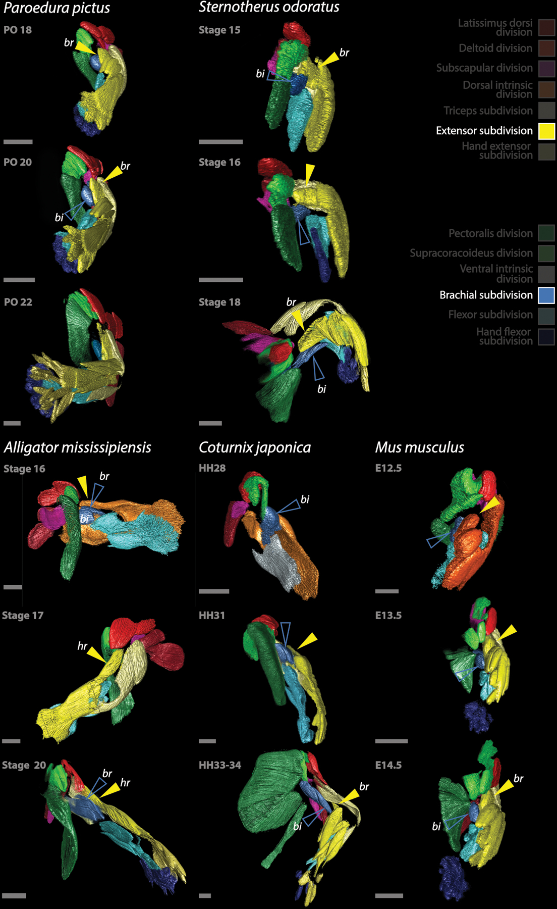
Limb muscles are remarkably complex and evolutionarily labile. Although their anatomy is of great interest for studies of the evolution of form and function, their homologies among major amniote clades have remained obscure. Studies of adult musculature are inconclusive owing to the highly derived morphology of modern amniote limbs but correspondences become increasingly evident earlier in ontogeny. We followed the embryonic development of forelimb musculature in representatives of six major amniote clades and found, contrary to current consensus, that these early splitting patterns are highly conserved across Amniota. Muscle mass cleavage patterns and topology are highly conserved in reptiles including birds, irrespective of their skeletal modifications: the avian flight apparatus results from slight early topological modifications that are exaggerated during ontogeny. Therian mammals, while conservative in their cleavage patterns, depart drastically from the ancestral amniote musculoskeletal organization in terms of topology. These topological changes occur through extension, translocation and displacement of muscle groups later in development. Overall, the simplicity underlying the apparent complexity of forelimb muscle development allows us to resolve conflicting hypotheses about homology and to trace the history of each individual forelimb muscle throughout the amniote radiations.


Conflict of interest statement

Conflict of interest

The authors declare no conflict of interest.

Figures

Extended Data Fig. 1
Extended Data Fig. 1. Homologies of the Dorsal musculature of the forelimb in Amniota
Homologies of the Dorsal musculature of the forelimb in Amniota. Muscles in individual rows are inferred to be homologous across clades based on their embryological origin and cleavage pattern from the divisions and subdivisions identified in the species studied. Rows containing more than one muscle per taxa reference cases in which the distinction of the muscles are not clear or individual portions of one muscle can be homologized to individual muscles of another group. Each muscle is named according to the nomenclature proposed by the authors listed under the clade names. Names proposed for reptilian musculature by Abdala and Diogo, 2010, are listed below if the nomenclature is not the same as in the other studies or they refer to a muscle complex.
Extended Data Fig. 2
Extended Data Fig. 2. Homologies of the Ventral musculature of the forelimb in Amniota
Homologies of the Ventral musculature of the forelimb in Amniota. Muscles in individual rows are inferred to be homologous across clades based on their embryological origin and cleavage pattern from the divisions and subdivisions identified in the species studied. Rows containing more than one muscle per taxa reference cases in which the distinction of the muscles are not clear or individual portions of one muscle can be homologized to individual muscles of another group. Each muscle is named according to the nomenclature proposed by the authors listed under the clade names. Names proposed for reptilian musculature by Abdala and Diogo, 2010, are listed below if the nomenclature is not the same as in the other studies or they refer to a muscle complex.
Extended Data Fig. 3
Extended Data Fig. 3. Development of the forelimb musculature of reptiles, with focus on the ventral extrinsic musculature
Development of the forelimb musculature of reptiles, with focus on the ventral extrinsic musculature derived from the Pectoralis (dark green) and Supracoracoideus (light green) divisions. All views ventral, except Paroedura PO 22 that is lateral and Coturnix HH33–34 that is a medial view. The Supracoracoideus division usually lays anterior to the Pectoralis division; in Coturnix in turn, it has shifted deep to it and extends into the humerus forming a pronounced curve. The proximal portion forms M. supracoracoideus (empty light green arrow) while the distal portion, beyond the curve, forms M. tensor propatagialis (light green arrow). PD: Pectoralis division, sc: M. supracoracoideus, SCD: Supracoracoid division, tpp: M. tensor propatagialis. All scale bars are 500 μm.
Extended Data Fig. 4
Extended Data Fig. 4. Development of the forelimb musculature of reptiles, with focus on the dorsal extrinsic musculature
Development of the forelimb musculature of reptiles, with focus on the dorsal extrinsic musculature derived from the Latissimus (dark red), Deltoid (red) and Subscapular (magenta) divisions. All images show lateral view, except for the second row of Coturnix showing medial views of the forelimb musculature. The empty red arrows and the red arrows point at the development of the scapular and clavicular lobes of the Deltoid division respectively. In Sternotherus, like in some other turtles, the scapular portion does not form. The empty dark-red arrow points at the anterior lobe formed from the Latissimus division. In Alligator, this gives rise to the Teres major muscle (tm). In Chrisemys, as described by Walker 1947, this also gives rise to M. teres major. In Sternotherus, this muscle does not seem to develop, though in stage 16 an incipient anterior lobe of the Latissimus division, comparable to that of Alligator can be observed (tm?). In Coturnix, the Latissimus division divides (although slightly later) into two lobes, giving rise to the anterior (tm?) and posterior heads of the latissimus muscle. This sort of lobation is not observed in Paroedura and no comparable muscle develops in lizards. The empty magenta arrow points at M. subscapularis (M. scapulohumeralis caudalis of birds). dc: M. deltoideus clavicularis, DD: Deltoid division, ds: M. deltoideus scapularis, ld: M. latissimus dorsii, ldp: M. latissimus dorsii posterior, LD: Latissimus division, shc: M. scapulohumeralis caudalis, sbcc: M. subcoracoideus, sbsc: M. subscapularis, SSD: Subscapular division, tm: M. teres major, *: M. scapulohumeralis anterior. All scale bars are 500 μm.
Extended Data Fig. 5
Extended Data Fig. 5. Development of the forelimb musculature of therian mammals with focus on the ventral extrinsic musculature
Development of the forelimb musculature of therian mammals, Mus and Monodelphis, with focus on the ventral extrinsic musculature derived from the Pectoral (dark green) and Supracoracoideus (light green) divisions. The Pectoralis division originates M. panniculus carnosus (white bordered dark green arrow), M. pectoralis minor (empty dark green arrow) and the deep portion of M. pectoralis major (dark green arrow). The ventral portion of the Supracoracoideus division forms the superficial portion of M. pectoralis major (empty light green arrow) and the clavicular deltoid (light green arrow). The dorsal extension of the Supracoracoideus division invades the dorsal aspect of the scapula (*) and bifurcates around the scapular spine (**) originating M. supraspinatus (yellow bordered light green arrow) and M. infraspinatus (red bordered light green arrow). The upper series of mouse embryos depicts a medial view of stage E12.0, a lateral view of stage E12.5 and ventral views of E13.5 and 14.5. Bottom series of Monodelphis and Mus show the developing embryos with all muscle groups except for those deriving from the Pectoral and Supracoracoideus divisions removed, and the developing skeleton stained with either Sox9 or Col II antibodies. M. panniculus carnosus was removed from Monodelphis MC 33 and 33+ and Mus E14.5. dc: M. deltoideus clavicularis, isp: M. infraspinatus, pc: M. panniculus carnosus, PD: Pectoralis division, pmi: M. pectoralis minor, pmp: M. pectoralis major profundus, pms: M. pectoralis major superficialis, ssp: M. supraspinatus. All scale bars are 500 μm.
Extended Data Fig. 6
Extended Data Fig. 6. Development of the forelimb musculature of therian mammals with focus on the dorsal extrinsic musculature
Development of the forelimb musculature of therian mammals, Mus and Monodelhpis, with focus on the dorsal extrinsic musculature derived from the Latissimus (dark red), Deltoid (bright red) and Subscapular (magenta) divisions. As in reptiles, the Deltoid division of mice cleaves early into two lobes, forming M. scapulodeltoid (red arrow) and M. teres minor (empty red arrow). In Monodelphis a Teres minor muscle was not observed. The posterior portion of the Subscapular division extends along the posterior margin and lateral surface of the scapula (*), forming M. teres major (empty magenta arrow). The upper series shows Mus embryos between E12.0 and 14.5, all in lateral view. The bottom series of Monodelphis and Mus show the development of the muscles and skeleton in lateral view. M. latissimus dorsii is not shown in mice E14.5. DD: Deltoid division, ds: M. deltoideus scapularis, ld: M. latissimus dorsii, LD: Latissimus division, ssc: M. subscapularis, SSD: Subscapular division, tm: M. teres major, tmi: M. teres minor. All scale bars are 500 μm.
Extended Data Fig. 7
Extended Data Fig. 7. Development of the forelimb musculature of amniotes, with focus on the dorsal intrinsic musculature
Development of the forelimb musculature of amniotes, with focus on the dorsal intrinsic musculature derived from the Triceps (light yellow) and Extensor (yellow) divisions. Note the distal extension of the Triceps division along the ulna, originating the posterior most extensor muscle of the forearm (empty light-yellow arrow), and the humeral lobe of the Extensor division, extending proximally along the humerus (yellow arrow) and forming M. brachialis of lizards, turtles, birds and mammals, homologous to M. humeroradialis of crocodylians. Mm. dorsoepitrochlearis (light yellow arrow) of mammals derives from the Triceps subdivision. c: central lobe (posterior-most lobe) of the Extensor division, dep: M. dorsoepitrochlearis, h: humeral lobe of the Extensor subdivision, r: radial lobe of the Extensor subdivision, u: “ulnar” lobe of the forearm musculature, derived from the Triceps subdivision. All scale bars are 500 μm.
Extended Data Fig. 8
Extended Data Fig. 8. Development of the forelimb musculature of amniotes, with focus on the brachial muscles that act as flexors of the forearm
Development of the forelimb musculature of amniotes, with focus on the brachial muscles that act as flexors of the forearm. From the Extensor division, on the dorsal forearm, a muscle extends proximally over the humerus (yellow arrow) forming M. brachialis of Paroedura, Sternotherus, Coturnix and Mus, homologous to M. humeroradialis of Alligator. On the other hand, the biceps muscle and the crocodylians M. brachialis derive from the Brachial subdivision (empty blue arrow). bi: M. biceps brachii, br: M. brachialis (note that the muscle termed “brachialis” derive from different origins than in the other species in Alligator), hr: M. humeroradialis (note that the humeroradialis muscle derives from the same portion of the Brachial subdivision than the brachialis muscles of the other species). All scale bars are 500 μm.
Extended Data Fig. 9
Extended Data Fig. 9. Development of the forelimb musculature of amniotes, with focus on the intrinsic musculature deriving from the Flexor division
Development of the forelimb musculature of amniotes, with focus on the intrinsic musculature deriving from the Flexor division (light blue). The Flexor division forms a deep lobe, and superficially a central lobe (empty light blue arrow), flanked by an ulnar lobe on the ulnar side of the forearm (bottom portion of each arm in the images) and a radial lobe on the radial side (top). Note that the central lobe of the Flexor subdivision (pointed at by the empty light-blue arrow and originating M. flexor digitorum longus/communis) is the source of the so-called M. flexor carpi radialis of mice, otherwise originated from the radial lobe in other species. M. palmaris longus of Sternotherus, derives from the central lobe too, while its homonym derives from the ulnar lobe in mice. c: central lobe of the Flexor subdivision, d: deep lobe of the Flexor subdivision, epa: M. epitrochleoanconeus, fcra: M. flexor carpi radialis, fcu: M. flexor carpi ulnaris, fdc: M. flexor digitorum comunis, fdl: M. flexor digitorum longus, pal: M. palmaris longus, prop: M. pronator profundus, pros: M. pronator superficialis, prot: M. pronator teres, r: radial lobe of the Flexor subdivision, u: ulnar lobe of the Flexor subdivision. All scale bars are 500 μm.
Extended Data Fig. 10
Extended Data Fig. 10. Development of the forelimb musculature of amniotes, with focus on the ventral hand musculature deriving from the Hand flexor division
Development of the forelimb musculature of amniotes, with focus on the ventral hand musculature deriving from the Hand flexor division (dark blue). The hand flexor musculature derives from the distal portion of the Flexor division and stratifies into layers; a superficial one (white bordered blue arrow) forms two layers of muscles, usually grouped as the Mm. flexores digitorum breves. M. lumbricales (*) of the mouse seem to develop from this portion of the Hand flexor division. A deeper layer (light blue bordered dark blue arrow), also stratified, forms the deepest sets of muscles, termed lumbricales and interossei dorsales and ventrales. In the mouse, the most-superficial set of hand flexor muscles (colored white in E15.5) elongates and translocates proximally into the forearm to form M. flexor digitorum superficialis. Deriving from the HF, a group of muscles extends dorsally in between the metacarpals in Monodelphis stages MC33+ and 34 (red bordered blue arrows), likely precursors of M. flexor digitorum breves profundi and/or M. intermetacarpales. Note that the Hand extensor musculature fails to develop as observed in a dorsal view of the mouse forearm in E14.5 and of the short-tailed opossum MC stages 33+ and 34. Also, both the dorsal and ventral hand musculature is conspicuously reduced in Coturnix, developing from a non-stratified muscle division and forming considerable fewer muscles than in other reptiles. DL: deep layer of the Hand flexor subdivision, fbp: M. flexores digitroum breves profundi, fdl: humeral head of M. flexor digitorum longus, fdlu: ulnar head of M. flexor digitorum longus, fds: M. flexor digitorum superficialis, SL: superficial layer of Hand flexor subdivision, *: M. lumbricales. All scale bars are 500 μm.
Figure 1:
Figure 1:
Raw data visualization and 3D segmentation. A, B: individual z-slices obtained from zeiss LSM880 imaging of a PO20 Paroedura pictus embryo, visualized with FiJi. C: 3D maximum projection of the entire z-stack, generated with FiJI. D, E: Individual z-slices visualized in VGStudio Max3.4. Regions of Interest (ROI) are selected in each z-slice and the reconstructed 3D volume can be visualized as the rendered volume (F). Scale bars A, B, C and F 500 μm. D and E show 650 μm scalebar of VGStudio.
Figure 2:
Figure 2:
Stereotypical early cleavage pattern of the forelimb musculature in amniote embryos. In all species studied, the Dorsal and Ventral muscle masses cleave into the same distinct anatomical units, originating the individual muscles of the arm. The only exception to this sequence and arrangement of divisions seems to be the failure to develop of the Hand extensor subdivision (HE) in therian mammals (only the mouse is shown). All scale bars are 500 μm.
Figure 3:
Figure 3:
Diagram representing the stereotyped cleavage pattern of the amniote forelimb musculature. **: This subdivision fails to develop in therian mammals.
Figure 4:
Figure 4:
Unique aspects of mammal anatomy derive from unique aspects of embryology. While reptile musculoskeletal anatomy can vary drastically, these different anatomies derive from fairly similar and conservative developmental steps. In contrast, the unique aspects of mammal adult anatomy departing drastically from the ancestral amniote pattern (highlighted in yellow) derive from embryological processes likely unique to mammals as are the expansion of a broad scapula accomodating muscles of ventral origin extending towards the dorsal aspect of the shoulder, the displacement of the Deltoid musculature by the Supracoracoideus division, or the absence of intrinsic extensor musculature on the dorsal aspect of the hand, leading to the re-arrangement of the forearm extensor musculature and the hand tendinous anatomy. ⴕ denote extinct fossil taxa: Captorhinus, Dimetrodon, Ophiacodon. Drawings modified from Romer, 1922, Holmes1977, Romer 1950 and Haines 1939.
Figure 5:
Figure 5:
The developing Brachial plexus and forelimb musculature in Alligator mississipiensis. The influence and instructing interactions of surrounding tissues is relevant for the developmental pattern of forelimb musculature. As observed in other species (not shown) the nerves of the brachial plexus (SN VII to XI in Alligator) extend in between the Divisions of the forelimb developing musculature, being possibly even responsible or in part involved in the separation. In early stages, the proximal base of the Dorsal cord separates the deep Subscapular division from the rest of the Dorsal muscle mass, and ventrally, the Ventral intrinsic division is separated from the Pectoralis and Supracoracoideus divisions that remain together. In later stages the nerves also extend to lie in between the separated Deltoid, Latissimus and Dorsal intrinsic divisions dorsally and separating the Pecotralis and Supracoracoideus divisions. The correspondence between the passage of the nerves and the separation of the muscle masses is stronger in earlier stages, and in older embryos the nerves associate less intimately with them, indicating that probably in later stages the separation of individual muscles relies on different cues than in the earlier cleavage steps. Scale bars 500 μm.

Similar articles

Cited by

References

    1. Howell AB Morphogenesis of the shoulder architecture. Part IV. Reptilia. The Quarterly Review of Biology 11, 183–208 (1936).
    1. Howell AB Phylogeny of the distal musculature of the pectoral appendage. Journal of Morphology 60, 287–315 (1936).
    1. Howell AB Morphogenesis of the shoulder architecture: Aves. The Auk 54, 364–375 (1937).
    1. Howell AB Morphogenesis of the shoulder architecture. Part VI. Therian Mammalia. The Quarterly Review of Biology 12, 440–463 (1937).
    1. Howell AB Morphogenesis of the shoulder architecture. Part V. Monotremata. The Quarterly Review of Biology 12, 191–205 (1937).

Publication types