. 2022 Mar 21;13(1):1521.
doi: 10.1038/s41467-022-29233-4.

Endothelial pannexin-1 channels modulate macrophage and smooth muscle cell activation in abdominal aortic aneurysm formation

Affiliations

Endothelial pannexin-1 channels modulate macrophage and smooth muscle cell activation in abdominal aortic aneurysm formation

Amanda C Filiberto et al. Nat Commun. .

Abstract

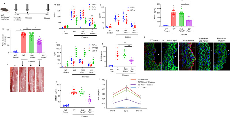
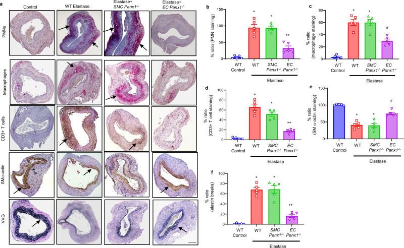
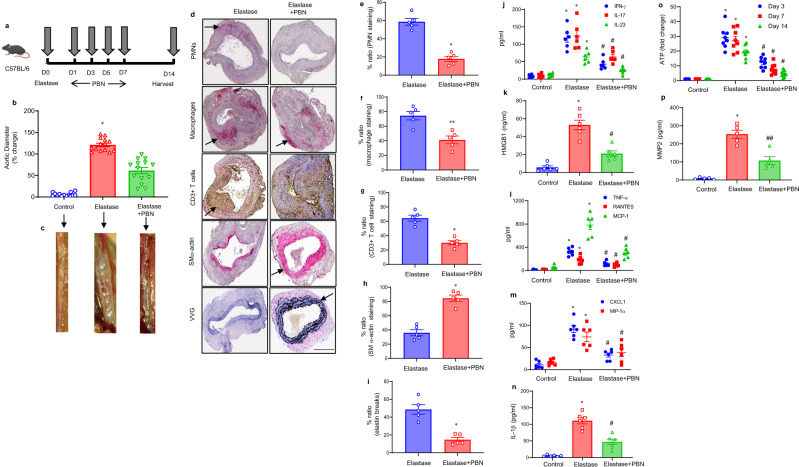
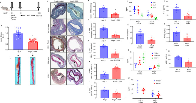
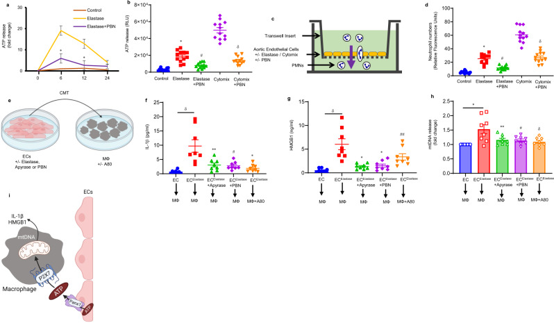
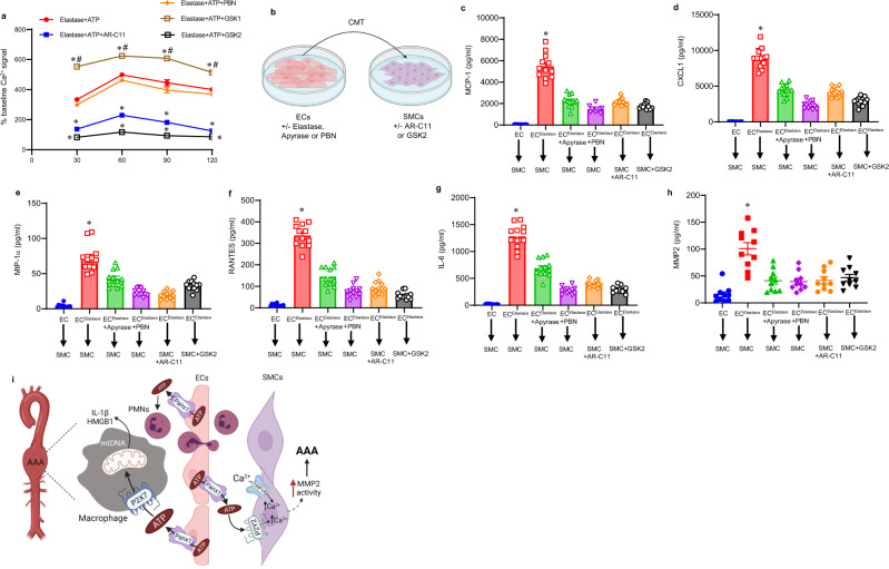
Pannexin-1 (Panx1) channels have been shown to regulate leukocyte trafficking and tissue inflammation but the mechanism of Panx1 in chronic vascular diseases like abdominal aortic aneurysms (AAA) is unknown. Here we demonstrate that Panx1 on endothelial cells, but not smooth muscle cells, orchestrate a cascade of signaling events to mediate vascular inflammation and remodeling. Mechanistically, Panx1 on endothelial cells acts as a conduit for ATP release that stimulates macrophage activation via P2X7 receptors and mitochondrial DNA release to increase IL-1β and HMGB1 secretion. Secondly, Panx1 signaling regulates smooth muscle cell-dependent intracellular Ca2+ release and vascular remodeling via P2Y2 receptors. Panx1 blockade using probenecid markedly inhibits leukocyte transmigration, aortic inflammation and remodeling to mitigate AAA formation. Panx1 expression is upregulated in human AAAs and retrospective clinical data demonstrated reduced mortality in aortic aneurysm patients treated with Panx1 inhibitors. Collectively, these data identify Panx1 signaling as a contributory mechanism of AAA formation.


Conflict of interest statement

The authors declare no competing interests.

Figures

Fig. 1
Fig. 1. Panx1 on endothelial cells regulates AAA formation and vascular inflammation.
a Schematic description of the topical elastase-treatment model is shown. Inducible knockout male mice were treated with tamoxifen for 10 days prior to elastase treatment. WT or inducible knockout male mice were then treated with topical elastase, or heat-inactivated elastase (control), on day 0, and aortic diameter was measured on day 14. b Aortic diameter is significantly reduced in elastase-treated EC-Panx1−/−, but not SMC-Panx1−/− mice, compared with elastase-treated WT mice. *P < 0.0001; #P < 0.0001 vs. WT elastase; ns, P = 0.09; n = 20 mice/group. c Representative images of aortic phenotype in all groups. dh Pro-inflammatory cytokine expression in aortic tissue is significantly attenuated in elastase-treated EC-Panx1−/−, but not SMC-Panx1−/− mice, compared with elastase-treated WT mice. *P < 0.0001 vs. WT control; #P = 0.0004 vs. WT elastase; δP = 0.01 vs. SMC-Panx1−/− elastase; ##P = 0.001 vs. WT elastase and SMC-Panx1−/− elastase; **P = 0.002 vs. SMC-Panx1−/− elastase; n = 6/group. i Aortic tissue content of ATP was significantly attenuated in elastase-treated EC-Panx1−/−, but not SMC-Panx1−/− mice, compared with elastase-treated WT mice on days 3, 7, and 14. *P < 0.008 vs WT elastase and SMC-Panx1−/− elastase; n = 5/group. j Expression of MMP2 in aortic tissue was significantly attenuated in elastase-treated EC-Panx1−/− mice compared with elastase-treated WT and SMC-Panx1−/− mice. *P < 0.0001 vs. WT control; #P = 0.005 vs. WT elastase and **P = 0.01 vs. SMC-Panx1−/− elastase; n = 5/group. All data above are represented as mean values ± SEM and comparative statistical analyses were done by one-way ANOVA followed by multiple comparisons. k Aortic tissue from elastase-treated WT mice demonstrated a marked increase in Panx1 expression on day 14 compared with elastase-treated EC-Panx1−/− mice. *: lumen; red: Panx1; green: elastic lamina and blue, blue: DAPI. Scale bar is 50 μm; n = 5/group. Representative images from independent experimental replicates are depicted.
Fig. 2
Fig. 2. Panx1 deletion on endothelial cells mitigates leukocyte trafficking in AAA.
a Comparative histology and immunohistochemistry performed on day 14 indicates a marked decrease in CD3 + T cell, neutrophil (PMN), and macrophage (Mac-2) immunostaining, increase in smooth-muscle-cell α-actin (SMα-actin) expression, and decrease in elastic-fiber disruption in aortic tissue (Verhoeff–Van Gieson, VVG staining for elastin) of elastase-treated EC-Panx1−/− mice compared with elastase-treated WT and SMC-Panx1−/− mice. n = 5/group. Arrows indicate areas of immunostaining. Scale bar is 200 μm. bf Quantification of immunohistochemical staining demonstrating a significant decrease in neutrophils (PMNs), macrophages, CD3 + T cells, and elastin degradation (VVG) staining, as well as increase in smooth muscle α-actin expression in elastase-treated EC Panx1−/− aortic tissue compared with elastase-treated WT and SMC-Panx1−/− mice. No significant differences were observed between elastase-treated WT and SMC-Panx1−/− mice. *P < 0.0001 vs. WT control; **P < 0.0001 vs. WT elastase and SMC-Panx1−/−; #P = 0.0003 vs. WT elastase and SMC-Panx1−/−. Data is represented as mean values ± SEM and comparative statistical analyses were done by one-way ANOVA.
Fig. 3
Fig. 3. Pharmacological inhibition of Panx1 mitigates AAA formation in the elastase-treatment model.
a Schematic depicting treatment protocol for probenecid (PBN) in the elastase model of AAA. b PBN treatment attenuates aortic diameter in elastase-treated male WT mice compared with elastase treatment alone. *P = 0.0001 vs. all other groups; n = 14/group. c Representative images of aortic phenotype in the respective groups. di Comparative histology displayed a marked decrease in leukocyte infiltration and elastin-fiber disruption, as well as increase in SMα-actin expression in PBN-treated mice compared with elastase-treated mice alone.*P = 0.007; **P = 0.01; n = 5/group. Arrows indicate areas of immunostaining. Scale bar is 400 μm. jn Pro-inflammatory cytokine expression in aortic tissue is significantly attenuated in elastase-treated WT mice after PBN administration compared with elastase-treated mice alone. *P < 0.0001 vs. respective controls; #P < 0.001 vs. elastase.; n = 6/group. o A significant decrease in aortic tissue ATP content was observed in PBN-treated mice compared with elastase-treated mice alone. *P < 0.0001 vs. controls; #P < 0.0001 vs. elastase; n = 8 mice/group. p Expression of MMP2 in aortic tissue was significantly attenuated in PBN-treated mice compared with elastase-treated mice alone. *P < 0.0001 vs. control; #P = 0.0003 vs. elastase; n = 5/group. Data are represented as mean values ± SEM and comparative statistical analyses were done by one-way ANOVA followed by multiple comparisons. Source data are provided as a Source Data file.
Fig. 4
Fig. 4. Inhibition of Panx1 channels attenuates AAA formation in the Ang-II model.
a Schematic depicting the Ang-II model of AAA in ApoE−/− mice. Osmotic pumps with either Ang II or saline (control) were inserted into the subcutaneous tissue of mice with/without treatment with PBN. Aortic diameter was measured on day 28, and tissue was harvested for further analysis. b Ang-II mice treated with PBN demonstrated significantly decreased aortic diameter compared with angiotensin treated mice alone. *P = 0.001; n = 15 mice/group. c Representative images of aortic phenotype in all groups. di Comparative histology performed on day 28 demonstrates that Ang-II-treated ApoE−/− mice administered with PBN have decreased polymorphonuclear neutrophil (PMNs), macrophage (Mac-2), CD3 + T-cell infiltration, and elastic-fiber disruption (Verhoeff–Van Gieson staining), and increase in SM-α-actin expression compared with mice treated with Ang II alone. Arrows indicate areas of immunostaining. *P = 0.007; n = 5 mice/group. Scale bar is 200 μm. jn Aortic inflammation is mitigated by Panx1 antagonism in the Ang II model of AAA. Aortic tissue from Ang-II-treated ApoE−/− mice after PBN administration showed a significant attenuation in pro-inflammatory cytokine/chemokine production compared with Ang-II-treated mice alone. *P = 0.007 vs. respective Ang-II controls; **P = 0.01; n = 5 mice/group. o A significant decrease in aortic tissue ATP content was observed in PBN-treated mice compared with Ang-II-treated mice alone. *P = 0.01; n = 5 mice/group. p Expression of MMP2 in aortic tissue was significantly attenuated in PBN-treated mice compared with elastase-treated mice alone. *P = 0.02; n = 4/group. Data are represented as mean values ± SEM and comparative statistical analyses were done by two-tailed t-test.
Fig. 5
Fig. 5. Panx1-dependent ATP release from ECs modulates neutrophil transmigration and macrophage activation.
a Transient elastase-treatment-induced eATP release from EC cultures is significantly mitigated by PBN treatment at 6- and 12 h. *P < 0.0001 vs. elastase; n = 12/group. b Elastase- and/or cytomix-induced eATP release by ECs is attenuated by Panx1 inhibition via PBN treatment compared with untreated controls. * P < 0.0001 vs. control; #P = 0.0009 vs. elastase; δP < 0.0001 vs. cytomix; n = 12/group. c Schematic showing the in vitro transwell model to demonstrate transendothelial migration of polymorphonuclear neutrophils (PMNs). Fluorescent-labeled primary murine-derived PMNs were included in the top chamber containing ECs and cocultured for 24 h. d Exposure to elastase or cytomix showed a significant increase in neutrophil transmigration, which was significantly attenuated by pretreatment with PBN. *P < 0.0001 vs. control; #P = 0.0002 vs. elastase; δP < 0.0001 vs. cytomix; n = 12/group. e Schematic depicting conditioned media transfer (CMT)-based experiments from elastase-treated ECs to macrophages with/without pretreatment with inhibitors. f, g CMT from elastase-treated ECs to macrophages induces a significant upregulation of IL-1β and HMGB1 secretion, which was blocked by pretreatment of ECs with apyrase or PBN, as well as pretreatment of macrophages with P2X7 inhibitor (A80). δP < 0.0001 vs. EC → MФ; *P = 0.0001, **P = 0.0009, #P = 0.0005 and ##P = 0.03 vs. ECelastase → MФ; n = 8/group. All comparative data above are represented as mean values ± SEM and statistical analyses were done by one-way ANOVA. h Panx1-dependent ATP release from ECs stimulates mtDNA release that is prevented by PBN and P2X7R inhibition. CMT from elastase-induced ECs was performed on macrophages. DNA was isolated from cytosolic fractions of macrophages and the levels of mitochondrial (mt)DNA were analyzed by quantitative RT-PCR. CMT from elastase-treated ECs to macrophages induces a significant upregulation of mtDNA release, which was blocked by pretreatment of ECs with apyrase or PBN, as well as pretreatment of macrophages with P2X7 inhibitor. *P = 0.0005; **P = 0.02, #P = 0.01, and δP = 0.004 vs. ECelastase → MФ; n = 8/group. Data are presented as fold change relative to EC → MФ and compared using two-tailed Wilcoxon test. i Schematic displaying the crosstalk between ECs and macrophages via Panx1-dependent release from elastase-induced ECs that stimulates P2X7R and mtDNA release from macrophages.
Fig. 6
Fig. 6. Panx1-dependent ATP release from ECs results in SMC activation that is inhibited by P2Y2 and TRPV4 antagonism.
a An increase in Ca2+-influx was observed in elastase and recombinant ATP- (1 μM) treated SMCs that was inhibited by P2Y2 (AR-C11) and TRPV4 antagonist (GSK2) blockade, but increased after TRPV4 agonism by GSK1 treatment. The Ca2+ signal after treatment was expressed as % change intensity compared with baseline in untreated cells. *P < 0.0001 vs. elastase+ATP; #P < 0.0001 vs. elastase+ATP + GSK2; n = 12/group. b Schematic depicting conditioned media-transfer (CMT)-based experiments from elastase-treated ECs to SMCs with/without pretreatment with inhibitors. cg CMT from elastase-treated ECs to primary SMCs induces a significant upregulation of MCP-1, CXCL1, MIP-1α, RANTES, and IL-6 secretion, which was blocked by pretreatment of ECs with apyrase or PBN, as well as pretreatment of SMCs with P2Y2 or TRPV4 inhibitors, respectively. *P < 0.0001 vs. all other groups; n = 12/group. h Primary SMCs were treated with CMT from elastase-treated ECs and MMP2 activity secretion was analyzed in cell culture supernatants after 24 h. A multifold increase in MMP2 activity was observed in SMCs after CMT from elastase-treated ECs compared with controls, and was significantly attenuated by pretreatment of ECs with apyrase or PBN, as well as pretreatment of SMCs with P2Y2 or TRPV4 inhibitors, respectively. *P < 0.0001 vs. other groups; n = 8/group. i Schematic representation of signaling events mediated by Panx1 signaling during the pathogenesis of AAA. ATP release from Panx1 channels in aortic ECs modulates macrophage activation via P2X7 receptors, and stimulates mtDNA release, leading to IL-1β and HMGB1 secretion thereby causing aortic inflammation. Also, Panx1-mediated ATP release from ECs activates P2Y2 receptors and TRPV4 channels on SMCs to stimulate pro-inflammatory cytokine secretion and destabilization of intracellular Ca2+ homeostasis that facilitates increased MMP2 activity resulting in aortic remodeling. Panx1/ATP signaling also contributes to leukocyte trafficking in the aortic wall, as observed by transendothelial migration of neutrophils. HMGB1, high-mobility-group box1; PMNs, neutrophils; ECs, endothelial cells; SMCs, smooth-muscle cells; mtDNA, mitochondrial DNA. Comparative data are represented as mean values ± SEM and statistical analyses were done by one-way ANOVA.

Similar articles

Cited by

References

    1. Nordon IM, Hinchliffe RJ, Loftus IM, Thompson MM. Pathophysiology and epidemiology of abdominal aortic aneurysms. Nat. Rev. Cardiol. 2011;8:92–102. - PubMed
    1. Norman PE, et al. Population based randomised controlled trial on impact of screening on mortality from abdominal aortic aneurysm. BMJ. 2004;329:1259. - PMC - PubMed
    1. Sakalihasan N, et al. Abdominal aortic aneurysms. Nat. Rev. Dis. Prim. 2018;4:34. - PubMed
    1. Harris LM, Faggioli GL, Fiedler R, Curl GR, Ricotta JJ. Ruptured abdominal aortic aneurysms: factors affecting mortality rates. J. Vasc. Surg. 1991;14:812–818. - PubMed
    1. Golledge J. Abdominal aortic aneurysm: update on pathogenesis and medical treatments. Nat. Rev. Cardiol. 2019;16:225–242. - PubMed

MeSH terms

LinkOut - more resources