Review
. 2022 Apr 27;122(8):7690-7719.
doi: 10.1021/acs.chemrev.1c00857. Epub 2022 Mar 22.

Mass Spectrometry Methods for Measuring Protein Stability

Affiliations
Review

Mass Spectrometry Methods for Measuring Protein Stability

Daniel D Vallejo et al. Chem Rev. .

Abstract

Mass spectrometry is a central technology in the life sciences, providing our most comprehensive account of the molecular inventory of the cell. In parallel with developments in mass spectrometry technologies targeting such assessments of cellular composition, mass spectrometry tools have emerged as versatile probes of biomolecular stability. In this review, we cover recent advancements in this branch of mass spectrometry that target proteins, a centrally important class of macromolecules that accounts for most biochemical functions and drug targets. Our efforts cover tools such as hydrogen-deuterium exchange, chemical cross-linking, ion mobility, collision induced unfolding, and other techniques capable of stability assessments on a proteomic scale. In addition, we focus on a range of application areas where mass spectrometry-driven protein stability measurements have made notable impacts, including studies of membrane proteins, heat shock proteins, amyloidogenic proteins, and biotherapeutics. We conclude by briefly discussing the future of this vibrant and fast-moving area of research.


Figures
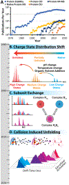
Figure 1
Figure 1
A) The total number of publications in the pubmed.gov database for the search terms indicated in legend. B) The majority of early protein stability measurements conducted by MS (pre-2000) relied on the observation of CSD shifts. The blue CSD represents a “native” protein possessing lower charge states. Upon denaturation, the protein unfolds presenting a CSD centered at higher charge (lower m/z), occupying partially unfolded structures (orange CSD) enroute to a fully unfolded population (red CSD). C) Native MS emerged in the early 2000s, and one of the initial biological systems studied by this approach were α-crystallin proteins. MS is able to track the exchange between co-incubated homo-oligomers, which eventually leads to the formation of hetero-oligomers. D) Building upon these initial measurements, CIU employs gas phase activation in conjunction with IM, to observe the gas phase change in structure due to collisional heating. This approach is growing rapidly and is being applied to a broad variety of biological systems.
Figure 2.
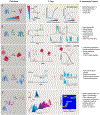
Figure 2.. Mass-Spectrometry Techniques for Protein Conformational Stability Measurements.
For each technique described, there is a corresponding summary figure, ii) data and iii) information content column. A) Variable Temperature. A protein experiences a temperature gradient as it is introduced into the mass spectrometer. A shift in CSD indicates conformational changes. By monitoring IM conformers, insights into thermodynamic values can be made. B) Footprinting uses selective or non-selective reagents (green stars) to monitor changes to the solvent-accessible regions of a protein. C) Electron Capture Dissociation is a fragmentation technique where proteins are exposed to low energy electrons, which are captured and produce backbone fragmentation. ECD fragmentation patterns can changed based on the precursor conformation and charge state. D) Collision Induce Dissociation allows the evaluation of protein complex subunit stoichiometry and composition. By monitoring protein ejection from protein complex precursors, dissociation thresholds can be determined and related to the stabilities of subunits and interfaces. E) Collision Induced Unfolding is an IM-MS technique where a protein conformation is monitored as its internal energy is increased using collisional activation. A shift in voltage required for eliciting the unfolding transitions observed is indicative of stability shifts when comparing between fingerprints of different states (e.g. apo vs holo).
Figure 3:
Figure 3:
Scaling the dimensionality of MS-based protein stability measurements. A) One-dimensional assays of protein stability can include variable temperature, mass spectrometry, ion mobility, and collision voltage scanning. B) Hyphenation of the 1-D techniques in A provides access to powerful 2-D techniques such as vt-MS, IM-MS, and CIU that can separate and generate a wide range of unfolded protein structures. C) Further enhancements can be achieved with 3-D hyphenated techniques based on those shown in A and B above. D) A logical extension of the techniques shown in C leads to a “4-D” assay of protein stability governed by MS methods, capable of measuring changes in protein stability across multiple phases/conditions simultaneously.
Figure 4.
Figure 4.
Data and information content that can be expected from variable temperature, MS and IM datasets for the following biotherapeutic modalities: A) mAbs, B) biosimilars, C) fusion proteins, D) antibody-drug conjugates, and E) bispecific antibodies. Generally, for variable temperature experiments shifts to lower Tm values indicate a decrease in stability and higher values indicate an increase in stability. Changes in mass spectrometry generally indicate different structures or stoichiometries. For IM shifts to lower CCS values indicate more compact structures while larger values indicate larger, often unfolded structures. By applying activation energy and monitoring unfolding, ex. Biosimilar IM, shifts in stability can be monitored by shifts in the IM peak relative to the activation energy.
Figure 5.
Figure 5.. CIU applications for biotherapeutic mAbs.
A) Differentiation of monoclonal IgG subclasses by disulfide bonding patterns and difference in CIU unfolding due to domain exchange. B) Biosimilar antibodies have qualitatively similar fingerprints, but contemporary CIU analyses are able to quantitate subtle differences in stability. C) Bispecific antibodies present CIU characteristics centered between the precursor structures. D) Shifts in CCS and stability can be quantified as a function of increasing drug load in ADC biotherapeutics.
Figure 6.
Figure 6.. Summary of various MS-related measurements associated with MP stability assessments.
A) identification of endogenous lipid binding B) thermodynamics of lipid binding to membrane protein C) oligomeric state assignment D) evaluation of disease state mutations in amino acid sequence E) site-selective ligand binding events F) resolving multiple simultaneous ligand bound states G) oxidative labeling.
Figure 7.
Figure 7.
Overview of select MS techniques that allow direct stability measurements with challenging amyloidogenic proteins A) Native MS usually produces a narrow range of charge states compare to non-native MS. Native MS can retain the native structures of amyloidogenic proteins and even the non-covalent complexes formed through ligand or protein binding through gentle ionization parameters. B) nESI needle is filled with a mixture of different oligomers of an amyloidogenic protein. IM-MS is able to separate the complex population of oligomers in drift time space based on their size, charge and shape. C) CIU shifts otherwise known as CIU50 values can be obtained through a series of IM-MS experiments at increasing collision energy. If a ligand binding event caused an amyloid protein to increase in stability, CIU50 values will reflect this increase. D) HDX-MS can capture localized information, in as little as few milliseconds or as long as days, allowing us to take snapshots of an amyloid aggregation process
Figure 8.
Figure 8.. Subunit Exchange between sHSP Oligomers Illuminated through MS.
A) Two sHSP homo-oligomers (R and B, numbers indicated the number of subunits within the example oligomers) with suspected hetero-oligomeric interactions are co-incubated in solution, and MS data is collected at various time points (t1, t2, t3 etc.). B) The MS data is quantified versus time to measure the decay of homo-oligomers and the growth in abundance of hetero-oligomeric sHSP species. The rate of this conversion from homo-oligomers to hetero-oligomers can be measured as a function of time, and can serve as an indication or stability. C) When hetero-oligomeric sHSP complexes are stable their subunit exchange is expected to occur quickly. D) Conversely, when hetero-oligomeric complexes are unstable, the equilibrium shifts in the direction of homo-oligomers.

Similar articles

Cited by

References

    1. Eggerth AH; Bellows M The Flocculation of Bacteria by Proteins. J. Gen. Physiol 1922, 4, 669–680. - PMC - PubMed
    1. Eggerth AH Changes In The Stability And Potential Of Cell Suspensions I. The Stability And Potential Of Bacterium Coli. J. Gen. Physiol 1923, 6, 63–71. - PMC - PubMed
    1. Northrop JH; Kunitz M Crystalline Trypsin I. Isolation And Tests Of Purity. J. Gen. Physiol 1932, 16, 267–294. - PMC - PubMed
    1. Northrop JH Crystalline Trypsin Iv. Reversibility Of The Inactivation And Denaturation Of Trypsin By Heat. J. Gen. Physiol 1932, 16, 323–337. - PMC - PubMed
    1. Herriott RM Isolation, Crystallization, And Properties Of Swine Pepsinogen. J. Gen. Physiol 1938, 21, 501–540. - PMC - PubMed

Publication types