. 2022 Apr 6;144(13):5820-5826.
doi: 10.1021/jacs.1c11706. Epub 2022 Mar 22.

Programmable Cell-Free Transcriptional Switches for Antibody Detection

Affiliations

Programmable Cell-Free Transcriptional Switches for Antibody Detection

Aitor Patino Diaz et al. J Am Chem Soc. .

Abstract

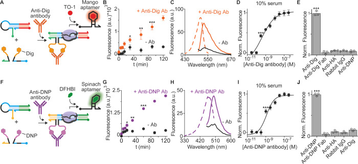
We report here the development of a cell-free in vitro transcription system for the detection of specific target antibodies. The approach is based on the use of programmable antigen-conjugated DNA-based conformational switches that, upon binding to a target antibody, can trigger the cell-free transcription of a light-up fluorescence-activating RNA aptamer. The system couples the unique programmability and responsiveness of DNA-based systems with the specificity and sensitivity offered by in vitro genetic circuitries and commercially available transcription kits. We demonstrate that cell-free transcriptional switches can efficiently measure antibody levels directly in blood serum. Thanks to the programmable nature of the sensing platform, the method can be adapted to different antibodies: we demonstrate here the sensitive, rapid, and cost-effective detection of three different antibodies and the possible use of this approach for the simultaneous detection of two antibodies in the same solution.


Conflict of interest statement

The authors declare no competing financial interest.

Figures

Figure 1
Figure 1
Programmable antibody-responsive transcriptional switches. (A) The antibody-responsive transcriptional switch is composed of two modules: the transcriptional switch module and the antibody-responsive module. The first is a double-stranded DNA switch designed to adopt a stem-loop hairpin conformation that prevents efficient transcription of an RNA light-up aptamer due to the incomplete formation of the promoter sequence. The second module is composed of two antigen-conjugated DNA strands (split input strands) that, upon bivalent binding to a target antibody, are brought into close proximity and can hybridize to form a functional bimolecular complex. Such a complex induces a conformational change on the switch and makes the promoter sequence accessible for transcription (right). (B) The so-activated transcriptional switch can transcribe, in the presence of RNA polymerase and nucleotides, a reporter light-up RNA aptamer that signals the presence of the target antibody.
Figure 2
Figure 2
Design of the co-localization-induced transcriptional switch. (A) Scheme of the transcriptional switch with relevant functional domains indicated. (B) Table of the different variants tested and their corresponding length of the accessible and hidden promoter domain. (C) Three representative transcriptional switch variants (#1, #6, and #9). (D) Scheme of the strand displacement reaction between the transcriptional switch and a unimolecular input strand. (E) Fluorescence signal obtained with the different variants in the absence and in the presence (30 nM) of the unimolecular input strand. (F) Ratio between the end-point fluorescence signals in the presence and absence of the input strand with the different variants. (G) Scheme of the co-localization-induced hybridization of the split input strands in the presence of an Ab-mimic DNA strand. (H) Fluorescence signal in the absence and presence (100 nM) of the Ab-mimic DNA strand at different split input strand concentrations. (I) Transcription kinetic traces in the absence (gray) and presence (black) of the Ab-mimic strand with a 30 nM concentration of split input strands. The experiments here were conducted at 25 °C in a 20 μL solution of a commercial transcription kit supplemented with the transcriptional switch module (100 nM), and the split input strands (30 nM) and the input strand (or Ab-mimic) were indicated. The transcription reaction was allowed to proceed for 120 min (or shorter time as indicated in panel I), and then, an aliquot was transferred to 100 μL of 10 mM Tris–HCl and 75 mM KCl, pH 7.4 solution containing 300 nM of TO-1, and the fluorescence signal measured after 15 min at 545 nm. The experimental values in this and in the following figures represent averages of at least three separate measurements, and the error bars reflect the standard deviations.
Figure 3
Figure 3
Antibody detection using programmable antibody-responsive transcriptional switches. (A) Scheme of the transcriptional switch for the detection of Anti-Dig antibodies. (B) Time course experiments showing the signal of the Mango aptamer-binding fluorophore (TO-1) obtained upon cell-free transcription experiments carried out in the absence and presence (100 nM) of the Anti-Dig antibody. (C) Emission and excitation spectra at the end-point (120 min) of reactions shown in panel (B). (D) Fluorescence signals in a 10% diluted bovine serum solution supplemented with increasing Anti-Dig antibody concentrations. (E) Fluorescence signal obtained with Anti-Dig antibodies and different non-specific targets (all at 100 nM) in 10% bovine serum. (F) Scheme of the transcriptional switch activated by the Anti-DNP antibody using DNP-conjugated DNA strands. (G) Time course experiments showing the signal of the Spinach aptamer-binding fluorophore (DFHBI) obtained upon cell-free transcription experiments carried out in the absence and presence (100 nM) of the Anti-DNP antibody. (H) Emission and excitation spectra at the end-point (120 min) of the reactions shown in panel (G). (I) Fluorescence signals in a 10% diluted bovine serum solution supplemented with increasing Anti-DNP antibody concentrations. (J) Fluorescence signal obtained with Anti-DNP antibodies and different non-specific targets (all at 100 nM) in 10% bovine serum. The experiments here were conducted at 25 °C in a 20 μL solution of a commercial transcription kit supplemented with the transcriptional switch module (100 nM), the antibody-responsive module (each at 30 nM), and the antibody (100 nM). For binding experiments (D, I), the antibody concentrations were varied from 0 to 300 nM, while the other components were kept at the same concentration as mentioned before. The transcription reaction was allowed to proceed for 120 min (or shorter time as indicated in panels B, G), and then, an aliquot was transferred to 100 μL of 10 mM Tris–HCl and 75 mM KCl, pH 7.4 solution containing the relevant dye, and the fluorescence signal measured after 15 min.
Figure 4
Figure 4
Modular transcriptional switch for Anti-HA antibody detection. (A) Modular version of the transcriptional switch to use peptide-PNA chimera strands as recognition elements of Anti-HA antibodies. (B) Fluorescence signal as a function of the Anti-HA concentration in a 10% diluted bovine serum solution. (C) Antibody quantification assessed by spiking blank serum samples with different known concentrations of the Anti-HA antibody (2, 7, 17, and 25 nM). Median values (black value and horizontal line) and 95% confidence intervals (gray values in parenthesis and red bars) are indicated. The experiments here were conducted at 25 °C in a 20 μL solution of a commercial transcription kit supplemented with the transcriptional switch module (100 nM), split input strands (each at 30 nM), HA-PNA chimera strands (100 nM), and different concentrations of the Anti-HA antibody (as indicated). The transcription reaction was allowed to proceed for 120 min, and then, an aliquot was transferred to 100 μL of 10 mM Tris–HCl and 75 mM KCl, pH 7.4 solution containing 300 nM of TO-1, and the fluorescence signal measured after 15 min.

Similar articles

Cited by

References

    1. Wiersinga W. J.; Rhodes A.; Cheng A. C.; Peacock S. J.; Prescott H. C. Pathophysiology, Transmission, Diagnosis, and Treatment of Coronavirus Disease 2019 (COVID-19): A Review. JAMA 2020, 324, 782.10.1001/jama.2020.12839. - DOI - PubMed
    1. Moody A. Rapid Diagnostic Tests for Malaria Parasites. Clin. Microbiol. Rev. 2002, 15, 66–78. 10.1128/CMR.15.1.66-78.2002. - DOI - PMC - PubMed
    1. Li Z.; Yi Y.; Luo X.; Xiong N.; Liu Y.; Li S.; Sun R.; Wang Y.; Hu B.; Chen W.; Zhang Y.; Wang J.; Huang B.; Lin Y.; Yang J.; Cai W.; Wang X.; Cheng J.; Chen Z.; Sun K.; Pan W.; Zhan Z.; Chen L.; Ye F. Development and Clinical Application of a Rapid IgM-IgG Combined Antibody Test for SARS-CoV-2 Infection Diagnosis. J. Med. Virol. 2020, 92, 1518–1524. 10.1002/jmv.25727. - DOI - PMC - PubMed
    1. Loizou S.; McCrea J. D.; Rudge A. C.; Reynolds R.; Boyle C. C.; Harris E. N. Measurement of Anti-Cardiolipin Antibodies by an Enzyme-Linked Immunosorbent Assay (ELISA): Standardization and Quantitation of Results. Clin. Exp. Immunol. 1985, 62, 738–745. - PMC - PubMed
    1. Pardee K.; Green A. A.; Takahashi M. K.; Braff D.; Lambert G.; Lee J. W.; Ferrante T.; Ma D.; Donghia N.; Fan M.; Daringer N. M.; Bosch I.; Dudley D. M.; O’Connor D. H.; Gehrke L.; Collins J. J. Rapid, Low-Cost Detection of Zika Virus Using Programmable Biomolecular Components. Cell 2016, 165, 1255–1266. 10.1016/j.cell.2016.04.059. - DOI - PubMed

Publication types

MeSH terms