. 2022 Mar 22;15(726):eabg5203.
doi: 10.1126/scisignal.abg5203. Epub 2022 Mar 22.

Biased agonists of the chemokine receptor CXCR3 differentially signal through Gαi:β-arrestin complexes

Affiliations

Biased agonists of the chemokine receptor CXCR3 differentially signal through Gαi:β-arrestin complexes

Kevin Zheng et al. Sci Signal. .

Abstract

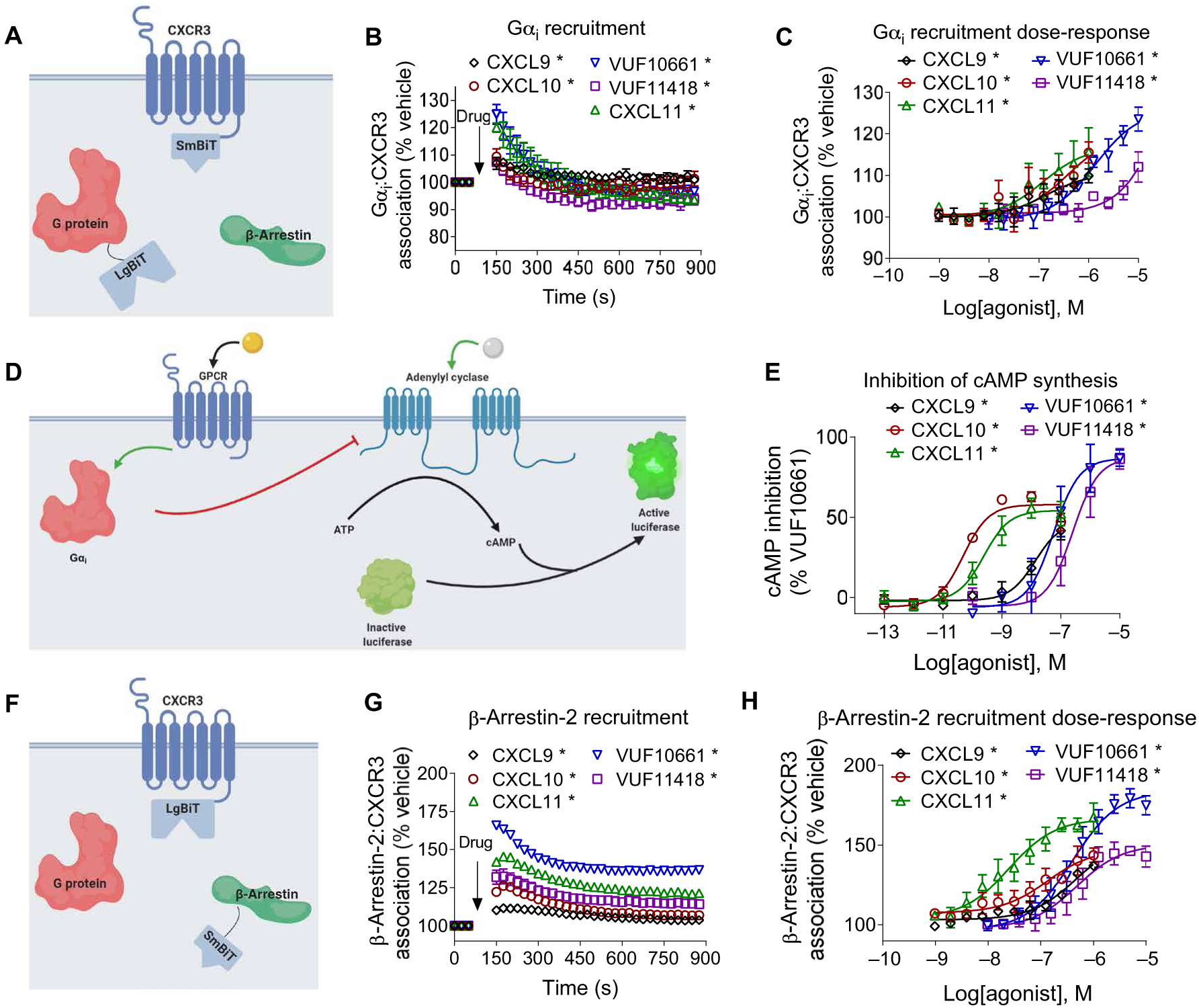
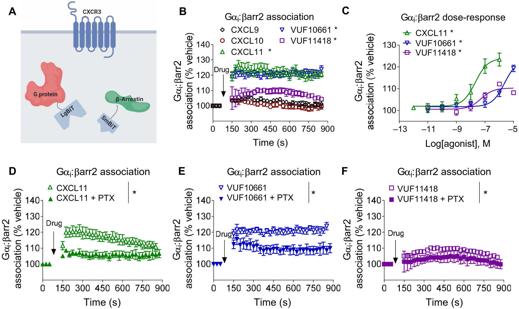
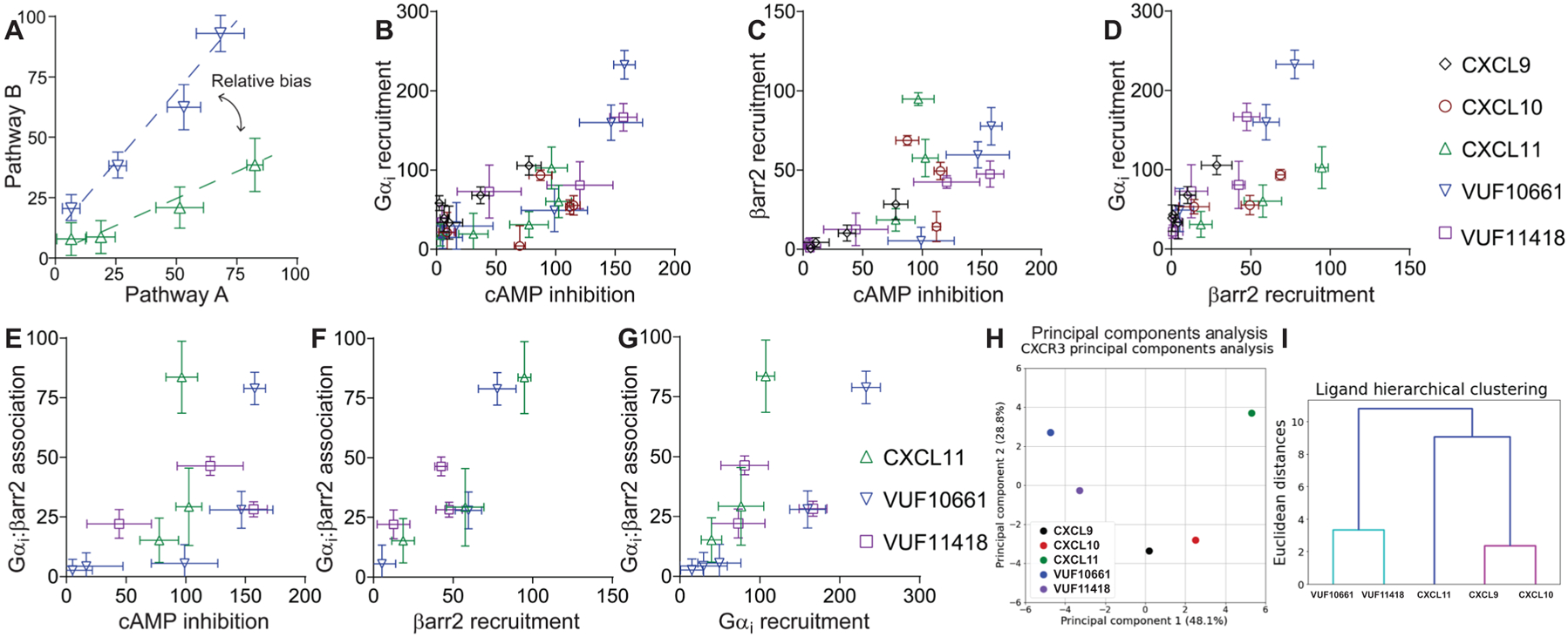
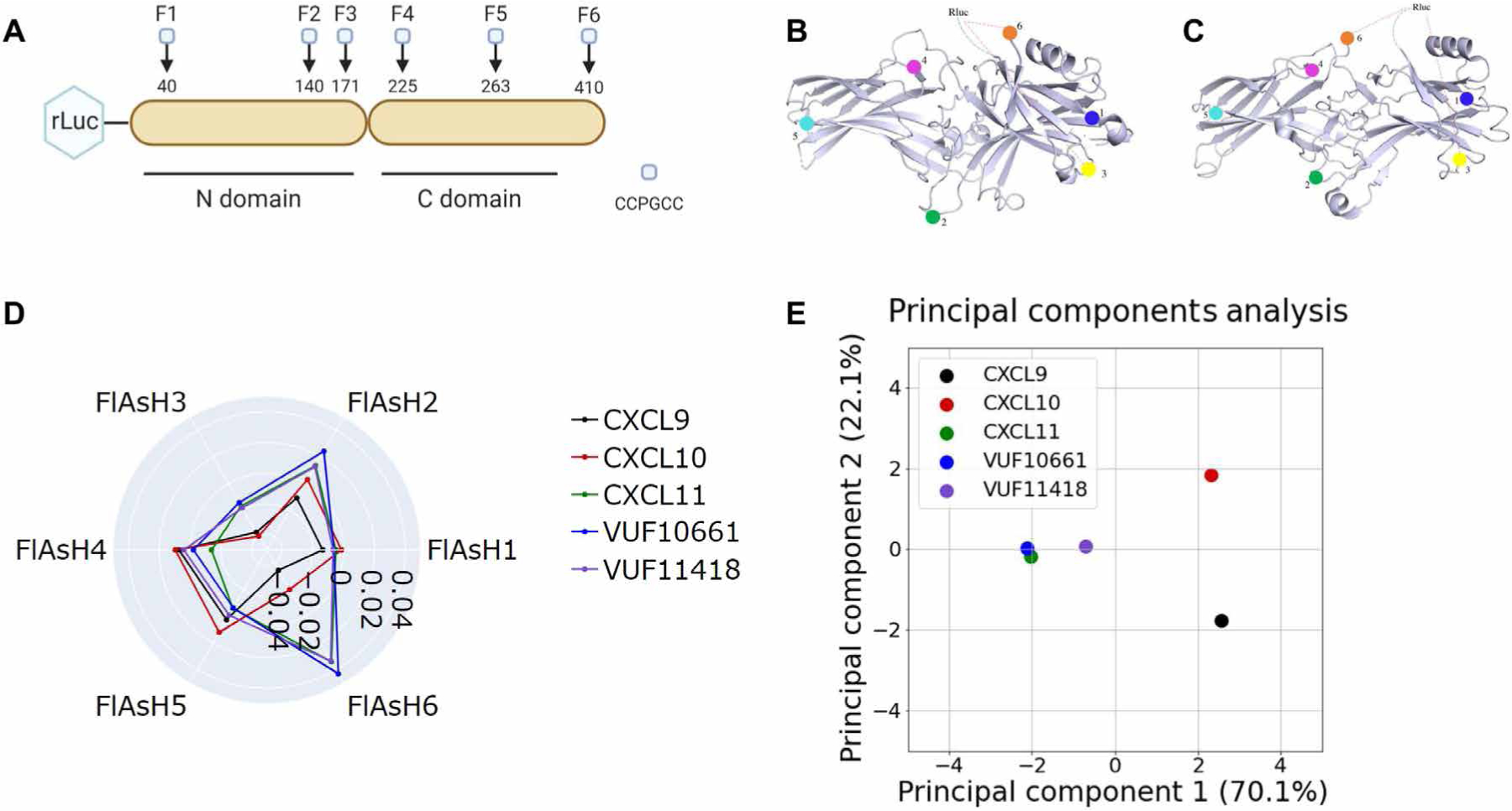
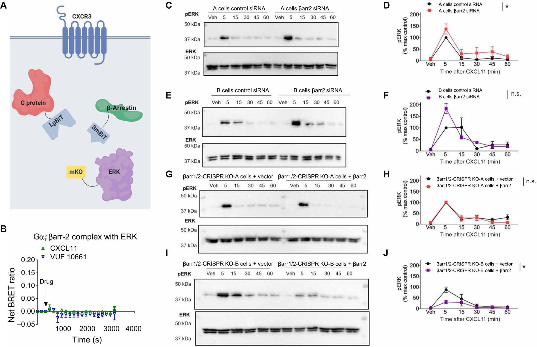
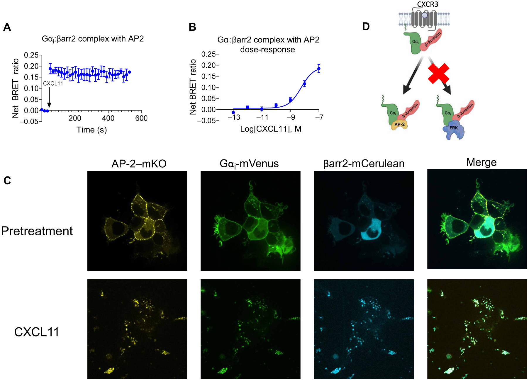
G protein-coupled receptors (GPCRs) are the largest family of cell surface receptors and signal through the proximal effectors, G proteins and β-arrestins, to influence nearly every biological process. The G protein and β-arrestin signaling pathways have largely been considered separable; however, direct interactions between Gα proteins and β-arrestins have been described that appear to be part of a distinct GPCR signaling pathway. Within these complexes, Gαi/o, but not other Gα protein subtypes, directly interacts with β-arrestin, regardless of the canonical Gα protein that is coupled to the GPCR. Here, we report that the endogenous biased chemokine agonists of CXCR3 (CXCL9, CXCL10, and CXCL11), together with two small-molecule biased agonists, differentially formed Gαi:β-arrestin complexes. Formation of the Gαi:β-arrestin complexes did not correlate well with either G protein activation or β-arrestin recruitment. β-arrestin biosensors demonstrated that ligands that promoted Gαi:β-arrestin complex formation generated similar β-arrestin conformations. We also found that Gαi:β-arrestin complexes did not couple to the mitogen-activated protein kinase ERK, as is observed with other receptors such as the V2 vasopressin receptor, but did couple with the clathrin adaptor protein AP-2, which suggests context-dependent signaling by these complexes. These findings reinforce the notion that Gαi:β-arrestin complex formation is a distinct GPCR signaling pathway and enhance our understanding of the spectrum of biased agonism.


Conflict of interest statement

Competing interests: J.S.S., T.P., and S.R. are inventors on patent application no. 16/876,934 submitted by Duke University that covers Complex BRET Technique for Measuring Biological Interactions. All other authors declare that they have no competing interests.

Figures

Fig. 1.
Fig. 1.. Gαi protein recruitment, inhibition of cAMP synthesis, and β-arrestin recruitment by biased agonists of CXCR3.
(A) Arrangement of the nanoBiT luciferase fragments used to assess G protein recruitment. (B) HEK293T transiently expressing CXCR3-smBiT and Gαi-LgBiT were treated with 100 nM CXCL9, 100 nM CXCL10, 100 nM CXCL11, 1 μM VUF10661, 1 μM VUF11418, or vehicle, and the resulting luminescence was analyzed for 750 s after an initial preread. (C) Gαi recruitment to CXCR3 after treatment with the indicated concentrations of agonists 1 min after treatment. (D) Scheme illustrating the Gαi-regulated cAMP assay. Before the cells were treated with biased agonists of CXCR3, the cellular cAMP concentration was increased by treatment with 10 μM forskolin. (E) HEK293T cells transiently expressing cAMP-activated modified firefly luciferase and CXCR3 were treated with vehicle, 100 nM CXCL9, 100 nM CXCL10, 100 nM CXCL11, 1 μM VUF10661, or 1 μM VUF11418. The inhibition of cAMP synthesis is displayed as a percentage of that in VUF10661-treated cells. (F) Arrangement of the luciferase fragments CXCR3-LgBiT and smBiT–β-arrestin2 to assess β-arrestin2 recruitment to CXCR3. (G) HEK293T transiently expressing CXCR3-LgBiT and smBiT–β-arrestin2 were treated with 100 nM CXCL9, 100 nM CXCL10, 100 nM CXCL11, 1 μM VUF10661, 1 μM VUF11418, or vehicle and then were analyzed for 750 s after an initial preread. (H) β-arrestin2 recruitment after treatment with the indicated concentrations of agonist 6 min after treatment. Data are means ± SEM of three or four independent experiments. *P < 0.05 by two-way ANOVA; Dunnett’s post hoc analysis showed a significant difference relative to pretreatment.
Fig. 2.
Fig. 2.. Biased agonists of CXCR3 differentially promote the formation of Gαi:β-arrestin complexes.
(A) Scheme of the assay used to assess Gαi:β-arrestin complex formation. (B) HEK293T cells transiently expressing CXCR3, smBiT–β-arrestin2 (βarr2), and Gαi-LgBiT were treated with 100 nM CXCL9, 100 nM CXCL10, 100 nM CXCL11, 1 μM VUF10661, 1 μM VUF11418, or vehicle, and luminescence was monitored for 15 min. (C) Transfected HEK293T cells expressing CXCR3, smBiT–β-arrestin2, and Gαi-LgBiT were treated with the indicated concentrations of CXCL11, VUF10661, VUF11418, or vehicle and were assayed 6 min after treatment. (D to F) HEK293T cells transiently expressing CXCR3, smBiT–β-arrestin2, and Gαi-LgBiT were pretreated with PTX (200 ng/m) and subsequently treated with (D) CXCL11, (E) VUF10661, or (F) VUF11418 before luminescence was measured to determine the formation of Gαi:β-arrestin complexes. Data are means ± SEM of three to five independent experiments. *P < 0.05 by two-way ANOVA. Data in (B) and (C) included Dunnett’s post hoc analysis to show significant differences between treatments.
Fig. 3.
Fig. 3.. Bias plots of biased ligands of CXCR3 in G protein recruitment, G protein signaling, β-arrestin2 recruitment, and Gαi:β-arrestin2 complex formation.
(A) Example of the interpretation of a bias plot. A difference in the two response-response curves (at the same agonist concentration) is indicative of a relative bias between agonists. (B to G) Bias plots of cAMP inhibition and Gαi recruitment (B); cAMP inhibition and β-arrestin2 recruitment (C); β-arrestin2 recruitment and Gαi recruitment (D); cAMP inhibition and Gαi:β-arrestin2 association, β-arrestin2 recruitment, and Gαi:β-arrestin association (F); and Gαi recruitment and Gαi:β-arrestin association (G). Because CXCL11 is the only full endogenous agonist for both the G protein and β-arrestin pathways, it was used as the reference ligand. (H) PCA was used to assess CXCR3 agonist similarity based on signaling assays; computed principal components are visualized with the top two principal components. Principal component 1 contributes to 48% of the observed variation, and principal component 2 contributes to 29% of observed variation. Points denote the composite response of a single ligand at varying concentrations. (I) Dendrogram of hierarchical clustering to determine the number of clusters and the relationship between the ligand signaling profiles. PCA and dendrogram analyses were of means from three to five independent experiments for each signaling assay.
Fig. 4.
Fig. 4.. Biased agonists that induce Gαi:β-arrestin complex formation induce similar conformations of β-arrestin.
(A) The RLuc–β-arrestin2–FlAsH1 to RLuc–β-arrestin2–FlAsH6 reporters (F1 to F6) have the amino acid motif CCPGCC inserted after residues 40, 140, 171, 225, 263, and 410 of β-arrestin2. This motif acts as a dipole acceptor to assess conformational changes in β-arrestin2. (B and C) The positions of the FlAsH binding motifs are highlighted in the inactive (B) and active (C) structures of β-arrestin1. (D) HEK293N cells were transfected with plasmids encoding CXCR3 and one of the RLuc–β-arrestin2–FlAsH reporters (FlAsH1 to FlAsH6). The cells were then treated with 100 nM CXCL9, 100 nM CXCL10, 100 nM CXCL11, 1 μM VUF10661, 1 μM VUF11418, or vehicle. The radar plot depicts the intramolecular net BRET ratio calculated from subtracting the vehicle from treatment groups. (E) PCA was used to assess biased agonist–induced β-arrestin conformation similarities; computed principal components are visualized with the top two principal components. Principal component 1 contributes to 70% of the observed variation, and principal component 2 contributes to 22% of the observed variation. Points denote the composite response of a single ligand at varying doses. Data are means of three to five independent experiments.
Fig. 5.
Fig. 5.. Formation of a Gαi:β-arrestin:CXCR3 ternary complex.
(A) Arrangement of the luciferase fragments and the monomeric Kusabira Orange (mKO) acceptor fluorophore for complex BRET on Gαi (LgBiT), β-arrestin2 (SmBiT), and CXCR3 (mKO). (B) HEK293T cells transiently transfected with plasmids encoding CXCR3-mKO, Gαi-LgBiT, and SmBiT–β-arrestin2 were stimulated with 100 nM CXCL11, 1 μM VUF10661, 1 μM VUF11418, or vehicle. Complex BRET ratios were calculated for the Gαi:β-arrestin:CXCR3 ternary complex after the indicated treatments. Data are means ± SEM of three to five independent experiments. *P < 0.05 by two-way ANOVA, with Dunnett’s post hoc analysis showing a significant difference relative to vehicle control.
Fig. 6.
Fig. 6.. β-Arrestin is not necessary for CXCR3-dependent ERK activation, and no Gαi:β-arrestin:ERK complex is observed.
(A) Arrangement of the luciferase fragments and mKO acceptor fluorophore for complex BRET on Gαi (LgBiT), β-arrestin2 (SmBiT), and ERK2 (mKO). (B) Complex BRET ratio for Gαi:β-arrestin2:ERK after treatment with 100 nM CXCL11, 1 μM VUF10661, or vehicle. Data were normalized to both vehicle treatment and cytosolic mKO. (C to G) Western blotting analysis of the time course of ERK phosphorylation in A and B parental and β-arrestin1/2 CRISPR KO HEK293 cells stimulated with 100 nM CXCL11. (C) Western blotting analysis of phospho-ERK (pERK) and (D) its quantification in A parental cells transfected with control siRNA or β-arrestin2–specific siRNA. (E) Western blotting analysis of phospho-ERK and (F) its quantification in B parental cells transfected with control siRNA or β-arrestin2–specific siRNA. (G) Western blotting analysis of phospho-ERK and (H) its quantification in A β-arrestin1/2 CRISPR KO cells transfected with the control or β-arrestin2 rescue plasmid. (I) Western blotting analysis of phospho-ERK and (J) its quantification in B β-arrestin1/2 CRISPR KO cells transfected with the control or β-arrestin2 rescue plasmid. *P < 0.05 by two-way ANOVA to determine the main effect of either the siRNA or rescue. Data are from three experiments per condition. n.s., not significant.
Fig. 7.
Fig. 7.. Formation of a Gαi:β-arrestin:AP-2 ternary complex.
(A to D) Transfected HEK293T cells expressing CXCR3, smBiT–β-arrestin2, Gαi-LgBiT, and AP-2-mKO or cytosolic mKO were treated with vehicle or the indicated concentrations of CXCL11. (A) Complex BRET ratio for Gαi:β-arrestin2:AP-2 after treatment with 100 nM CXCL11 or vehicle. (B) Transfected HEK293T cells were treated with vehicle or the indicated concentrations of CXCL11. Net BRET was measured 6 min after treatment. (C) Confocal microscopy analysis of CXCL11-induced complexes of Gαi:β-arrestin:AP-2 in HEK293T cells transfected with mVenus-tagged Gαi, mKO-tagged AP-2, and mCerulean-tagged β-arrestin2. Cells were imaged before (basal) and 20 min after treatment. Data are representative of three experiments per condition. For BRET experiments, data were normalized to both vehicle treatment and cytosolic mKO transfection conditions. (D) Scheme demonstrating the selective formation of Gαi:β-arrestin:AP-2 rather than Gαi:β-arrestin:ERK in response to the activation of CXCR3 by CXCL11.

Similar articles

Cited by

References

    1. Santos R, Ursu O, Gaulton A, Bento AP, Donadi RS, Bologa CG, Karlsson A, Al-Lazikani B, Hersey A, Oprea TI, Overington JP, A comprehensive map of molecular drug targets. Nat. Rev. Drug Discov 16, 19–34 (2017). - PMC - PubMed
    1. Hamm HE, The many faces of G protein signaling. J. Biol. Chem 273, 669–672 (1998). - PubMed
    1. Smith JS, Rajagopal S, The β-arrestins: Multifunctional regulators of G protein-coupled receptors. J. Biol. Chem 291, 8969–8977 (2016). - PMC - PubMed
    1. Manglik A, Lin H, Aryal DK, McCorvy JD, Dengler D, Corder G, Levit A, Kling RC, Bernat V, Hübner H, Huang X-P, Sassano MF, Giguère PM, Löber S, Duan D, Scherrer G, Kobilka BK, Gmeiner P, Roth BL, Shoichet BK, Structure-based discovery of opioid analgesics with reduced side effects. Nature 537, 185–190 (2016). - PMC - PubMed
    1. Cahill TJ, Thomsen ARB, Tarrasch JT, Plouffe B, Nguyen AH, Yang F, Huang L-Y, Kahsai AW, Bassoni DL, Gavino BJ, Lamerdin JE, Triest S, Shukla AK, Berger B, Little J, Antar A, Blanc A, Qu C-X, Chen X, Kawakami K, Inoue A, Aoki J, Steyaert J, Sun J-P, Bouvier M, Skiniotis G, Lefkowitz RJ, Distinct conformations of GPCR-β-arrestin complexes mediate desensitization, signaling, and endocytosis. Proc. Natl. Acad. Sci. U.S.A 114, 2562–2567 (2017). - PMC - PubMed

Publication types

MeSH terms