. 2022 Apr 22;7(8):e151509.
doi: 10.1172/jci.insight.151509.

ADAMTS2 and ADAMTS14 can substitute for ADAMTS3 in adults for pro-VEGFC activation and lymphatic homeostasis

Affiliations

ADAMTS2 and ADAMTS14 can substitute for ADAMTS3 in adults for pro-VEGFC activation and lymphatic homeostasis

Laura Dupont et al. JCI Insight. .

Abstract

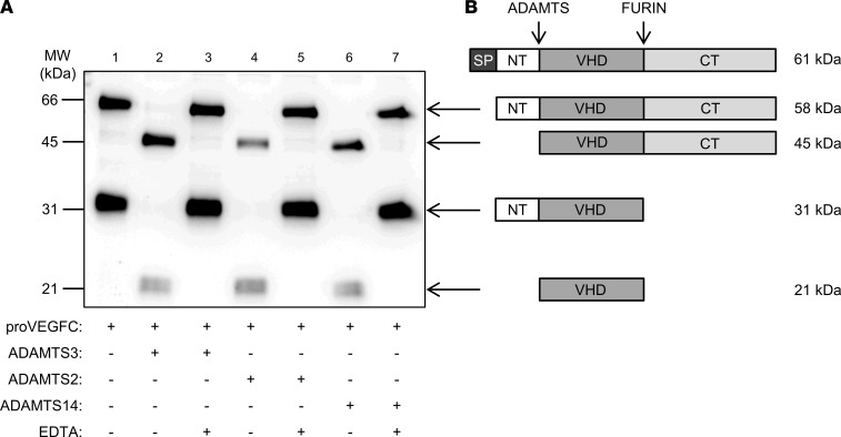
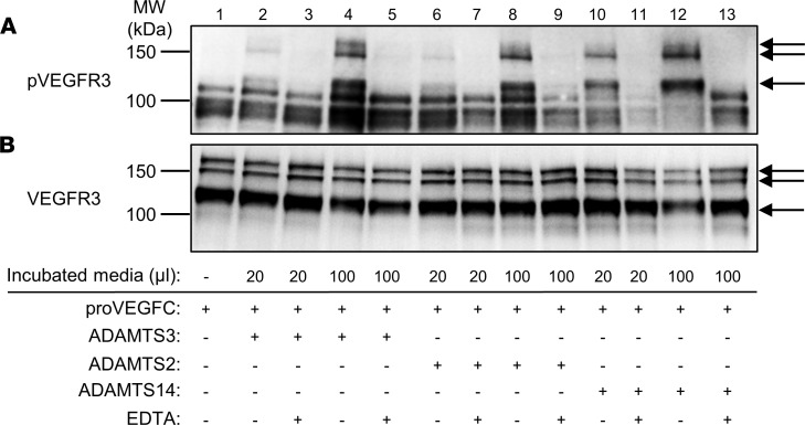
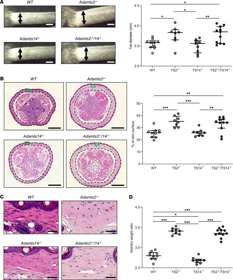
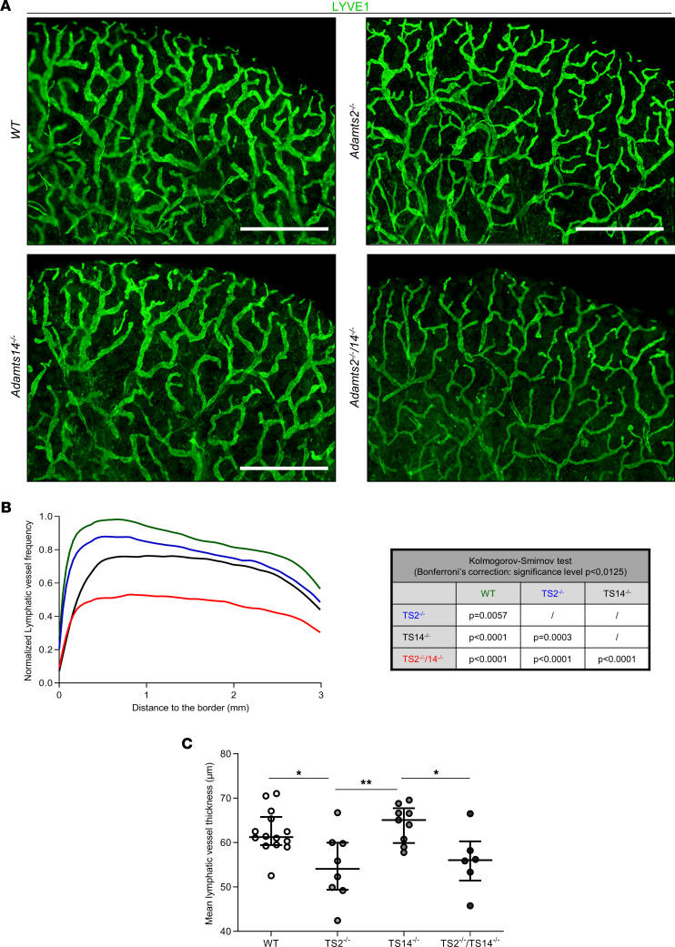
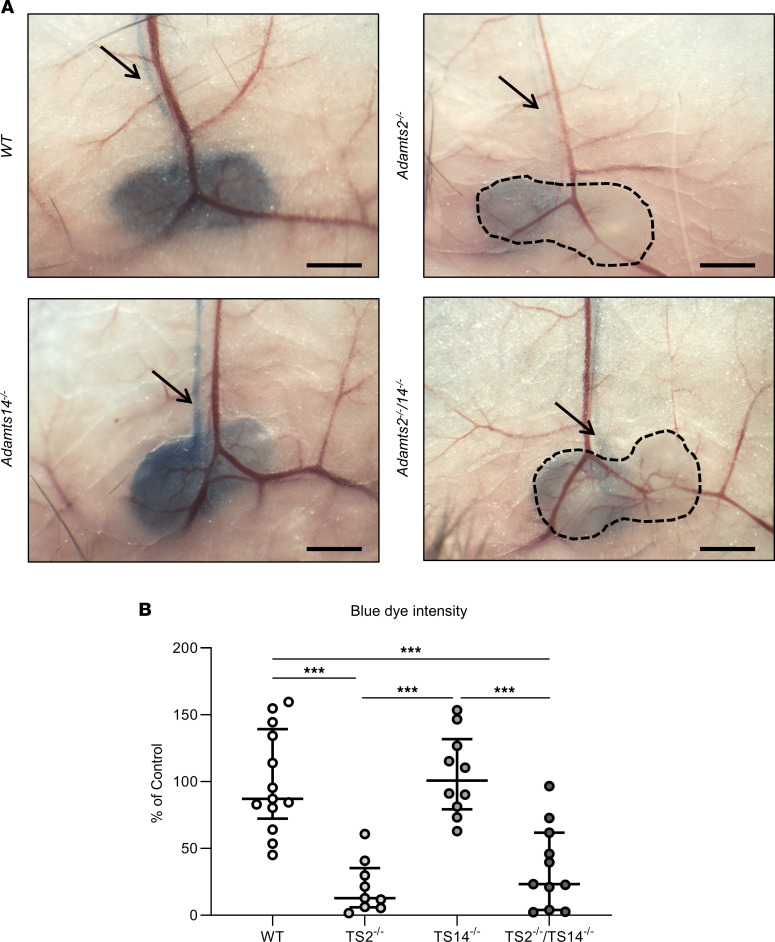
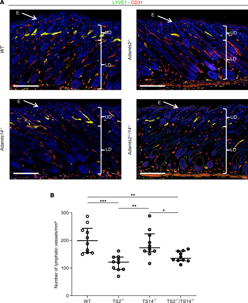
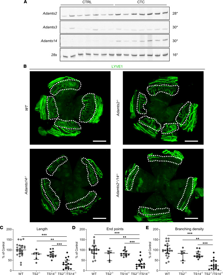
The capacity of ADAMTS3 to cleave pro-VEGFC into active VEGFC able to bind its receptors and to stimulate lymphangiogenesis has been clearly established during embryonic life. However, this function of ADAMTS3 is unlikely to persist in adulthood because of its restricted expression pattern after birth. Because ADAMTS2 and ADAMTS14 are closely related to ADAMTS3 and are mainly expressed in connective tissues where the lymphatic network extends, we hypothesized that they could substitute for ADAMTS3 during adulthood in mammals allowing proteolytic activation of pro-VEGFC. Here, we demonstrated that ADAMTS2 and ADAMTS14 are able to process pro-VEGFC into active VEGFC as efficiently as ADAMTS3. In vivo, adult mice lacking Adamts2 developed skin lymphedema due to a reduction of the density and diameter of lymphatic vessels, leading to a decrease of lymphatic functionality, while genetic ablation of Adamts14 had no impact. In a model of thermal cauterization of cornea, lymphangiogenesis was significantly reduced in Adamts2- and Adamts14-KO mice and further repressed in Adamts2/Adamts14 double-KO mice. In summary, we have demonstrated that ADAMTS2 and ADAMTS14 are as efficient as ADAMTS3 in activation of pro-VEGFC and are involved in the homeostasis of the lymphatic vasculature in adulthood, both in physiological and pathological processes.

Keywords: Angiogenesis; Cardiovascular disease; Growth factors; Vascular Biology.


Figures

Figure 1
Figure 1. Processing of human pro-VEGFC by ADAMTS3, ADAMTS2, and ADAMTS14 proteases.
(A) Conditioned medium from HEK293 cells expressing full-length pro-VEGFC was incubated with buffer (as negative control, lane1), ADAMTS3 (as positive control, lane 2), ADAMTS2, or ADAMTS14, in the presence or absence of EDTA used as inhibitor. The electrophoretic pattern of VEGFC was analyzed by Western blotting in reducing conditions. In absence of active enzymes (lane 1, 3, 5, and 7), VEGFC can be detected as a 58 kDa form (full-length pro-VEGFC without signal peptide) and a 31 kDa form generated by C-terminal processing by furin. In the presence of active ADAMTS3 (lane 2), ADAMTS2 (lane 4), and ADAMTS14 (lane 6), the 58 kDa form was totally converted into a 45 kDa polypeptide, whereas the 31 kDa form was processed into the fully mature 21 kDa VEGFC, which is in line with N-terminal processing of VEGFC proteins. (B) Schematic illustration of the different VEGFC forms, with their molecular weights provided (SP, signal peptide; NT, N-terminal propeptide; VHD, VEGF homology domain; CT, C-terminal propeptide).
Figure 2
Figure 2. Phosphorylation of VEGFR3 by pro-VEGFC activated or not by ADAMTS3, ADAMTS2, or ADAMTS14.
Conditioned medium from HEK293 cells expressing full-length pro-VEGFC was first incubated for 18 hours with buffer alone (lane 1, negative control), ADAMTS3 (as positive control), ADAMTS2, or ADAMTS14, in the presence or absence of EDTA used as inhibitor. These different pretreated media were then added (20 μL or 100 μL) into 1 mL of serum-free EBM-2 on LEC cultures. (A) After 5 minutes, cells were lysed, and phosphorylated VEGFR3 (pVEGFR3) was visualized by Western blotting. (B) After stripping of the antibodies, the same membrane was then used to visualize total VEGFR3. Treatment of the pro-VEGFC–rich conditioned medium with active ADAMTS3, ADAMTS2, and ADAMTS14 induced the phosphorylation of the 3 bands corresponding to VEGFR3 (arrows) in a dose-dependent manner, while the total amount of VEGFR3 was not affected, demonstrating that processing of pro-VEGFC by ADAMTS2, ADAMTS3, or ADAMTS14 leads to the activation of pro-VEGFC in a similar manner.
Figure 3
Figure 3. Tail swelling in absence of Adamts2.
(A) Representative images of tails from 8-week-old WT, TS2–/–, TS14–/–, and TS2–/–TS14–/– mice. Base tail diameters were measured (double arrows). Scale bar: 2 mm. As compared with those in WT mice, diameters were increased in TS2–/– and TS2–/–TS14–/– mice, suggesting potential lymphedema. (B) Hematoxylin and eosin staining of paraffin-embedded sections of tails was performed to further characterize the tissue compartment responsible for the increased diameter. Scale bar: 1 mm. The percentage of surface covered by the dermis (delimited by black dotted lines) was determined (ImageJ software) and was found to be increased in TS2–/– and TS2–/–TS14–/– mice. (C) Higher-magnification images of boxed regions in B show that the dermis is less stained in TS2–/– and TS2–/–TS14–/– mice because it is swollen and less dense. Scale bar: 50 μm. (D) Tail skins were removed and weighed (wet weight) and then dried for 72 hours in an oven at 60°C before being reweighed (dry weight) to determine the ratios of wet-to-dry weight. An increase of water content was confirmed in TS2–/– and TS2–/–TS14–/– mice. Statistical analyses were performed using Kruskal-Wallis test followed by Holm-Šidák post hoc test for multiple comparisons. *P < 0.05; **P < 0.01; ***P < 0.001.
Figure 4
Figure 4. Absence of Adamts2 and/or Adamts14 affects the lymphatic network under physiological conditions in adult mice.
(A) Whole mounts of dorsal ear skin were stained with an antibody specific for LYVE1 for immunofluorescence visualization of lymphatics. Scale bar: 1 mm. (B) For computerized quantification, lymphatic vessel frequencies were determined according to the distance from the border of the ear, because the structure and density of the lymphatic network vary according to the distance from the ear edge. The shapes of the distribution curves were found to be statistically different (Kolmogorov-Smirnov test; significance levels were adjusted with Bonferroni’s correction for multicomparison). The most dramatic reduction was observed in TS2–/–TS14–/– mice, but it was also found to be reduced in TS2–/– and TS14–/– mice as compared with WT mice. By sharp contrast, no difference was seen regarding blood vessels, showing that these alterations affect lymphatics specifically (Supplemental Figure 5). (C) Computerized quantifications of mean diameter of the lymphatic vessels were also performed and demonstrated a smaller diameter in TS2–/– and TS2–/–TS14–/– mice. Statistical analyses were performed using Kruskal-Wallis test followed by Holm-Šidák post hoc test for multiple comparisons. *P < 0.05; **P < 0.01.
Figure 5
Figure 5. Impaired lymphatic function in absence of Adamts2.
(A) Evans blue dye was injected into the footpad. After 30 minutes, mice were sacrificed and inguinal lymph nodes were visualized. Scale bar: 1 mm. In WT and TS14–/– mice, lymph nodes and the efferent lymphatic vessels (arrows) had a marked blue color. By contrast, they were barely detectable in TS2–/– and TS2–/–TS14–/– mice, demonstrating a delayed draining from the site of injection. The images were acquired with a camera-equipped dissection microscope (Optika). (B) Computer-assisted quantification of the blue dye intensity in inguinal lymph nodes of WT, TS2–/–, TS14–/–, and TS2–/–TS14–/– mice expressed as a percentage of WT control. Outliers were excluded based on Dixon’s test for extreme values. Statistical analyses were performed using Kruskal-Wallis test followed by Holm-Šidák post hoc test for multiple comparisons. ***P < 0.001.
Figure 6
Figure 6. Absence of Adamts2 affects lymphatic formation in postnatal day 6 mice.
Skin sections were stained using antibodies for LYVE1 (green) and CD31 (red). Scale bar: 200 μm. (A) Because of coexpression of LYVE1 and CD31, lymphatics appear as yellow structures, mainly in the upper dermis and in close association with the bulge region of the hair follicle (18). LYVE1-positive cells identified in the lower dermis and in the adipose tissue were mainly macrophages (Supplemental Figure 6). (B) Computerized quantification of LYVE1 staining in the upper dermis. As compared with WT mice, fewer lymphatics were observed in TS2–/– and TS2–/–TS14–/– mice. E, epidermis (white arrows); UD, upper dermis; LD, lower dermis. Statistical analyses were performed using Kruskal-Wallis test followed by Holm-Šidák post hoc test for multiple comparisons. *P < 0.05; **P < 0.01; ***P < 0.001.
Figure 7
Figure 7. Absence of Adamts2 or Adamts14 inhibits inflammatory corneal lymphangiogenesis.
(A) Expression of Adamts2 (28 cycles), Adamts3 (30 cycles), Adamts14 (30 cycles), and 28s (as control, 16 cycles) by RT-PCR on corneas collected from uninjured (CTRL) WT mice (n = 7) or from WT mice 7 days after corneal thermal cauterization (CTC) (n = 7). The expressions of Adamts2 and Adamts14, but not of Adamts3, were increased during the healing process of the injured cornea. (B) Lymphatic vessel visualization using LYVE1 immunofluorescence on whole-mount corneas 7 days after thermal cauterization in WT, TS2–/–, TS14–/–, and TS2–/–TS14–/– mice. Scale bar: 1 mm. A decrease of lymphatic vessels invading the injured corneas (white dotted lines) were observed in KO mice as compared with WT mice. Computerized quantification of the (C) length, (D) end point numbers, and (E) branching density of lymphatic vessels in corneas 7 days after thermal cauterization in WT, TS2–/–, TS14–/–, and TS2–/–TS14–/– mice expressed as a percentage of WT control (n = 23 WT, n = 6 TS2–/–, n = 12 TS14–/–, and n = 16 TS2–/–TS14–/– corneas). Statistical analyses were performed using Kruskal-Wallis test followed by Holm-Šidák post hoc test for multiple comparisons. **P < 0.01; ***P < 0.001.

Similar articles

Cited by

References

    1. Petrova TV, Koh GY. Biological functions of lymphatic vessels. Science. 2020 Jul 10;369(6500):eaax4063. doi: 10.1126/science.aax4063. - DOI - PubMed
    1. Alitalo K. The lymphatic vasculature in disease. Nat Med. 2011;17(11):1371–1380. doi: 10.1038/nm.2545. - DOI - PubMed
    1. Brouillard P, et al. Genetics of lymphatic anomalies. J Clin Invest. 2014;124(3):898–904. doi: 10.1172/JCI71614. - DOI - PMC - PubMed
    1. Baldwin ME, et al. Vascular endothelial growth factor D is dispensable for development of the lymphatic system. Mol Cell Biol. 2005;25(6):2441–2449. doi: 10.1128/MCB.25.6.2441-2449.2005. - DOI - PMC - PubMed
    1. Künnapuu J, et al. Proteolytic cleavages in the VEGF family: generating diversity among angiogenic VEGFs, essential for the activation of lymphangiogenic VEGFs. Biology (Basel) 2021;10(2):167. - PMC - PubMed

Publication types

Substances