. 2022 May 9;7(9):e156344.
doi: 10.1172/jci.insight.156344.

UVB-mediated DNA damage induces matrix metalloproteinases to promote photoaging in an AhR- and SP1-dependent manner

Affiliations

UVB-mediated DNA damage induces matrix metalloproteinases to promote photoaging in an AhR- and SP1-dependent manner

Daniel J Kim et al. JCI Insight. .

Abstract

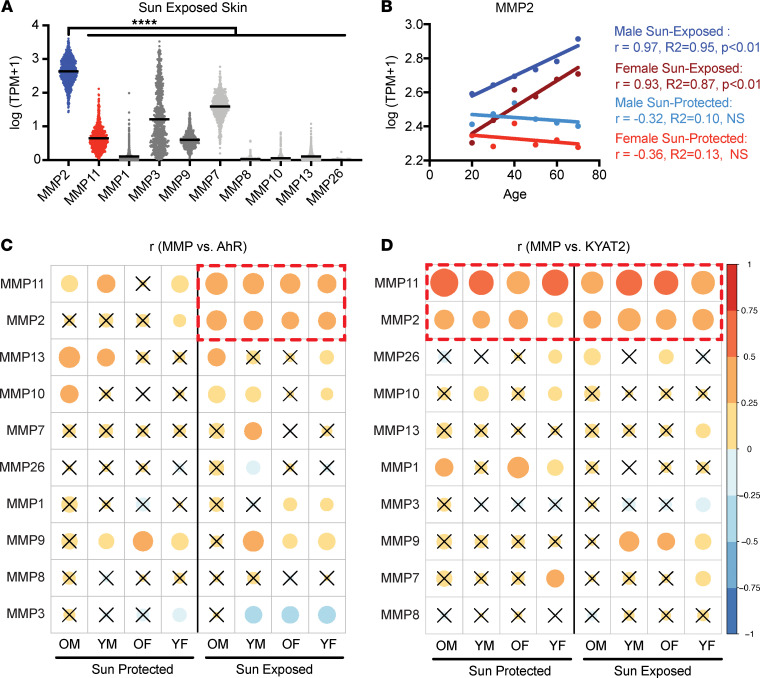
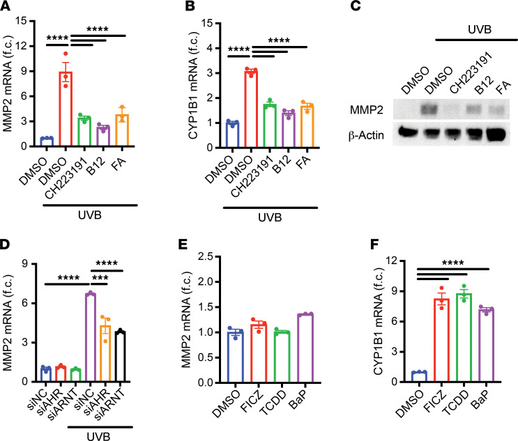
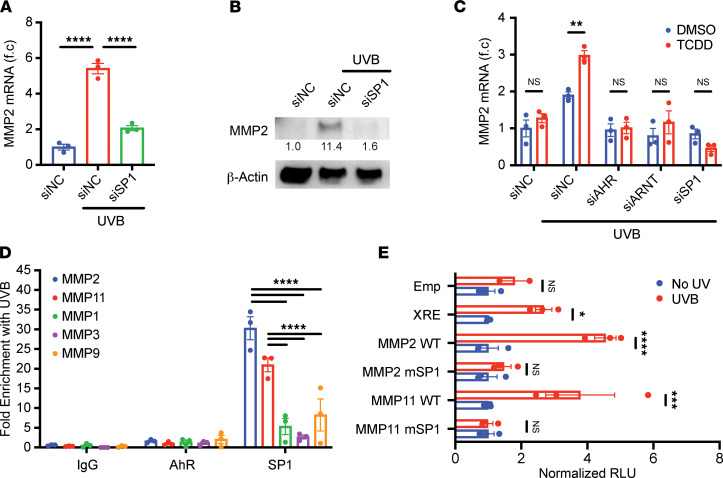
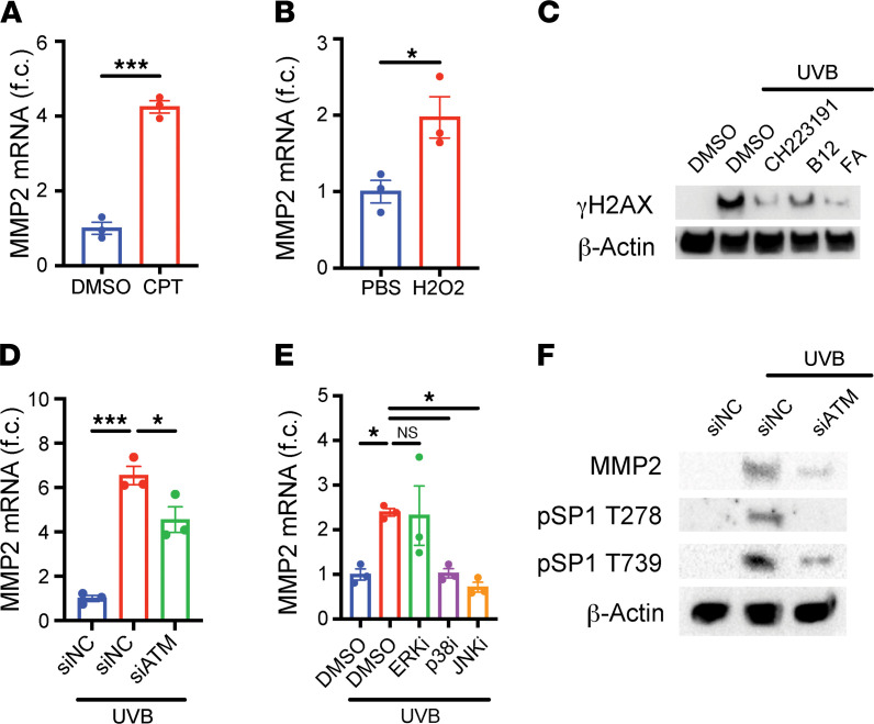
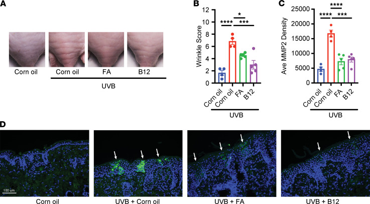
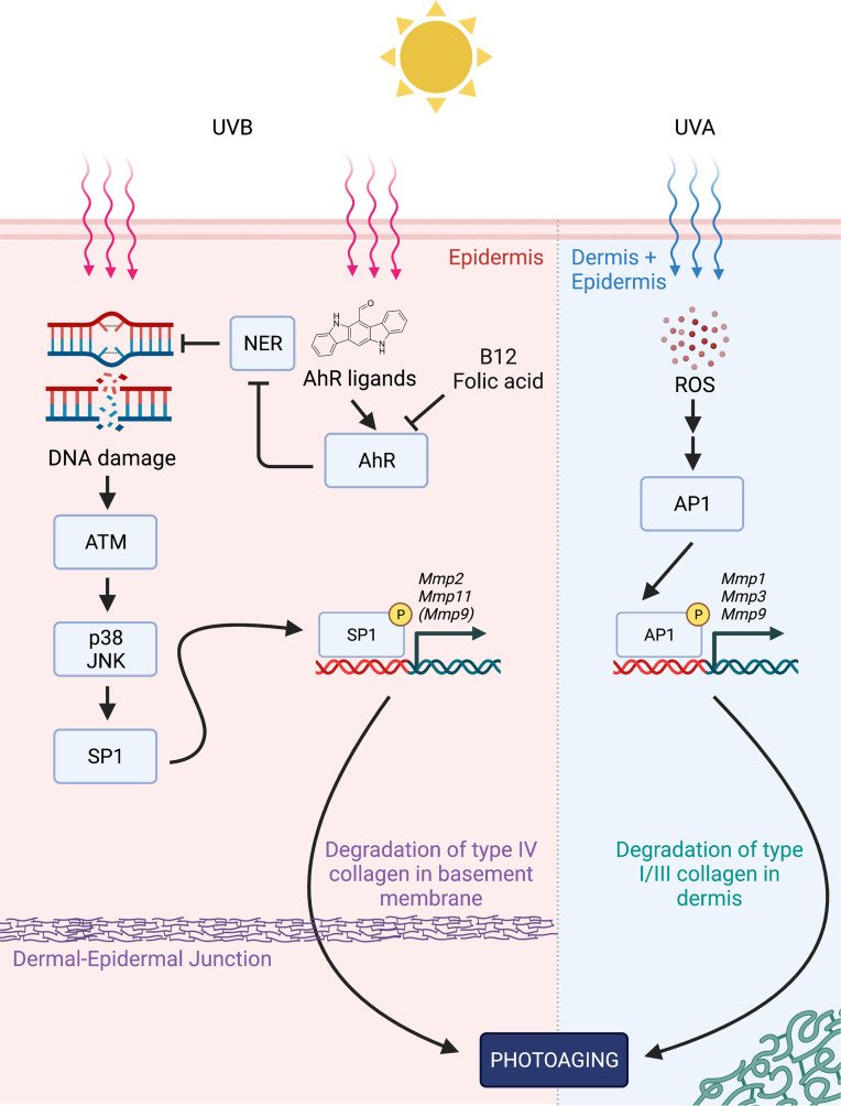
It is currently thought that UVB radiation drives photoaging of the skin primarily by generating ROS. In this model, ROS purportedly activates activator protein-1 to upregulate MMPs 1, 3, and 9, which then degrade collagen and other extracellular matrix components to produce wrinkles. However, these MMPs are expressed at relatively low levels and correlate poorly with wrinkles, suggesting that another mechanism distinct from ROS and MMP1/3/9 may be more directly associated with photoaging. Here we show that MMP2, which degrades type IV collagen, is abundantly expressed in human skin, increases with age in sun-exposed skin, and correlates robustly with aryl hydrocarbon receptor (AhR), a transcription factor directly activated by UV-generated photometabolites. Through mechanistic studies with HaCaT human immortalized keratinocytes, we found that AhR, specificity protein 1 (SP1), and other pathways associated with DNA damage are required for the induction of both MMP2 and MMP11 (another MMP implicated in photoaging), but not MMP1/3. Last, we found that topical treatment with AhR antagonists vitamin B12 and folic acid ameliorated UVB-induced wrinkle formation in mice while dampening MMP2 expression in the skin. These results directly implicate DNA damage in photoaging and reveal AhR as a potential target for preventing wrinkles.

Keywords: Aging; Collagens; Dermatology; Skin; Transcription.


Conflict of interest statement

Conflict of interest: The authors have declared that no conflict of interest exists.

Figures

Figure 1
Figure 1. Mmp2 mRNA expression increases with age in sun-exposed human skin and correlates with AhR pathway expression.
(A) MMP mRNA expression in sun-exposed human skin. (B) Mmp2 mRNA expression with increasing age in human skin. Correlations between MMP expression and (C) Ahr and (D) Kyat2 mRNA in human skin across donor demographics (OM, older males; YM, younger males; OF, older females; YF, younger females). Color represents correlation coefficients. Size represents P values. Crossed-out circles indicate nonsignificant correlations (P > 0.05). All data are log10-transformed TPM values from GTEx. n = 234 male sun-exposed samples, 467 female sun-exposed samples, 193 male sun-protected samples, and 411 female sun-protected samples. P values were calculated by Pearson’s correlation, unpaired 2-tailed Mann-Whitney test, or 1-way ANOVA; ****P < 0.0001.
Figure 2
Figure 2. AhR is required, but not sufficient, for UVB-mediated induction of Mmp2.
(AC) HaCaT keratinocytes were irradiated with 200 mJ/cm2 UVB and subsequently treated with DMSO, 10 μM AhR antagonist CH223191, 50 ng/mL vitamin B12, or 20 ng/mL FA for 24 hours. (A) Mmp2 and (B) Cyp1b1 mRNA expression were measured by RT-qPCR and normalized to Hprt1 and DMSO-treated samples (n = 3). (C) Protein levels of MMP2 and β-Actin were measured by Western blot. (D) HaCaT keratinocytes were transfected with negative control siRNA (siNC), siAHR, or siARNT and subsequently irradiated with 100 mJ/cm2 UVB. Cells were lysed for analysis 24 hours later. Mmp2 mRNA was measured by RT-qPCR and normalized to Hprt1 and siNC-transfected samples (n = 3). (E and F) HaCaT keratinocytes were treated with DMSO, 10 nM FICZ, 5 nM TCDD, or 5 μM BaP for 24 hours. (E) Mmp2 and (F) Cyp1b1 mRNA were measured by RT-qPCR and normalized to Hprt1 and DMSO-treated samples (n = 3). Data are means ± SEM. P values were calculated by Pearson’s correlation or 1-way ANOVA; ***P < 0.001, ****P < 0.0001.
Figure 3
Figure 3. SP1 binding to promoter region is required for UVB-mediated induction of Mmp2 and Mmp11.
(A and B) HaCaT keratinocytes were transfected with siNC or siSP1 and irradiated with 100 mJ/cm2 UVB. Cells were lysed 24 hours later for analysis. (A) Mmp2 mRNA was measured by RT-qPCR and normalized to Hprt1 and siNC-transfected samples (n = 3). (B) Protein levels of MMP2 and β-Actin were measured by Western blot. Relative densitometry measurements are shown for MMP2. (C) HaCaT keratinocytes were transfected with indicated siRNA, irradiated with UVB, and subsequently treated with DMSO or 25 nM TCDD. Cells were lysed 24 hours later for analysis. Mmp2 mRNA was measured by RT-qPCR and normalized to Hprt1 (n = 3). (D) HaCaT cells were irradiated with UVB and lysed 24 hours later. Enrichment of AhR and SP1 at MMP promoter regions was measured by ChIP-PCR, normalizing to input samples and nonirradiated samples (n = 3). (E) HaCaT cells were cotransfected with Renilla luciferase construct and one of the following: an empty firefly luciferase construct (Emp), a firefly luciferase construct containing AhR response elements (XRE), WT MMP2 promoter (MMP2 WT), SP1 binding site mutant MMP2 promoter (MMP2 mSP1), WT MMP11 promoter (MMP11 WT), or SP1 binding site mutant MMP11 promoter (MMP11 mSP1). Cells were irradiated with UVB and lysed 24 hours later. Fold induction of relative luciferase units (RLU) was calculated by normalizing firefly luciferase activity to Renilla luciferase activity and nonirradiated samples (n = 3). Data are means ± SEM. P values were calculated by 1-way ANOVA or 2-way ANOVA. *P < 0.05, **P < 0.01, ***P < 0.001, ****P < 0.0001.
Figure 4
Figure 4. DNA damage, ATM, p38, and JNK are implicated in the induction of Mmp2 mRNA.
(A) HaCaT keratinocytes were treated with DMSO or 5 μM camptothecin (CPT) for 24 hours. Mmp2 mRNA was measured by RT-qPCR and normalized to Hprt1 and DMSO-treated samples (n = 3). (B) HaCaT keratinocytes were treated with PBS or 1 mM H2O2 for 24 hours. Mmp2 mRNA was measured by RT-qPCR and normalized to Hprt1 and PBS-treated samples (n = 3). (C) HaCaT keratinocytes were irradiated with 200 mJ/cm2 UVB and subsequently treated with DMSO, 10 μM CH-223191, 50 ng/mL vitamin B12, or 20 ng/mL FA for 24 hours. Protein levels of γH2AX and β-Actin were measured by Western blot. (D and F) HaCaT keratinocytes were transfected with siNC or siATM and irradiated with UVB. Cells were lysed 24 hours later for analysis. (D) Mmp2 mRNA was measured by RT-qPCR and normalized to Hprt1 and siNC-transfected samples (n = 3). (F) Protein levels of MMP2, phosphorylated SP1 (Thr-278 and Thr-739 residues), and β-Actin were measured by Western blot. (E) HaCaT keratinocytes were irradiated with UVB and treated with DMSO, 50 μM PD98059 (ERK inhibitor), 10 μM SB203580 (p38 inhibitor), or 25 μM SP600125 (JNK inhibitor) for 24 hours. Mmp2 mRNA was measured by RT-qPCR and normalized to Hprt1 and DMSO-treated samples (n = 3). Data are means ± SEM. P values were calculated by unpaired 2-tailed Student’s t test and 1-way ANOVA. *P < 0.05, ***P < 0.001.
Figure 5
Figure 5. Treatment with vitamin B12 or FA ameliorates UVB-induced wrinkles and MMP2 expression.
SKH1 hairless mice were irradiated 3 times a week for 16 weeks with gradually increasing doses. After each irradiation, corn oil, B12, or FA was topically applied to the dorsum of mice. At week 13, mice were evaluated for wrinkle formation. (A) Representative images of wrinkles from the lower dorsum of mice. (B) Wrinkles were graded by blinded investigators using the scale provided by Inomata et al. (11). (C and D) Sections perpendicular to the wrinkles were paraffin-fixed and stained with DAPI and anti-MMP2 antibody with fluorescence IHC. Bands of MMP2 expression for each section were identified at 200× original magnification, and fluorescence was quantified by ImageJ (NIH). (C) Average fluorescence intensity of MMP2 bands by mouse. (D) Representative images of MMP2 bands (white arrows) found on IHC (200× original magnification). Blue indicates DAPI staining; green indicates MMP2 staining. n = 4–5 mice for each treatment group. Data are means ± SEM. P values were calculated by 1-way ANOVA. *P < 0.05, ***P < 0.001, ****P < 0.0001.
Figure 6
Figure 6. Proposed model for MMP induction in photoaging.
In the epidermis, first, UVB-generated aromatic ligands activate AhR which suppress NER activity; second, enhanced DNA damage is detected by ATM kinase, which subsequently activates p38 and JNK MAPK pathways; and third, phosphorylated SP1 binds the GC-rich promoter regions of Mmp2 and Mmp11 (possibly alongside Mmp9) to promote their transcription. At the same time, in both the dermis and epidermis, UVA-generated ROS ultimately leads to the activation of AP1, leading to the upregulation of Mmp1, Mmp3, and Mmp9 (previously shown). Both sets of MMP genes could then contribute to the signs of photoaging in skin with MMP2, primarily degrading type IV collagen in the basement membrane, and other MMPs degrading types I/III collagen in the dermis. By antagonizing AhR and promoting clearance of UVB-generated DNA damage, B12 and FA could potentially counteract photoaging. Diagram generated using BioRender.

Similar articles

Cited by

References

    1. Khan AQ, et al. Roles of UVA radiation and DNA damage responses in melanoma pathogenesis. Environ Mol Mutagen. 2018;59(5):438–460. doi: 10.1002/em.22176. - DOI - PMC - PubMed
    1. Rabe JH, et al. Photoaging: mechanisms and repair. J Am Acad Dermatol. 2006;55(1):1–19. doi: 10.1016/j.jaad.2005.05.010. - DOI - PubMed
    1. Poon F, et al. Mechanisms and treatments of photoaging. Photodermatol Photoimmunol Photomed. 2015;31(2):65–74. doi: 10.1111/phpp.12145. - DOI - PubMed
    1. Lucas RM, et al. The consequences for human health of stratospheric ozone depletion in association with other environmental factors. Photochem Photobiol Sci. 2015;14(1):53–87. doi: 10.1039/C4PP90033B. - DOI - PubMed
    1. Cadet J, Douki T. Formation of UV-induced DNA damage contributing to skin cancer development. Photochem Photobiol Sci. 2018;17(12):1816–1841. doi: 10.1039/C7PP00395A. - DOI - PubMed

Publication types

Substances