Review
. 2022 Mar;2(3):e394.
doi: 10.1002/cpz1.394.

Validation-Based Insertional Mutagenesis (VBIM), A Powerful Forward Genetic Screening Strategy

Affiliations
Review

Validation-Based Insertional Mutagenesis (VBIM), A Powerful Forward Genetic Screening Strategy

Sarmishtha De et al. Curr Protoc. 2022 Mar.

Abstract

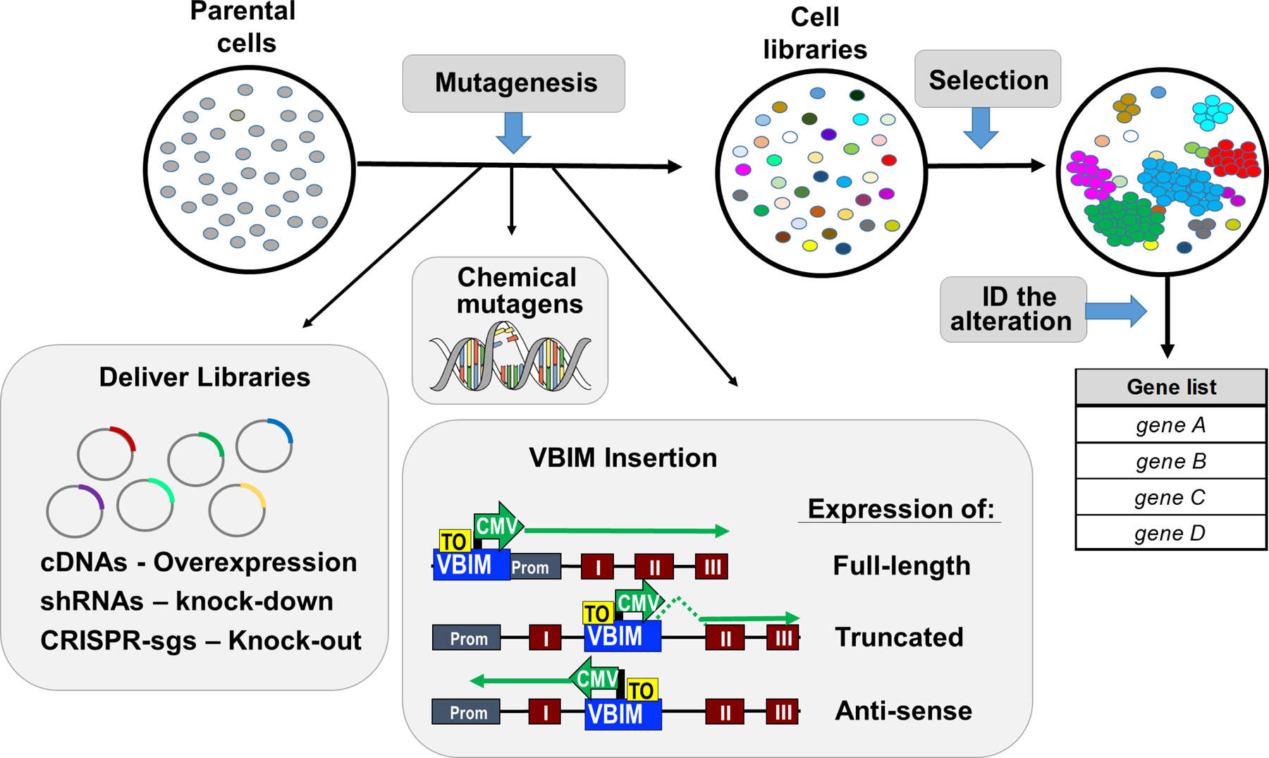
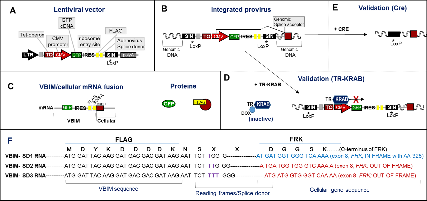
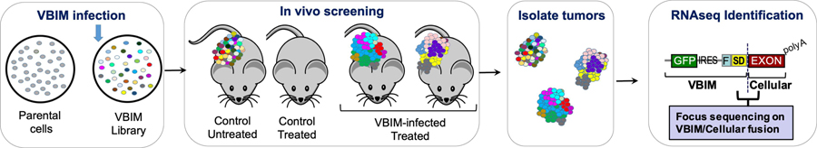
Forward genetics begins with a biological phenotype and attempts to identify genetic changes that influence that phenotype. These changes can be induced in a selected group of genes, for instance, by using libraries of cDNAs, shRNAs, CRISPR guide RNAs, or genetic suppressor elements (GSEs), or randomly throughout the genome using chemical or insertional mutagens, with each approach creating distinct genetic changes. The Validation-Based Insertional Mutagenesis (VBIM) strategy utilizes modified lentiviruses as insertional mutagens, placing strong promoters throughout the genome. Generating libraries with millions of cells carrying one or a few VBIM promoter insertions is straightforward, allowing selection of cells in which overexpression of VBIM-driven RNAs or proteins promote the phenotype of interest. VBIM-driven RNAs may encode full-length proteins, truncated proteins (which may have wild-type, constitutive, or dominant-negative activity), or antisense RNAs that can disrupt gene expression. The diversity in VBIM-driven changes allows for the identification of both gain-of-function and loss-of-function mutations in a single screen. Additionally, VBIM can target any genomic locus, regardless of whether it is expressed in the cells under study or known to have a biological function, allowing for true whole-genome screens without the complication and cost of constructing, maintaining, and delivering a comprehensive library. Here, we review the VBIM strategy and discuss examples in which VBIM has been successfully used in diverse screens to identify novel genes or novel functions for known genes. In addition, we discuss considerations for transitioning the VBIM strategy to in vivo screens. We hope that other laboratories will be encouraged to use the VBIM strategy to identify genes that influence their phenotypes of interest. © 2022 Wiley Periodicals LLC.

Keywords: cell libraries; drug resistance; forward genetics; in vivo screens; insertional mutagenesis; lentiviral vectors.


Conflict of interest statement

CONFLICT OF INTEREST STATEMENT

The authors declare no conflicts of interest.

Figures

Figure 1.
Figure 1.. Overview of forward genetics screening protocols.
In screens using human or mouse cells, genetic changes can be induced by chemical mutagens, insertional mutagens, or delivery of libraries of cDNAs, shRNAs, or CRISPR-sgRNAs. Following mutagenesis, each cell in the library has a unique genetic change. The cell libraries can be screened for a mutant phenotype, in which rare mutations allowed some cells to survive, thrive, and grow when most other cells are eliminated from the population. The mutation can be identified, and the list of candidate genes can be prioritized and characterized using traditional reverse genetic approaches. Following infection with the VBIM lentiviruses, integration can result in a number of possible alterations (see “VBIM insertion” insert). Examples shown include: (i) insertion in or near the endogenous promoter, potentially causing the over expression of a full-length RNA that may give rise to a full-length protein (if in a coding region) or a full-length lncRNA (if in a non-coding gene); (ii) insertion within an intron of a coding or non-coding gene in the sense orientation, resulting in a truncated RNA that could encode a truncated protein or lncRNA (which may have normal, constitutive, or dominant-negative activity); and (iii) insertion within an intron of a coding or non-coding gene in the antisense orientation, probably reducing efficient transcription from the endogenous promoter. Other possible integrations, such as those in intergenic regions or repetitive transcribed elements, are not shown.
Figure 2.
Figure 2.. Components of the VBIM lentiviruses.
Illustration of the VBIM lentiviral vector backbone (A) and the components of the VBIM provirus, following integration into the infected cells genome (B). The 3’ self-inactivating (SIN) LTR was mutated to delete the viral polyadenylation signal (denoted by *) and a LoxP site was included. Following infection of target cells, reverse transcription copies the self-inactivating 3’LTR, such that the provirus LTRs integrated in the genome have no promoter activity and no polyadenylation signal; also, the entire VBIM cassette is flanked by LoxP sites. Transcription from the integrated VBIM CMV promoter creates an RNA that includes the GFP (green fluorescent protein) cDNA, an IRES (internal ribosome entry site), a FLAG epitope tag, and a splice donor that results in splicing of the VBIM RNA (GFP-IRES-FLAG-SD) into downstream exons. (C) If the VBIM integration occurs in an intron, the CMV-driven RNA results in a VBIM/cellular mRNA fusion that encodes GFP and, if the targeted gene is coding, a FLAG-tagged cellular protein; if the VBIM insertion is not in a coding region or occurs in the antisense orientation, only GFP would be expressed. (D) Addition of TR-KRAB, a tetracycline/doxycycline-regulated protein, allows for regulated expression of the VBIM CMV promoter and the VBIM/cellular mRNA and encoded proteins. (E) Upon addition of Cre recombinase, all but 238 nucleotides of the VBIM provirus will be deleted, leaving only a SIN LTR with no promoter or polyadenylation signal. (F) Three different reading frames of FLAG-SD were created as separate constructs, VBIM-SD1, VBIM-SD2, and VBIM-SD3, differing by 1 nucleotide each (note the purple T, TT, or TTT). The FLAG epitope is embedded just upstream of the splice donor site, so that one of the VBIM-SD reading frames can properly splice into each splice acceptor to give rise to a potential FLAG-tagged fusion protein. An example of a VBIM-SD1 insertion into intron 7 of FRK (Fyn-related Kinase) demonstrates how splicing would create an in-frame FLAG fusion with the C-terminus of the FRK protein (encoding FRK 338–512). VBIM-SD2 and VBIM-SD3 would splice properly with exon 8 of FRK, but would be out-of-frame with the FRK C-terminus. See text for details.
Figure 3.
Figure 3.. Identification of VBIM-targeted genes by RNA-Seq.
(A and B) RNA-Seq data from individual clones or populations of selected cells can be filtered for those containing known VBIM RNA sequences (depicted as blue regions of the RNA-seq reads) and the attached cellular sequence identified using Nucleotide BLAST. (C) The mutation can be predicted based on the identified cellular sequences. For example, when VBIM RNA-seq reads are attached to an exon of a coding or non-coding gene, the insertion would be predicted to be in the preceding intron, as shown for the VBIM-SD1 insertion into intron 7 of FRK (Fyn-related Kinase), also shown in Figure 2F.
Figure 4.
Figure 4.. Schematic of in vivo VBIM selection.
Following the generation of VBIM libraries, cells injected into mice can be selected. In the case of selection for resistance to standard-of-care or experimental therapeutics, the experimental considerations (drug dosage and timing, cell number, VBIM integration numbers, etc.) need to create substantial differences in tumor growth kinetics when comparing sensitive and resistant cells/tumors. Pilot experiments examining controls that consist of cells expressing a known resistance gene or a mixture of resistant cells with sensitive cells at known frequencies should provide convincing tumor growth differences. During the screen, control cells infected with a non-mutagenic lentivirus should be included to assess therapeutic efficiency; tumors in VBIM-infected tumors should be consistently larger than control tumors. Following selection, RNA-seq for VBIM-tagged RNAs will identify genes that have been targeted within the resistant populations, with relative RNA expression correlating with relative enrichment (i.e. genes that are most frequently represented within the RNA-seq data are stronger candidates to induce resistance than genes with fewer reads).
Figure 5.
Figure 5.. Assessing tumor cell loss in vivo.
Murine triple negative breast cancer (EMT6), pancreatic cancer (Pan02), and melanoma (B16.F10) cell lines were used to assess the potential loss of diversity (i.e. drop-out) of each line following inoculation in vivo. Infections of 100,000, 200,000, or 500,000 cells into either Balb/c (EMT6) or C57bl/6 mice (Pan02 and B16.F10) are shown; 5 tumors were assessed for each cell line and each cell number, with each injection represented as an individual growth curve. In vivo tumor growth was monitored 1–6 days after inoculation using bioluminescence (luciferase signal for each tumor is plotted). (A) The luminescence signal from 500,000 cells/injection at time 0 (immediately after injection) and after 24 or 48 hours indicates that the number of luciferase-positive Pan02 and B16.F10 cells is drastically reduced at these times; EMT6 populations remain more stable after inoculation. (B) The luminescence signal for all 3 cell numbers/injections are plotted. (C) Extrapolation of the data from the luminescence to illustrate library complexity in a VBIM screen indicates that the initial diversity of the VBIM libraries would be better maintained in the EMT6 models. The Pan02 and B16.F10 models show a dramatic, stochastic reduction of cell number following injection, suggesting that a dramatic loss of VBIM diversity would occur. Such a large loss of library complexity would make a suitable genetic screen in these cells challenging.

Similar articles

References

    1. Bartel CA, Parameswaran N, Cipriano R, & Jackson MW (2016). FAM83 proteins: Fostering new interactions to drive oncogenic signaling and therapeutic resistance. Oncotarget, 7(32), 52597–52612. doi:10.18632/oncotarget.9544 - DOI - PMC - PubMed
    1. Bose R, & Ma CX (2021). Breast Cancer, HER2 Mutations, and Overcoming Drug Resistance. N Engl J Med, 385(13), 1241–1243. doi:10.1056/NEJMcibr2110552 - DOI - PubMed
    1. Cipriano R, Graham J, Miskimen KL, Bryson BL, Bruntz RC, Scott SA, . . . Jackson MW (2012). FAM83B mediates EGFR- and RAS-driven oncogenic transformation. J Clin Invest, 122(9), 3197–3210. doi:10.1172/JCI60517 - DOI - PMC - PubMed
    1. Cipriano R, Miskimen KL, Bryson BL, Foy CR, Bartel CA, & Jackson MW (2014). Conserved oncogenic behavior of the FAM83 family regulates MAPK signaling in human cancer. Mol Cancer Res, 12(8), 1156–1165. doi:10.1158/1541-7786.MCR-13-0289 - DOI - PMC - PubMed
    1. Cipriano R, Miskimen KLS, Bryson BL, Foy CR, Bartel CA, & Jackson MW (2013). FAM83B-mediated activation of PI3K/AKT and MAPK signaling cooperates to promote epithelial cell transformation and resistance to targeted therapies. Oncotarget, 4(5), 729–738. Retrieved from http://www.ncbi.nlm.nih.gov/pmc/articles/PMC3742833/ - PMC - PubMed