Review
. 2022 Jul 8:45:273-294.
doi: 10.1146/annurev-neuro-110520-031137. Epub 2022 Mar 22.

Fluorescence Imaging of Neural Activity, Neurochemical Dynamics, and Drug-Specific Receptor Conformation with Genetically Encoded Sensors

Affiliations
Review

Fluorescence Imaging of Neural Activity, Neurochemical Dynamics, and Drug-Specific Receptor Conformation with Genetically Encoded Sensors

Chunyang Dong et al. Annu Rev Neurosci. .

Abstract

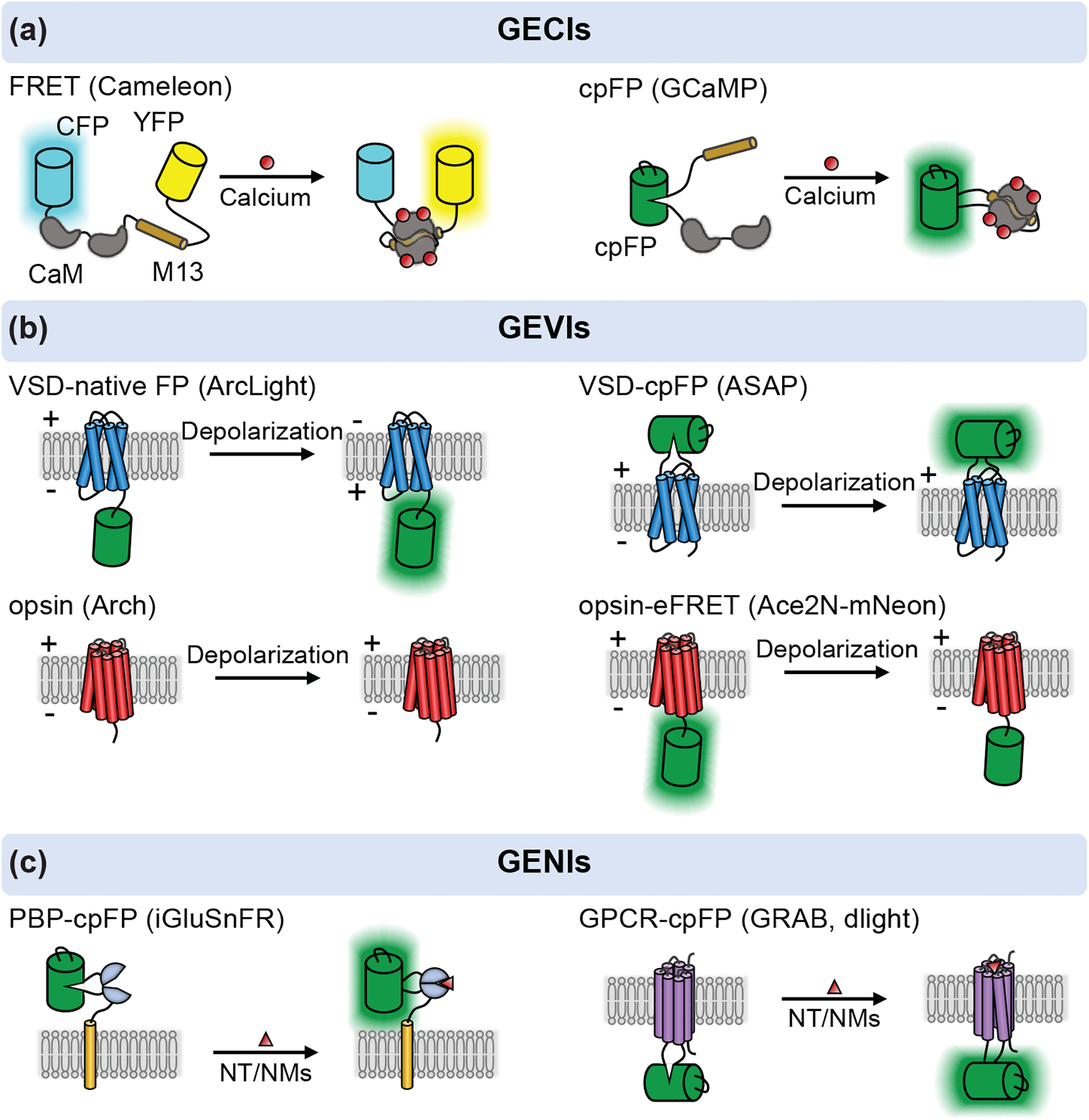
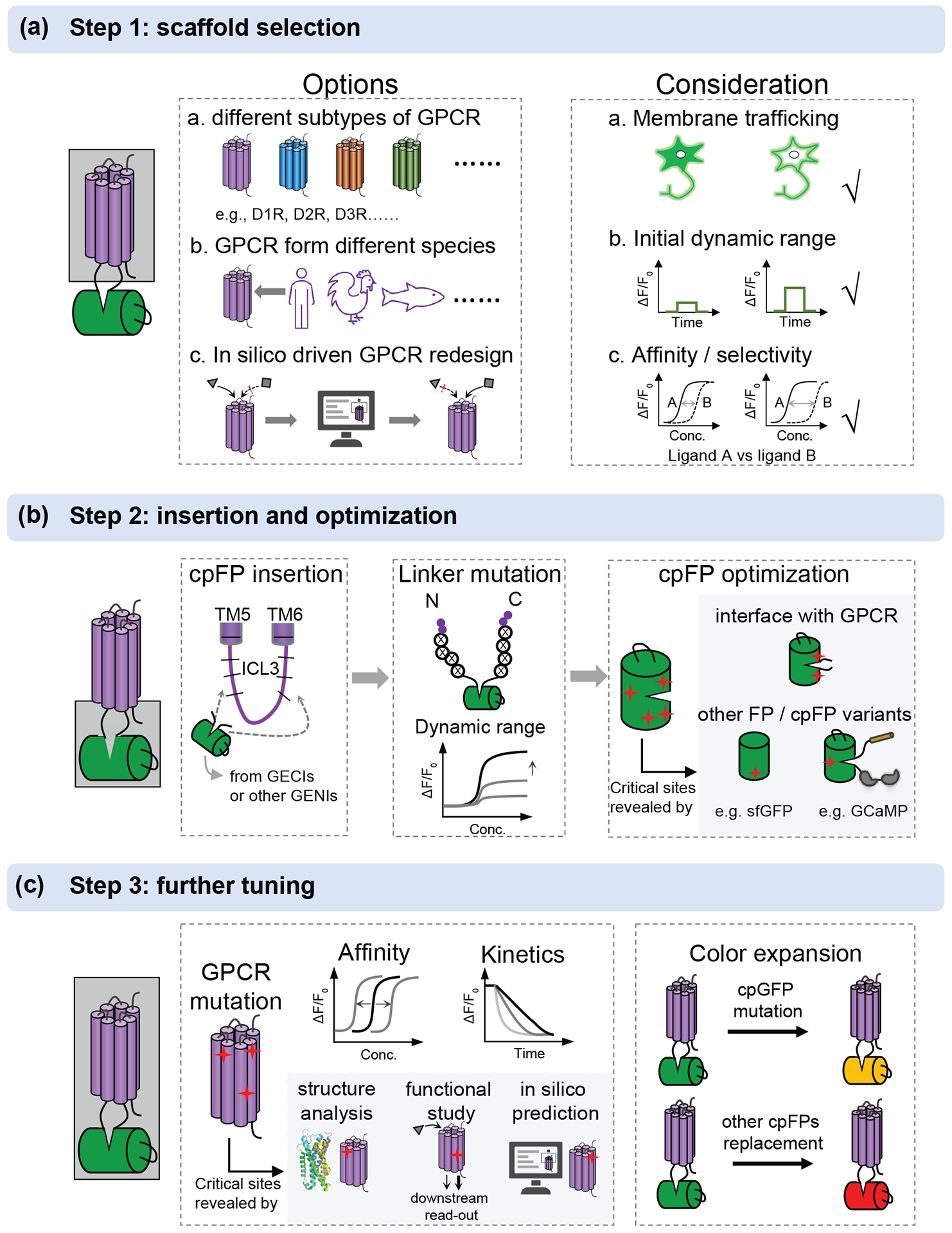
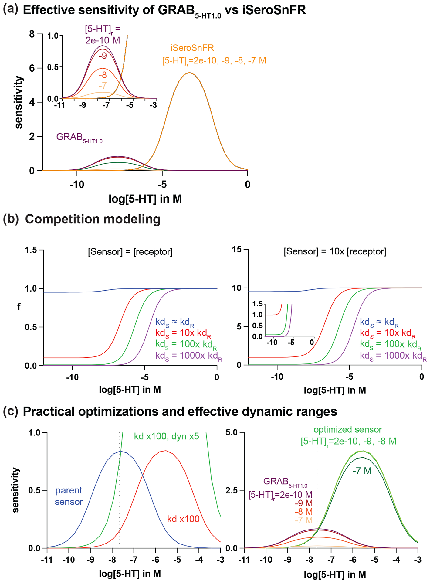
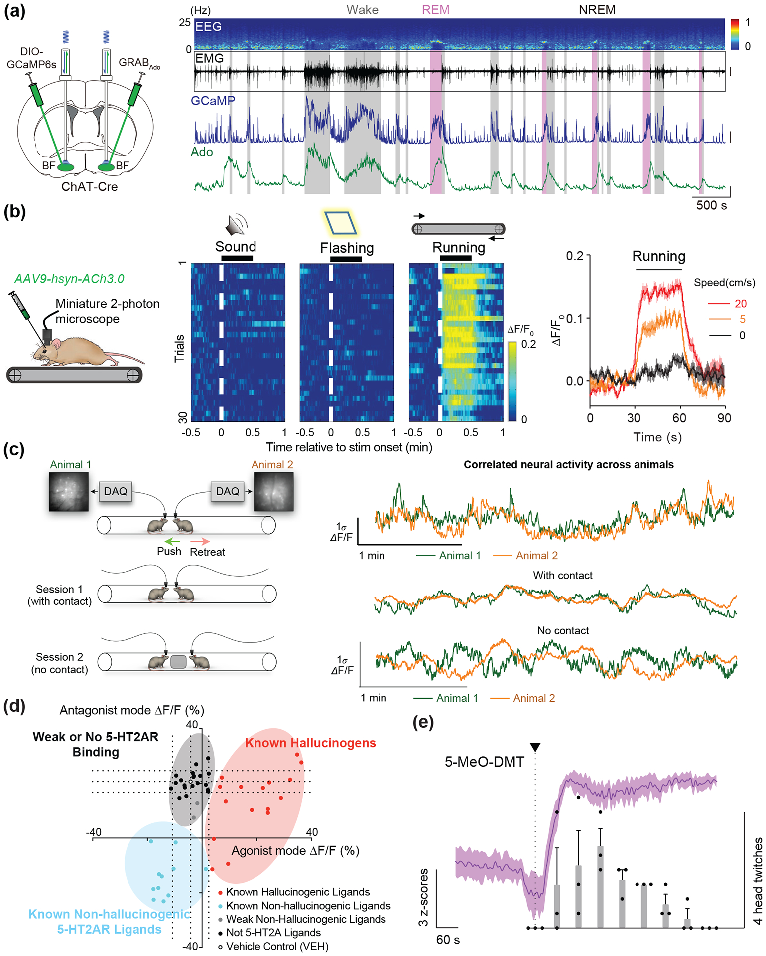
Recent advances in fluorescence imaging permit large-scale recording of neural activity and dynamics of neurochemical release with unprecedented resolution in behaving animals. Calcium imaging with highly optimized genetically encoded indicators provides a mesoscopic view of neural activity from genetically defined populations at cellular and subcellular resolutions. Rigorously improved voltage sensors and microscopy allow for robust spike imaging of populational neurons in various brain regions. In addition, recent protein engineering efforts in the past few years have led to the development of sensors for neurotransmitters and neuromodulators. Here, we discuss the development and applications of these genetically encoded fluorescent indicators in reporting neural activity in response to various behaviors in different biological systems as well as in drug discovery. We also report a simple model to guide sensor selection and optimization.

Keywords: biosensors; drug discovery; fluorescence imaging; genetically encoded indicators; neurocircuit imaging; neuromodulation.


Figures

Figure 1:
Figure 1:. A schematic of genetically encoded sensors for calcium (GECIs), voltage (GEVIs) and neurochemicals (GENIs).
(a) GECIs based on FRET and cpFP. The FERT based calcium sensor is developed by inserting the CaM-M13 in between a donor and acceptor fluorophores, such as CFP-YFP or BFP-GFP (e.g. Cameleon). The cpFP based sensor is intensiometric, in which a cpFP is inserted in between CaM and M13/RS20 (e.g. GCaMP). (b) GEVIs utilize the voltage-sensitive domain (VSD) or opsin as scaffold. The VSD based sensors are developed by attaching a native FP to the C-terminus of VSD (e.g. ArcLight) or inserting a cpGFP into the extracellular S3-S4 loop of VSD (e.g. ASAP series). The light-driven proton pumps (opsin) are functionally reversed to action as a voltage sensitive optical element (e.g. Arch). A bright FP is attached to the opsin to address the dimness of opsin based GEVIs via electrochromic FRET (eFRET), as shown in Ace2N-mNeon. The FP is replaced with HaloTag - Janelia fluro dyes to develop a hybrid voltage sensor, namely Voltron. (c) Two class of ligand-binding scaffolds, PBPs and GPCRs, are used to developed GENIs. In both cases, a cpFP is inserted into the hinge region of PBPs (e.g. iGluSnFR) or intracellular loop 3 of GPCRs (e.g. GRAB, dLight). Examples named in the figure are as follows: Cameleon (Miyawaki et al 1997), GCaMP (Nagai et al 2001), ArcLight (Jin et al 2012), ASAP series (St-Pierre et al 2014, Villette et al 2019, Yang et al 2016), Arch (Kralj et al 2011), Ace2N-mNeon (Gong et al 2015), Voltron (Abdelfattah et al 2019), iGluSnFR (Marvin et al 2013), GRAB (Sun et al 2018) and dLight (Patriarchi et al 2018).
Figure 2:
Figure 2:. A workflow for the development and optimization of GPCR based neurochemical sensors.
(a) The options and consideration for selection of an appropriate scaffold. GPCRs can be from different subtypes, different species and redesign in silicon. A good scaffold should have good membrane trafficking, high initial dynamic range after cpGFP insertion, appropriate affinity and high selectivity for ligand of interest. (b) After choosing a good scaffold, cpFP insertion, linker optimization and cpFP optimization can be performed sequentially. The critical sites in cpFP optimization are mainly on the interface with GPCR or learned from other FP or cpFP variants, as highlighted in gray. (c) Further tuning can be performed by mutating GPCR to tune the affinity and kinetics. The potential sites in GPCR can be obtained from the reported GPCR structures, previous function studies by downstream signaling detection and in silico prediction, as highlighted in gray. The color can be expanded by introducing mutation to cpGFP or replacing with other cpFPs, including commonly used cpmApple.
Figure 3:
Figure 3:. Computational modeling to guide sensor optimizations.
(a) Modeling of effective sensitivity of GRAB5-HT1.0 and iSeroSnFR at resting 5-HT concentration ([5-HT]r) of 200nM, 20nM, 2nM, and 200pM. (b) A 100-fold (green) or greater increase in sensor kd relative to that of the native receptor minimizes ligand buffering effect when sensor and receptor expression is equal (left) and when sensor expression is 10 times higher than receptor expression (right). (c) Left panel: a practical optimization for GRAB5-HT1.0 to maintain or enhance sensitivity while minimizing competition is to increase both the kd and the maximum dynamic range of the parent sensor (blue). According to the model, this can be achieved with a 100-fold increase in kd and a 5-fold increase in maximum dynamic range (green). Right panel: the optimized sensor is more tolerant to changes in [5-HT]r compared to the parent GRAB5-HT1.0.
Figure 4:
Figure 4:. Behavioral and pharmacological applications of NT/NM sensors.
(a) Long-term recording of adenosine release in cholinergic neurons using the GRABAdo sensor. Changes in adenosine-dependent fluorescence can be compared to cholinergic calcium activity and EEG/EMG signal across the full time-course of the sleep/wake cycle (Peng et al 2020). (b) Imaging GRABAch3.0 sensor using miniature 2-photon microscopy in a treadmill task in mice. Single-cell changes in ΔF/F0 response are tractable during different stages of the task, and across running speeds (Jing et al 2020). (c) Simultaneous calcium imaging in dmPFC of two mice during a social interaction test reveals correlations in neural activity during contact vs. no contact sessions (Kingsbury et al 2019). (d) psychLight based characterization of compounds based on 5-HT2AR binding and hallucinogenic potential (Dong, et al., 2021). (e) psychLight tracks in vivo action of 5-MeO-DMT administration over the course of the head-twitch response (Dong et al 2021).

Similar articles

Cited by

References

    1. Abdelfattah AS, Kawashima T, Singh A, Novak O, Liu H, et al. 2019. Bright and photostable chemigenetic indicators for extended in vivo voltage imaging. Science 365: 699–704 - PubMed
    1. Akerboom J, Carreras Calderon N, Tian L, Wabnig S, Prigge M, et al. 2013. Genetically encoded calcium indicators for multi-color neural activity imaging and combination with optogenetics. Front Mol Neurosci 6: 2. - PMC - PubMed
    1. Andreoni A, Davis CMO, Tian L. 2019. Measuring brain chemistry using genetically encoded fluorescent sensors. Current Opinion in Biomedical Engineering 12: 59–67
    1. Baird GS, Zacharias DA, Tsien RY. 1999. Circular permutation and receptor insertion within green fluorescent proteins. Proc Natl Acad Sci U S A 96: 11241–6 - PMC - PubMed
    1. Borden PM, Zhang P, Shivange AV, Marvin JS, Cichon J, et al. 2020. A fast genetically encoded fluorescent sensor for faithful in vivo acetylcholine detection in mice, fish, worms and flies. bioRxiv: 2020.02.07.939504

Publication types