

Curative resection with endoscopic submucosal dissection of early gastric cancer in Helicobacter pylori-negative Ménétrier's disease: A case report
- PMID: 35316958
- PMCID: PMC8905015
- DOI: 10.3748/wjg.v28.i5.594
Curative resection with endoscopic submucosal dissection of early gastric cancer in Helicobacter pylori-negative Ménétrier's disease: A case report
Abstract
Background: Adult-onset Ménétrier's disease is strongly associated with Helicobacter pylori (H. pylori) infection and an elevated risk of carcinogenesis. Cases of early-stage gastric cancer developed in H. pylori-negative Ménétrier's disease are extremely rare. We report a case of early gastric cancer in H. pylori-negative Ménétrier's disease that was curatively resected with endoscopic submucosal dissection (ESD).
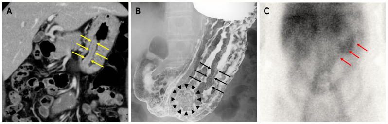
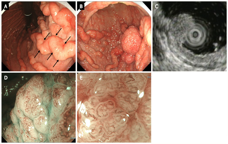
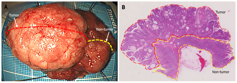
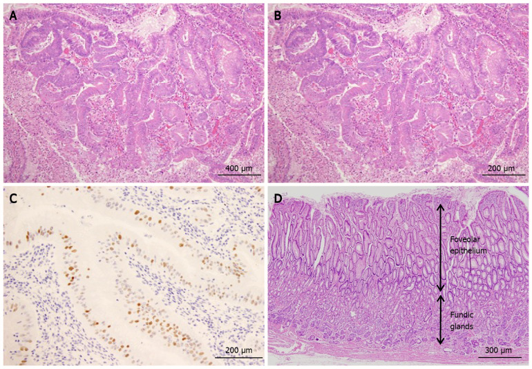
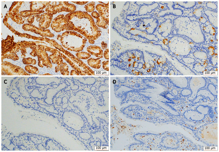
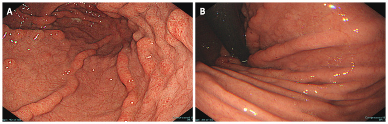
Case summary: A 60-year-old woman was referred to our hospital after her medical examination detected anemia. Contrast-enhanced upper gastrointestinal (UGI) radiography revealed translucency of the nodule-aggregating surface with giant rugae. Blood tests showed hypoproteinemia and were negative for serum H. pylori immunoglobulin G antibodies. The 99mTc-DTPA-human serum albumin scintigraphy showed protein loss from the stomach. UGI endoscopy showed a 40-mm protruding erythematous lesion on giant rugae of the greater curvature of lower gastric body, suggesting early-stage gastric cancer due to Ménétrier's disease. En bloc resection with ESD was performed for diagnosis and treatment. Histology of ESD showed well-differentiated tubular adenocarcinoma. The cancer was confined to the mucosa, and complete curative resection was achieved. Foveolar hyperplasia and atrophy of the gastric glands were observed in non-tumor areas, histologically corresponding to Ménétrier's disease. Three years after ESD, gastric cancer had not recurred, and Ménétrier's disease remained in remission with spontaneous regression of giant gastric rugae.
Conclusion: Complete curative resection was achieved through ESD in a patient with early-stage gastric cancer and H. pylori-negative Ménétrier's disease.
Keywords: Case report; Endoscopic resection; Endoscopic submucosal dissection; Gastric cancer; Helicobacter pylori; Ménétrier’s disease.
©The Author(s) 2022. Published by Baishideng Publishing Group Inc. All rights reserved.
Conflict of interest statement
Conflict-of-interest statement: The authors declare that they have no conflict of interest.
Figures
Similar articles
-
Survival Times of Patients With Menetrier's Disease and Risk of Gastric Cancer.Clin Gastroenterol Hepatol. 2021 Apr;19(4):707-712. doi: 10.1016/j.cgh.2020.03.017. Epub 2020 Mar 14. Clin Gastroenterol Hepatol. 2021. PMID: 32184187
-
Early gastric cancer in a patient with Menetrier's disease, lymphocytic gastritis and Helicobacter pylori.Eur J Gastroenterol Hepatol. 1995 Feb;7(2):187-90. Eur J Gastroenterol Hepatol. 1995. PMID: 7712313
-
Intestinal-type gastric adenocarcinoma without Helicobacter pylori infection successfully treated with endoscopic submucosal dissection.Clin J Gastroenterol. 2016 Aug;9(4):228-32. doi: 10.1007/s12328-016-0654-7. Epub 2016 Jun 3. Clin J Gastroenterol. 2016. PMID: 27259702
-
Menetrier's disease associated with Helicobacter pylori: three cases with sonographic findings and a literature review.Ann Trop Paediatr. 2011;31(2):141-7. doi: 10.1179/146532811X13006353133876. Ann Trop Paediatr. 2011. PMID: 21575319 Review.
-
Early gastric cancer arising from localized Ménétrier's disease.Gastroenterol Jpn. 1991 Apr;26(2):213-7. doi: 10.1007/BF02811083. Gastroenterol Jpn. 1991. PMID: 1645687 Review.
Cited by
-
Endoscopic submucosal dissection of a giant gastric polyp.VideoGIE. 2025 Mar 25;10(8):392-397. doi: 10.1016/j.vgie.2025.03.033. eCollection 2025 Aug. VideoGIE. 2025. PMID: 40704111 Free PMC article. No abstract available.
-
Upper gastrointestinal bleeding as an unusual manifestation of localized Ménétrier's disease with an underlying lipoma: A case report.World J Gastrointest Endosc. 2023 Jan 16;15(1):10-18. doi: 10.4253/wjge.v15.i1.10. World J Gastrointest Endosc. 2023. PMID: 36686066 Free PMC article.
References
-
- Ménétrier P. Des polyadenomes gastriques et de leurs rapports avec le cancer de l’estomac. Arch Physiol Norm Pathol. 1888;32:236–262.
-
- Hsu CT, Ito M, Kawase Y, Sekine I, Ohmagari T, Hashimoto S. Early gastric cancer arising from localized Ménétrier's disease. Gastroenterol Jpn. 1991;26:213–217. - PubMed
-
- Ozawa T, Wachi E, Yamashita N. A case of juvenile polyposis limited to the stomach accompanied by double gastric cancers and Ménétrier's disease. Nihon Shokakibyo Gakkai Zasshi. 2010;107:1641–1650. - PubMed
-
- Charton-Bain MC, Paraf F, Bruneval P. Superficial gastric carcinoma developed on localized hypertrophic lymphocytic gastritis: a variant of localized Ménétrier's disease? Pathol Res Pract. 2000;196:125–128. - PubMed
-
- Umegaki E, Sanomura M. Giant rugae. (in Japanese) Stomach and Intestine, Tokyo: Igakushoin, 2017; 52: 573.
Publication types
MeSH terms
LinkOut - more resources
Full Text Sources
Medical
Research Materials
Miscellaneous
