

Exercise stress echocardiography: Where are we now?
- PMID: 35316975
- PMCID: PMC8900523
- DOI: 10.4330/wjc.v14.i2.64
Exercise stress echocardiography: Where are we now?
Abstract
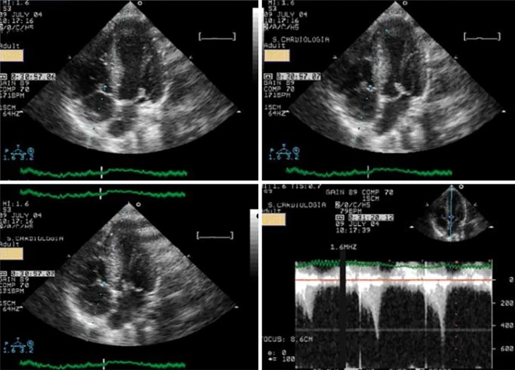
Exercise stress echocardiography (ESE) is a widely used diagnostic test in cardiology departments. ESE is mainly used to study patients with coronary artery disease; however, it has increasingly been used in other clinical scenarios including valve pathology, congenital heart disease, hypertrophic and dilated cardiomyopathies, athlete evaluations, diastolic function evaluation, and pulmonary circulation study. In our laboratories, we use an established methodology in which cardiac function is evaluated while exercising on a treadmill. After completing the exercise regimen, patients remain in a standing position or lie down on the left lateral decubitus, depending on the clinical questions to be answered for further evaluation. This method increases the quality and quantity of information obtained. Here, we present the various methods of exercise stress echocardiography and our experience in many clinical arenas in detail. We also present alternatives to ESE that may be used and their advantages and disadvantages. We review recent advances in ESE and future directions for this established method in the study of cardiac patients and underline the advantage of using a diagnostic tool that is radiation-free.
Keywords: Athletes; Children; Coronary artery disease; Exercise stress echocardiography; Intraventricular gradients; Valve disease.
©The Author(s) 2022. Published by Baishideng Publishing Group Inc. All rights reserved.
Conflict of interest statement
Conflict-of-interest statement: The authors have no Conflict of interest.
Figures


References
-
- Wann LS, Faris JV, Childress RH, Dillon JC, Weyman AE, Feigenbaum H. Exercise cross-sectional echocardiography in ischemic heart disease. Circulation . 1979;60:1300–1308. - PubMed
-
- Armstrong WF, O'Donnell J, Dillon JC, McHenry PL, Morris SN, Feigenbaum H. Complementary value of two-dimensional exercise echocardiography to routine treadmill exercise testing. Ann Intern Med . 1986;105:829–835. - PubMed
-
- Dimitrow PP, Bober M, Michałowska J, Sorysz D. Left ventricular outflow tract gradient provoked by upright position or exercise in treated patients with hypertrophic cardiomyopathy without obstruction at rest. Echocardiography . 2009;26:513–520. - PubMed
Publication types
LinkOut - more resources
Full Text Sources
