

Multiparametric Magnetic Resonance Imaging in Evaluation of Benign and Malignant Breast Masses with Pathological Correlation
- PMID: 35317029
- PMCID: PMC8934374
- DOI: 10.7759/cureus.22348
Multiparametric Magnetic Resonance Imaging in Evaluation of Benign and Malignant Breast Masses with Pathological Correlation
Abstract
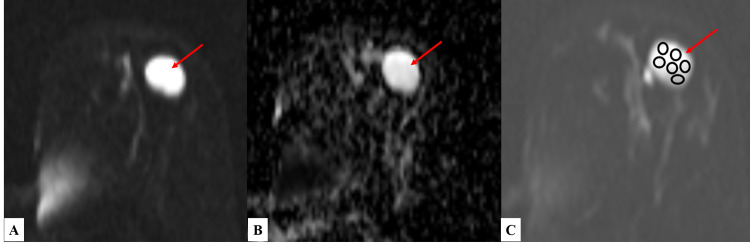
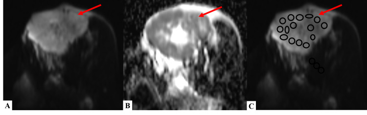
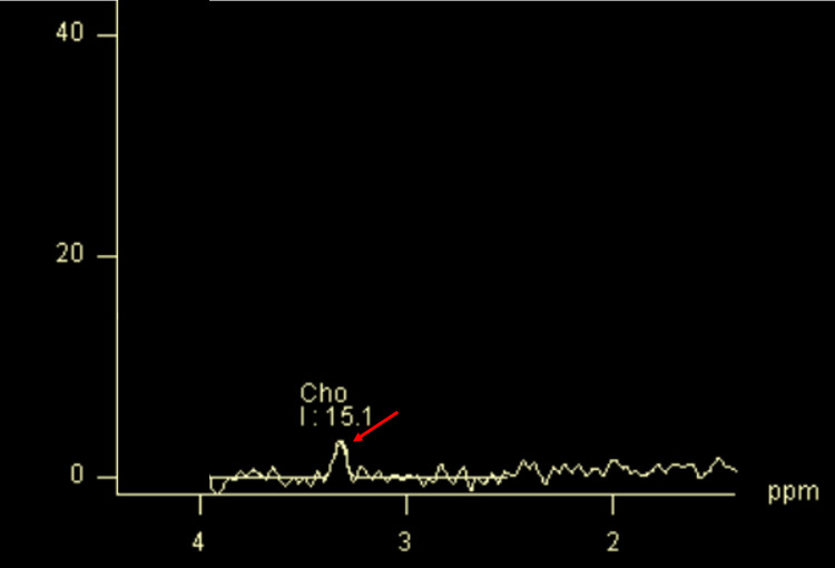
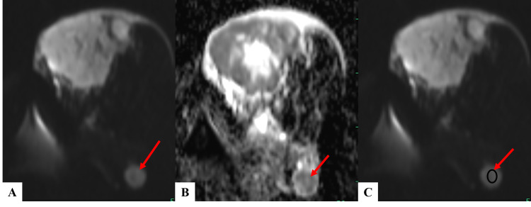
Background Dynamic contrast-enhanced (DCE) MRI sequences plays a vital role in diagnosing breast masses with high sensitivity and specificity as compared to other diagnostic modalities. The addition of diffusion-weighted imaging (DWI) and apparent diffusion coefficient (ADC) values significantly improves diagnostic accuracy. This study aimed to study the breast masses on DCE-MRI, restricted diffusion on DWI, ADC values, and choline peak on spectroscopy in breast cancer diagnosis. Material and methods This study was a prospective observational study which involved subjects with breast lumps. Baseline data was collected from the patients along with pertinent clinical history and relevant laboratory investigations. MR mammography (MRM) was performed on a 1.5 Tesla MR Scanner (MAGNETOM® Avanto, Siemens AG, Munich Germany) using a dedicated double breast coil. Results Forty-one subjects were included with a total of 54 breast masses in them. The mean age of the study population was 47.1±14.7 years. From the MRI final diagnosis, the majority (53.70%) were diagnosed as malignant lesions and 46.30% as benign. Out of 20 lesions diagnosed as benign on histopathology, only five percent had ADC value <1.3 ×10⁻³mm2/s, and the majority (95%) had ADC value >1.3 ×10⁻³mm2/s. All 20 lesions were circumscribed, ovoid, or round in shape showing no restricted diffusion on DWI, with corresponding ADC value of >1.3×10⁻³mm2/s, homogeneous post-contrast enhancement, or with dark internal septations, type I kinetic enhancement curve, and they showed no choline peak on spectroscopy. Out of 34 malignant lesions diagnosed on histopathology, the majority (85.29%) displayed restricted diffusion on DWI and had an ADC value of <1.3×10⁻³mm2/s, most of them had spiculated margins, type II/ III kinetic curve with choline peak on spectroscopy. Conclusion Multiparametric MR mammography, which included DCE-MRM, DWI, ADC values, and spectroscopy, correlated well with the histopathological diagnosis of benign and malignant breast masses.
Keywords: adc value; brest cancer; diffusion weighted imaging (dwi); kinetic curve; mr mammography.
Copyright © 2022, GR et al.
Conflict of interest statement
The authors have declared that no competing interests exist.
Figures

References
-
- Subtypes of benign breast disease as a risk factor for breast cancer: a systematic review and meta-analysis protocol. Zendehdel M, Niakan B, Keshtkar A, Rafiei E, Salamat F. https://pubmed.ncbi.nlm.nih.gov/29398746/ Iran J Med Sci. 2018;43:1–8. - PMC - PubMed
-
- World Health Organization. [ Jul; 2021 ];https://www.who.int 2022
-
- Efficacy of X-ray mammography, sonomammography and MR mammography for evaluation of breast lesions in women. Gupta P, Chatterjee S, Sharma V, Singh KK, Gupta D. Indian J Appl Res. 2017;7:26–30.
-
- Efficacy of MRI and mammography for breast-cancer screening in women with a familial or genetic predisposition. Kriege M, Brekelmans CT, Boetes C, et al. N Engl J Med. 2004;351:427–437. - PubMed
LinkOut - more resources
Full Text Sources