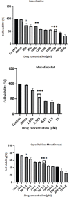
In vitro anti-proliferative effect of capecitabine (Xeloda) combined with mocetinostat (MGCD0103) in 4T1 breast cancer cell line by immunoblotting
- PMID: 35317122
- PMCID: PMC8917851
- DOI: 10.22038/IJBMS.2021.58393.12971
In vitro anti-proliferative effect of capecitabine (Xeloda) combined with mocetinostat (MGCD0103) in 4T1 breast cancer cell line by immunoblotting
Abstract
Objectives: Mouse breast cancer cell line 4T1 can accurately mimic the response to immune receptors and targeting therapeutic agents. Combined therapy has emerged as an important strategy with reduced side effects and maximum therapeutic effect. Mocetinostat (MGCD0103) is one of the members of Class I Histone Deacetylase Inhibitors (HDACi) and its mechanism of action has not been defined, yet. Capecitabine (Xeloda) is an antimetabolite and currently is widely utilized to treat a wide range of solid tumors. The aim of this study was to investigate the effects of the capecitabine, mocetinostat and their combined application on the 4T1 cell line.
Materials and methods: The effects of combined administration of mocetinostat and capecitabine on 4T1 cells were investigated by cell viability and migration assays, apoptosis analysis, and Western blotting technique.
Results: The concentrations of drugs that give a half-maximal response (IC50) were detected for capecitabine (1700 µM), mocetinostat (3,125 µM), and 50 µM Capecitabine+1,5 µM Mocetinostat for 48 hr. In capecitabine+mocetinostat combine group, we observed that cell migration decreased, DNA fragmentation increased compared to the control group. capecitabine + mocetinostat group induced apoptosis by decreasing Bcl-2, PI3K, Akt, c-myc protein levels, while increasing Bax, Caspase-3, PTEN, cleaved-PARP, Caspase-7, Caspase-9, p53, cleaved-Cas-9 protein levels in 4T1 cells.
Conclusion: Capecitabine and mocetinostat played a toxic role through inducing apoptosis on 4T1 cancer cells in a time- and concentration-dependent manner. These results showed that combined therapy with low concentrations were detected to be more effective than that with high-concentration alone drug treatment.
Keywords: Apoptosis; Breast neoplasms; Capecitabine; Drug synergism; Histone deacetylase - inhibitors.
Conflict of interest statement
The authors declare that no conflict of interest exists.
Figures


Similar articles
-
Investigation of the synergic effect of mocetinostat and capecitabine in a triple-negative breast neoplasms mouse model.J Investig Med. 2025 Mar;73(3):320-327. doi: 10.1177/10815589241309603. Epub 2025 Jan 14. J Investig Med. 2025. PMID: 39690705
-
Mocetinostat as a novel selective histone deacetylase (HDAC) inhibitor in the promotion of apoptosis in glioblastoma cell line C6 and T98G.Res Sq [Preprint]. 2024 Apr 1:rs.3.rs-4170668. doi: 10.21203/rs.3.rs-4170668/v1. Res Sq. 2024. PMID: 38645087 Free PMC article. Preprint.
-
Valproic acid potentiates the anticancer activity of capecitabine in vitro and in vivo in breast cancer models via induction of thymidine phosphorylase expression.Oncotarget. 2016 Feb 16;7(7):7715-31. doi: 10.18632/oncotarget.6802. Oncotarget. 2016. PMID: 26735339 Free PMC article.
-
Mocetinostat (MGCD0103): a review of an isotype-specific histone deacetylase inhibitor.Expert Opin Investig Drugs. 2011 Jun;20(6):823-9. doi: 10.1517/13543784.2011.577737. Epub 2011 May 10. Expert Opin Investig Drugs. 2011. PMID: 21554162 Free PMC article. Review.
-
Capecitabine: a review.Clin Ther. 2005 Jan;27(1):23-44. doi: 10.1016/j.clinthera.2005.01.005. Clin Ther. 2005. PMID: 15763604 Review.
Cited by
-
Comprehensive Investigation of a Tyrosine Kinase Inhibitor-Resistant Gene Zeste White 10 in Hepatocellular Carcinoma.World J Oncol. 2025 Apr;16(2):210-226. doi: 10.14740/wjon2514. Epub 2025 Mar 9. World J Oncol. 2025. PMID: 40162104 Free PMC article.
-
Examination of the effects of capecitabine treatment on the HT-29 colorectal cancer cell line and HCG 11, HCG 15, and HCG 18 lncRNAs in CRC patients before and after chemotherapy.Naunyn Schmiedebergs Arch Pharmacol. 2025 Jun;398(6):6929-6940. doi: 10.1007/s00210-024-03674-8. Epub 2024 Dec 19. Naunyn Schmiedebergs Arch Pharmacol. 2025. PMID: 39702604
-
Inhibition of histone deacetylases attenuates tumor progression and improves immunotherapy in breast cancer.Front Immunol. 2023 Mar 9;14:1164514. doi: 10.3389/fimmu.2023.1164514. eCollection 2023. Front Immunol. 2023. PMID: 36969235 Free PMC article. Review.
-
Metabolites in the Dance: Deciphering Gut-Microbiota-Mediated Metabolic Reprogramming of the Breast Tumor Microenvironment.Cancers (Basel). 2024 Dec 11;16(24):4132. doi: 10.3390/cancers16244132. Cancers (Basel). 2024. PMID: 39766032 Free PMC article. Review.
-
Cellular Epigenetic Targets and Epidrugs in Breast Cancer Therapy: Mechanisms, Challenges, and Future Perspectives.Pharmaceuticals (Basel). 2025 Feb 3;18(2):207. doi: 10.3390/ph18020207. Pharmaceuticals (Basel). 2025. PMID: 40006021 Free PMC article. Review.
References
LinkOut - more resources
Full Text Sources
Other Literature Sources
Research Materials
Miscellaneous
