. 2022 Feb 15:20:1177-1188.
doi: 10.1016/j.csbj.2022.02.011. eCollection 2022.

N-Acetyldopamine dimers from Oxya chinensis sinuosa attenuates lipopolysaccharides induced inflammation and inhibits cathepsin C activity

Affiliations

N-Acetyldopamine dimers from Oxya chinensis sinuosa attenuates lipopolysaccharides induced inflammation and inhibits cathepsin C activity

Ashutosh Bahuguna et al. Comput Struct Biotechnol J. .

Abstract

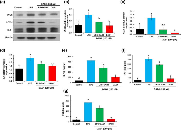
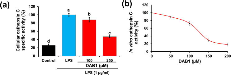
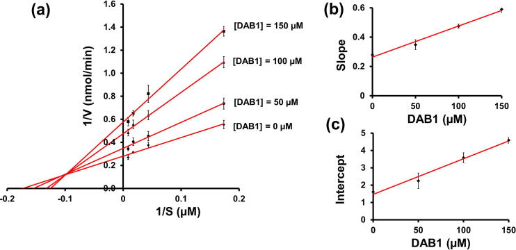
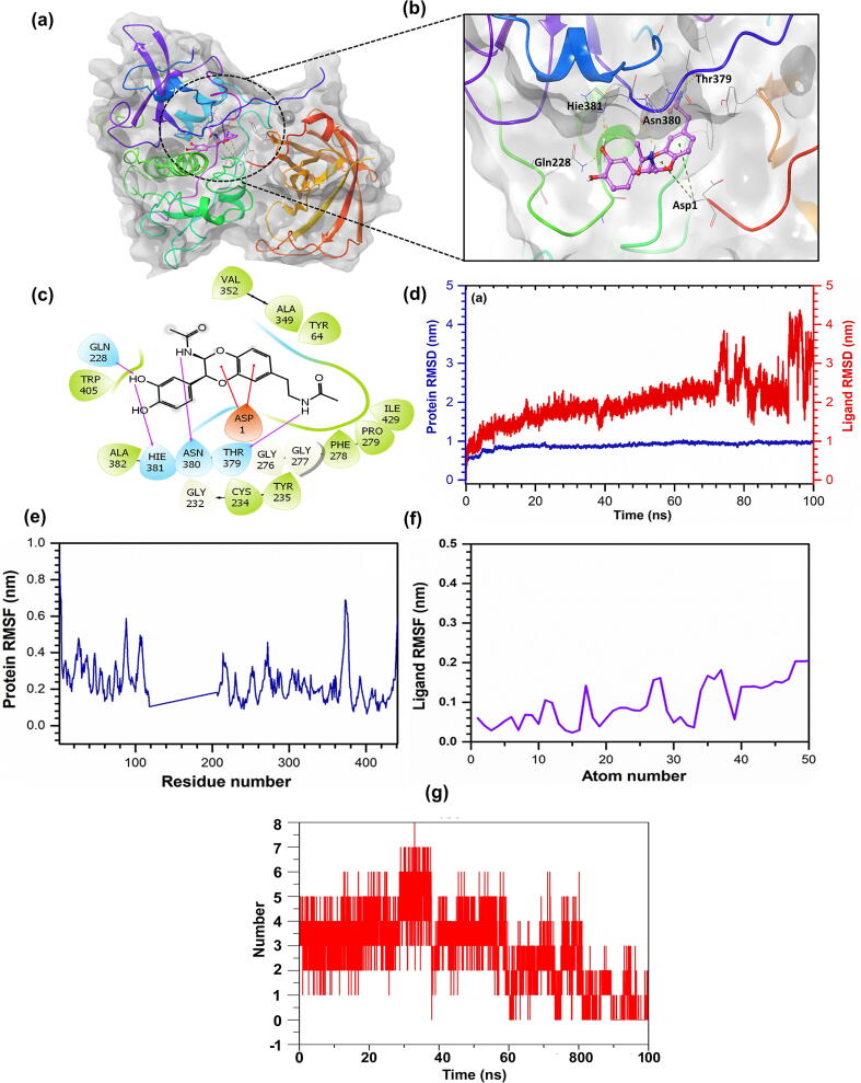
Oxya chinensis sinuosa (rice field grasshopper) is an edible insect with numerous health beneficial properties, traditionally being used to treat many ailments in Korea and other countries. O. chinensis sinuosa has been used from centuries, however, a little is known about the chemical functionality of its bioactive compounds. Therefore, this study examined the anti-inflammatory and cathepsin C inhibitory activities of N-acetyldopamine dimer (2R, 3S)-2-(3',4'-dihydroxyphenyl)-3-acetylamino-7-(N-acetyl-2″-aminoethyl)-1,4-benzodioxane (DAB1) isolated from O. chinensis sinuosa. Results showed that DAB1 reduced the expression of pro-inflammatory mediator (iNOS, COX-2) and cytokines (TNF-α, IL-1β, and IL-6), and curtailed the nuclear translocation of NF-κB by inhibiting the phosphorylation of IκBα in lipopolysaccharide stimulated macrophages. Additionally, DAB1 inhibited cathepsin C activity at the cellular level, supported by in vitro assay (Ki, 71.56 ± 10.21 µM and Kis, 133.55 ± 18.2 µM). Moreover, combinatorial molecular simulation and binding free energy analysis suggested a significant stability and binding affinity of cathepsin C-DAB1 complex via formation of hydrogen bond and hydrophobic interactions with the catalytic residues (Gln228, Thr379, Asn380, and Hie381). Also, essential dynamics analysis showed DAB1 induced non-functional motions in cathepsin C structure. Collectively, DAB1 was concluded as anti-inflammatory and cathepsin C inhibiting agent and could be used in the drug development against respective diseases.

Keywords: Cathepsin C; Essential dynamics; In silico; Inflammation; NF-κB; Oxya chinensis sinuosa.


Conflict of interest statement

The authors declare that they have no known competing financial interests or personal relationships that could have appeared to influence the work reported in this paper.

Figures

None
Graphical abstract
Fig. 1
Fig. 1
Chemical structures of two N-acetyldopamine dimers, (a) (2R,3S)-2-(3′,4′-dihydroxyphenyl)-3-acetylamino-7-(N-acetyl-2″-aminoethyl)-1,4-benzodioxane (DAB1) and (b) (2R,3S)-2-(3′,4′-dihydroxyphenyl)-3-acetylamino-7-(N-acetyl-2″-aminethylene)-1,4-benzodioxane (DAB2) from O. chinensis sinuosa and their effect on the viability of Raw 264.7 murine macrophages evaluated by MTT assay. (c) & (d) Cell viabilities in the presence of DAB1 and DAB2 (100–500 µM), respectively. Each value represents the mean ± standard deviation. In bar graphs letters with different alphabets represent the significant difference at p < 0.05, based on Duncan,s multiple comparison test.
Fig. 2
Fig. 2
Effect of compounds (a) DAB1 and (b) DAB2 (100–250 µM) on NO production in LPS (1 µg/ml) stimulated Raw 264.7 macrophages. Each value represents the mean ± standard deviation. In bar graphs letters with different alphabets represent the significant difference at p < 0.05, based on Duncan’s multiple comparison test.
Fig. 3
Fig. 3
Effect of DAB1 on the inflammatory markers and cytokines in the LPS stimulated Raw 264.7 murine macrophages. β-actin was used as a loading control. (a) Western blotting for iNOS, COX-2, and IL-6. (b), (c), and (d) are the densitometry analyses of iNOS, COX-2, and IL-6, respectively. (e) Quantification of secretory IL-1β, (f) quantification of secretory TNF-α, (g) quantification of PGE2. Each value represents the mean ± standard deviation. In bar graphs letters with different alphabets represent the significant difference at p < 0.05, based on Duncan,s multiple comparison test.
Fig. 4
Fig. 4
Effect of DAB1 in the (a) nuclear translocation of NF-κB and (b) IκBα degradation in LPS stimulated Raw 264.7 murine macrophages. Tubulin and β-actin were used as loading controls. In bar graphs letters with different alphabets represent the significant difference at p < 0.05.
Fig. 5
Fig. 5
Effect of DAB1 on the cellular and in vitro cathepsin C activity. (a) Cathepsin C activity in Raw 264.7 murine macrophages in the absence and presence of DAB1. (b)In vitro cathepsin C inhibition by DAB1 with purified enzyme. Each value represents the mean ± standard deviation. In bar graphs letters with different alphabets represent the significant difference at p < 0.05.
Fig. 6
Fig. 6
Cathepsin C inhibition kinetics by DAB1. (a) Lineweaver-Burk plots. (b) The secondary plot between slope and different concentrations of DAB1 for the determination of inhibition constant (Ki) (c) The plot of intercept versus the DAB1 concentrations for the determination of inhibition constant (Kis).
Fig. 7
Fig. 7
In silico interaction of DAB1 with cathepsin C. (a) and (b) 3D structure of the docked complex of cathepsin C-DAB1. (c) 2D mapping of the cathepsin C-DAB1 complex monitored within 4 Å space around the ligand in the active pocket of cathepsin C. Pink color arrows represent the hydrogen bonds, while green, blue, and grey color residues stand for the hydrophobic, polar, and glycine interactions between cathepsin C and DAB1. (d) RMSD value was extracted from the 100 ns molecular dynamics simulation trajectories for cathepsin C alpha carbon atom and ligand DAB1. (e) and (f) RMSF plot was generated for the cathepsin C-DAB1 docked complex and ligand DAB1, respectively. (g) Hydrogen-bond formation between protein and ligand as function of time.
Fig. 8
Fig. 8
PCA, and DCCM analysis for the molecular dynamics simulation trajectories of cathepsin C-DAB1 complex. (a) PCA, the percentage of overall mean square displacement of residue positional fluctuation noted in each dimension is expressed by corresponding eigen value (PCs). The color values blue to white to red representing the periodic jump during 100 ns simulation. (b) Dynamic cross correlation for cathepsin C docked with DAB1. The positive and negative correlation in the movement of residue is represented as cyan and red color, respectively.

Similar articles

Cited by

References

    1. Nathan C. Points of control in inflammation. Nature. 2002;420(6917):846–852. - PubMed
    1. Wan P., Xie M., Chen G., Dai Z., Hu B., Zeng X., et al. Anti-inflammatory effects of dicaffeoylquinic acids from Ilex kudingcha on lipopolysaccharide-treated RAW264.7 macrophages and potential mechanisms. Food Chem Toxicol. 2019;126:332–342. - PubMed
    1. Gabay C. Interleukin-6 and chronic inflammation. Arthritis Res Therapy. 2006;8(Suppl 2):S3. - PMC - PubMed
    1. Grivennikov S.I., Greten F.R., Karin M. Immunity, inflammation, and cancer. Cell. 2010;140(6):883–899. - PMC - PubMed
    1. Tasneem S., Liu B., Li B., Choudhary M.I., Wang W. Molecular pharmacology of inflammation: Medicinal plants as anti-inflammatory agents. Pharmacol Res. 2019;139:126–140. - PubMed