Review
. 2022 Mar 18;13(3):238-249.
doi: 10.5312/wjo.v13.i3.238.

Diagnosis, treatment and complications of radial head and neck fractures in the pediatric patient

Affiliations
Review

Diagnosis, treatment and complications of radial head and neck fractures in the pediatric patient

Arno A Macken et al. World J Orthop. .

Abstract

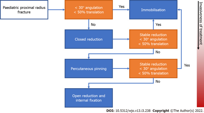
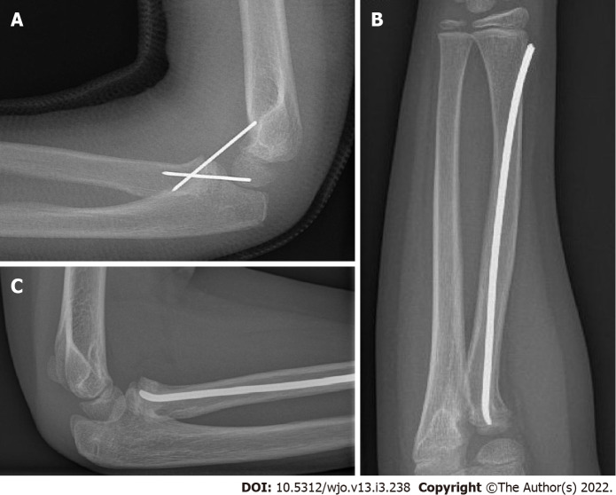
Radial head and neck fractures represent up to 14% of all pediatric elbow fractures and can be a difficult challenge in the pediatric patient. In up to 39% of proximal radius fractures, there is a concomitant fracture, which can easily be overlooked on the initial standard radiographs. The treatment options for proximal radius fractures in children range from non-surgical treatment, such as immobilization alone and closed reduction followed by immobilization, to more invasive options, including closed reduction with percutaneous pinning and open reduction with internal fixation. The choice of treatment depends on the degree of angulation and displacement of the fracture and the age of the patient; an angulation of less than 30 degrees and translation of less than 50% is generally accepted, whereas a higher degree of displacement is considered an indication for surgical intervention. Fractures with limited displacement and non-surgical treatment generally result in superior outcomes in terms of patient-reported outcome measures, range of motion and complications compared to severely displaced fractures requiring surgical intervention. With proper management, good to excellent results are achieved in most cases, and long-term sequelae are rare. However, severe complications do occur, including radio-ulnar synostosis, osteonecrosis, rotational impairment, and premature physeal closure with a malformation of the radial head as a result, especially after more invasive procedures. Adequate follow-up is therefore warranted.

Keywords: Closed fracture reduction; Fracture; Fracture fixation; Open reduction fracture; Osteonecrosis; Pediatrics; Proximal radius; Radial head; Synostosis.


Conflict of interest statement

Conflict-of-interest statement: All authors declare no conflict of interest for this article.

Figures

Figure 1
Figure 1
Fat pad sign. Lateral radiograph of a 13-year-old boy, showing an anterior and posterior fat pad sign without visible fracture. A proximal radius fracture was identified using computed tomography.
Figure 2
Figure 2
Measurement of angulation and translation of the proximal radius fracture. A: Angulation measurement. Angulation of a proximal radius fracture is measured by drawing a line perpendicular to the surface of the radial head (blue line) and a line through the middle of the radial shaft (orange line). The angle is measured at the intersection of the two lines (white arc); B: Translation measurement. Translation of a proximal radius fracture is calculated by dividing the length the uncovered part of the metaphysis (orange line) by the total width of the proximal radius (blue line), multiplying by one hundred provides the percentage of translation. Alternatively, the distance from the middle of the proximal part to the middle of the distal part can be measured in millimeters (continuous white line).
Figure 3
Figure 3
Anteroposterior radiograph of proximal radius fracture. A: Grade I fracture. Anteroposterior radiograph of a 5-year-old boy with a proximal radius fracture that is (nearly) nondisplaced. Judet grade I; Metaizeau grade I; O’Brien type I; B: Grade II fracture. Radiograph of a 9-year-old girl with a proximal radius fracture in 27 degrees of angulation and 17% translation. Judet grade II; Metaizeau grade II; O’Brien type I; C: Grade III fracture. Anteroposterior radiograph of a 10-year-old girl with a proximal radius fracture in 58 degrees of angulation and 55% translation. Judet grade III; Metaizeau grade III; O’Brien type II; D: Grade IV fracture. Anteroposterior radiograph of a 7-year-old girl with a proximal radius fracture in 87 degrees of angulation and 80% translation. Judet grade IVb; Metaizeau grade IVb; O’Brien type III.
Figure 4
Figure 4
Treatment flowchart. Treatment flowchart that plots the stepwise progression from conservative to increasingly invasive treatment of pediatric proximal radius fractures. Starting from the left, orange boxes represent points of decision-making and blue boxes represent treatment options. Boxes placed lower in the chart represent more invasive procedures than those placed above.
Figure 5
Figure 5
A fracture can be reduced percutaneously in several ways. A: Percutaneous Kirschner wire fixation. Lateral radiograph of a 7-year-old boy with a proximal radius fracture after percutaneous reduction and fixation using two Kirschner wires; B and C: Intramedullary nail: Anteroposterior radiograph of a 10-year-old girl with a proximal radius fracture that was reduced and fixated using a flexible intramedullary nail (Metaizeau technique) (B), Lateral radiograph of a 10-year-old girl with a proximal radius fracture that was reduced and fixated using a flexible intramedullary nail (Metaizeau technique) (C).

Similar articles

Cited by

References

    1. Radomisli TE, Rosen AL. Controversies regarding radial neck fractures in children. Clin Orthop Relat Res. 1998:30–39. - PubMed
    1. Nicholson LT, Skaggs DL. Proximal Radius Fractures in Children. J Am Acad Orthop Surg. 2019;27:e876–e886. - PubMed
    1. Kumar S, Mishra A, Odak S, Dwyer J. Treatment principles, prognostic factors and controversies in radial neck fractures in children: A systematic review. J Clin Orthop Trauma. 2020;11:S456–S463. - PMC - PubMed
    1. Landin LA. Fracture patterns in children. Analysis of 8,682 fractures with special reference to incidence, etiology and secular changes in a Swedish urban population 1950-1979. Acta Orthop Scand Suppl. 1983;202:1–109. - PubMed
    1. DeFroda SF, Hansen H, Gil JA, Hawari AH, Cruz AI Jr. Radiographic Evaluation of Common Pediatric Elbow Injuries. Orthop Rev (Pavia) 2017;9:7030. - PMC - PubMed