. 2022 Mar 4:10:804164.
doi: 10.3389/fcell.2022.804164. eCollection 2022.

A Novel Tissue Atlas and Online Tool for the Interrogation of Small RNA Expression in Human Tissues and Biofluids

Affiliations

A Novel Tissue Atlas and Online Tool for the Interrogation of Small RNA Expression in Human Tissues and Biofluids

Eric Alsop et al. Front Cell Dev Biol. .

Abstract

One promising goal for utilizing the molecular information circulating in biofluids is the discovery of clinically useful biomarkers. Extracellular RNAs (exRNAs) are one of the most diverse classes of molecular cargo, easily assayed by sequencing and with expressions that rapidly change in response to subject status. Despite diverse exRNA cargo, most evaluations from biofluids have focused on small RNA sequencing and analysis, specifically on microRNAs (miRNAs). Another goal of characterizing circulating molecular information, is to correlate expression to injuries associated with specific tissues of origin. Biomarker candidates are often described as being specific, enriched in a particular tissue or associated with a disease process. Likewise, miRNA data is often reported to be specific, enriched for a tissue, without rigorous testing to support the claim. Here we provide a tissue atlas of small RNAs from 30 different tissues and three different blood cell types. We analyzed the tissues for enrichment of small RNA sequences and assessed their expression in biofluids: plasma, cerebrospinal fluid, urine, and saliva. We employed published data sets representing physiological (resting vs. acute exercise) and pathologic states (early- vs. late-stage liver fibrosis, and differential subtypes of stroke) to determine differential tissue-enriched small RNAs. We also developed an online tool that provides information about exRNA sequences found in different biofluids and tissues. The data can be used to better understand the various types of small RNA sequences in different tissues as well as their potential release into biofluids, which should help in the validation or design of biomarker studies.

Keywords: cerebrospinal fluid; extracellular RNA; extracellular vesicle; plasma; saliva; small RNA; tissue atlas; urine.


Conflict of interest statement

KV and SD are members of the scientific advisory board at Dyrnamix. Their involvement in this company had no bearing on the work performed in this manuscript. KV is on the scientific advisory board of HTG. Her involvement has no bearing on this work. The remaining authors declare that the research was conducted in the absence of any commercial or financial relationships that could be construed as a potential conflict of interest.

Figures

FIGURE 1
FIGURE 1
Biofluid Profiles. For the biofluids, CSF-EVs, cell-free plasma, plasma-EVs, cell-free saliva and cell-free urine, the associated small RNAs were sequenced and aligned to the human genome (A). In Panel (A), the reads are aligned to the transcriptome, genome, rRNA, UniVec (laboratory contaminants), are categorized as unmapped, or reads that fail QC. In Panel (B), the transcriptome mapped reads are taken out and further broken down into small RNA biotypes; miRNA, tRNA, yRNA, piRNA, protein-coding fragments, and other (these are categories of RNA that annotate in GENCODE, lncRNA fragments, pseudogenes, etc).
FIGURE 2
FIGURE 2
Tissue Profiles. For thirty different tissues, PBMCs, monocytes, and red blood cells we isolated the RNA and sequenced their small RNA contents. In Panel (A), the reads are aligned to the transcriptome, genome, rRNA, UniVec (contaminants), are unmapped, or reads that fail QC. In Panel (B), the transcriptome mapped reads are removed and further broken down into small RNA biotypes; miRNA, tRNA, yRNA, piRNA, protein-coding fragments, and other (these are categories of RNA that annotate in GENCODE, lncRNA fragments, pseudogenes, etc).
FIGURE 3
FIGURE 3
Sequence Variation and Categorization. Panel (A) highlights the canonical miRNA sequence that is entered in miRbase for two miRNAs, miR-451a and miR-30a-5p. The other sequences are isomiRs of the mature sequence (highlighted) listed in miRbase. The table shows the normalized expression for the sequences in plasma from five different subjects and the average across the 179 cell-free plasma samples. In some cases, an isomiR has higher expression in plasma than the highlighted canonical sequence. miR-451a is highly expressed in red blood cells, as can be seen in the heat map in Panel (B). Panel (B) displays the canonical and isomiR sequences for miR-451a found in red blood cells, PBMCs, and monocytes. The most abundant sequence detected in urine samples is displayed in Figure 3c and is = similar to both a piRNA and a tRNA sequence.
FIGURE 4
FIGURE 4
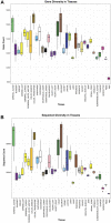
Tissue Diversity. Panel (A) displays the number of genes detected using small RNASeq in each tissue. In this case, all isomiRs, isoforms, and fragments are collapsed to the parent gene and counted once. In Panel (B), all unique sequences, isomiRs, isoforms, and fragments are counted separately and summed up.
FIGURE 5
FIGURE 5
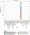
Tissue Elevation. miR-216-5p is elevated in pancreas. The expression of the mature sequence is highlighted in Panel. The stacked bar plot displays the relative proportion of other miR-216-5p isomiRs, all of which are elevated in pancreas.
FIGURE 6
FIGURE 6
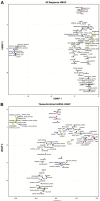
Tissue Clustering. We clustered the tissues using all sequences in Panel (A). Using tissue-elevated miRNA sequences in Panel (B), the tissue samples clustered into tighter groups. In both figures, the tissue replicates show low variability.
FIGURE 7
FIGURE 7
Biofluid Diversity. Panels (A, B) display the number of tissue-enriched genes identified in the small RNA sequenced data. The parent tissue-elevated genes that were detected in each biofluid are displayed in Panel (A). CSF had the lowest number of tissue-elevated genes and saliva had the highest. Panel (B) displays the number of tissue-elevated sequences that were detected in each biofluid.
FIGURE 8
FIGURE 8
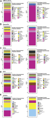
Tissue-elevated miRNA/isomiR Sequence Detection in Biofluids. The number of tissue-elevated sequences that were detected in each biofluid and their abundance are displayed in Panel. The number of tissue-elevated sequences detected for each tissue in cell-free plasma are shown in the first stacked bar plot Panel (A). Tissue-elevated sequences from 31 different tissue and cell types were detected (the numbers are found in Supplementary Table S5). The second stacked bar plot in Panel (A) displays the fraction of tissue-elevated reads going to each sample type. 69% of the tissue-elevated reads in cell-free plasma go to red blood cells. The number of tissue-elevated sequences detected and the fraction of tissue-elevated reads going to each tissue are displayed for plasma EVs [Panel (B)], saliva [Panel (C)], Urine [Panel (D)], and CSF [Panel (E)].
FIGURE 9
FIGURE 9
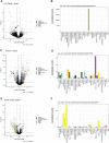
Differentially Expressed Tissue-Elevated Sequences. We examined three different conditions for differentially expressed sequences: (A) displays the differentially expressed tissue-elevated sequences found in blood samples from Hepatitis C patients with liver fibrosis stage 1 or stage 4. Panel (B) is a tissue-elevated isomiR that is highly expressed in liver. (C) are samples taken from participants before and after exercise, and the expression level of a muscle-elevated isomiR across tissues (D). The final example is from a comparison of plasma samples taken from individuals that had either an ischaemic stroke or a subarachnoid hemorrhage, Panel (E). In Panel (F), we selected a CNS-elevated isomiR of miR-181-5p to illustrate tissue-elevation.
FIGURE 10
FIGURE 10
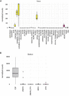
exRNA Expression Atlas. In (A) we display the expression of a tissue-elevated miRNA for the CNS (miR-1298) across all tissues. Panel (B) displays the detection of tissue-elevated miR-1298 in different biofluids. As can be seen in this figure, this enriched CNS miRNA is detectable in CSF, but poorly detected in the other biofluids. These data will provide insight regarding which biofluids hold which tissue-elevated sequences.

Similar articles

  • High Throughput Sequencing of Extracellular RNA from Human Plasma.
    Danielson KM, Rubio R, Abderazzaq F, Das S, Wang YE. Danielson KM, et al. PLoS One. 2017 Jan 6;12(1):e0164644. doi: 10.1371/journal.pone.0164644. eCollection 2017. PLoS One. 2017. PMID: 28060806 Free PMC article.
  • Charting Extracellular Transcriptomes in The Human Biofluid RNA Atlas.
    Hulstaert E, Morlion A, Avila Cobos F, Verniers K, Nuytens J, Vanden Eynde E, Yigit N, Anckaert J, Geerts A, Hindryckx P, Jacques P, Brusselle G, Bracke KR, Maes T, Malfait T, Derveaux T, Ninclaus V, Van Cauwenbergh C, Roelens K, Roets E, Hemelsoet D, Tilleman K, Brochez L, Kuersten S, Simon LM, Karg S, Kautzky-Willers A, Leutner M, Nöhammer C, Slaby O, Prins RW, Koster J, Lefever S, Schroth GP, Vandesompele J, Mestdagh P. Hulstaert E, et al. Cell Rep. 2020 Dec 29;33(13):108552. doi: 10.1016/j.celrep.2020.108552. Cell Rep. 2020. PMID: 33378673
  • exRNA Atlas Analysis Reveals Distinct Extracellular RNA Cargo Types and Their Carriers Present across Human Biofluids.
    Murillo OD, Thistlethwaite W, Rozowsky J, Subramanian SL, Lucero R, Shah N, Jackson AR, Srinivasan S, Chung A, Laurent CD, Kitchen RR, Galeev T, Warrell J, Diao JA, Welsh JA, Hanspers K, Riutta A, Burgstaller-Muehlbacher S, Shah RV, Yeri A, Jenkins LM, Ahsen ME, Cordon-Cardo C, Dogra N, Gifford SM, Smith JT, Stolovitzky G, Tewari AK, Wunsch BH, Yadav KK, Danielson KM, Filant J, Moeller C, Nejad P, Paul A, Simonson B, Wong DK, Zhang X, Balaj L, Gandhi R, Sood AK, Alexander RP, Wang L, Wu C, Wong DTW, Galas DJ, Van Keuren-Jensen K, Patel T, Jones JC, Das S, Cheung KH, Pico AR, Su AI, Raffai RL, Laurent LC, Roth ME, Gerstein MB, Milosavljevic A. Murillo OD, et al. Cell. 2019 Apr 4;177(2):463-477.e15. doi: 10.1016/j.cell.2019.02.018. Cell. 2019. PMID: 30951672 Free PMC article.
  • Circulating RNAs in prostate cancer patients.
    Mugoni V, Ciani Y, Nardella C, Demichelis F. Mugoni V, et al. Cancer Lett. 2022 Jan 1;524:57-69. doi: 10.1016/j.canlet.2021.10.011. Epub 2021 Oct 14. Cancer Lett. 2022. PMID: 34656688 Review.
  • Extracellular RNAs: A New Awareness of Old Perspectives.
    Sadik N, Cruz L, Gurtner A, Rodosthenous RS, Dusoswa SA, Ziegler O, Van Solinge TS, Wei Z, Salvador-Garicano AM, Gyorgy B, Broekman M, Balaj L. Sadik N, et al. Methods Mol Biol. 2018;1740:1-15. doi: 10.1007/978-1-4939-7652-2_1. Methods Mol Biol. 2018. PMID: 29388131 Free PMC article. Review.

Cited by

References

    1. Abdelhamid R. F., Plessy C., Yamauchi Y., Taoka M., de Hoon M., Gingeras T. R., et al. (2014). Multiplicity of 5′ Cap Structures Present on Short RNAs. PLoS One 9 (7), e102895. 10.1371/journal.pone.0102895 - DOI - PMC - PubMed
    1. Agarwal V., Bell G. W., Nam J.-W., Bartel D. P. (2015). Predicting Effective microRNA Target Sites in Mammalian mRNAs. Elife 4. 10.7554/eLife.05005 - DOI - PMC - PubMed
    1. Akat K. M., Lee Y. A., Hurley A., Morozov P., Max K. E. A., Brown M., et al. (2019). Detection of Circulating Extracellular mRNAs by Modified Small-RNA-Sequencing Analysis. JCI Insight 4. 10.1172/jci.insight.127317 - DOI - PMC - PubMed
    1. Arroyo J. D., Chevillet J. R., Kroh E. M., Ruf I. K., Pritchard C. C., Gibson D. F., et al. (2011). Argonaute2 Complexes Carry a Population of Circulating microRNAs Independent of Vesicles in Human Plasma. Proc. Natl. Acad. Sci. 108 (12), 5003–5008. 10.1073/pnas.1019055108 - DOI - PMC - PubMed
    1. Avendaño‐Vázquez S. E., Flores‐Jasso C. F. (2020). Stumbling on Elusive Cargo: How isomiRs challenge microRNA Detection and Quantification, the Case of Extracellular Vesicles. J. Extracellular Vesicles 9 (1), 1784617. 10.1080/20013078.2020.1784617 - DOI - PMC - PubMed