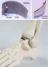
Clinical efficacy of the Ankle Spacer for the treatment of multiple secondary osteochondral lesions of the talus
- PMID: 35317406
- PMCID: PMC8891659
- DOI: 10.5312/wjo.v13.i2.178
Clinical efficacy of the Ankle Spacer for the treatment of multiple secondary osteochondral lesions of the talus
Abstract
Background: The Ankle Spacer was developed as a joint-sparing alternative to invasive end-stage surgeries. Currently, there are no clinical studies on the Ankle Spacer.
Aim: To describe the operative technique and the clinical efficacy of the Ankle Spacer for the treatment of multiple, cystic osteochondral lesions of the talus in patients with failed prior operative treatment.
Methods: This is a prospective study during which patients were assessed preoperatively, at 2- and 6 wk, and at 3, 6, 12 and 24 mo postoperatively. Patients with multiple, cystic or large (≥ 15 mm) osteochondral lesions of the talus after failed prior surgery were included. The primary outcome measure was the numeric rating scale (NRS) for pain during walking at 2 years postoperatively. Secondary outcome measures included the NRS in rest and during stair climbing, the American Orthopaedic Foot and Ankle Society Hindfoot Score, the Foot and Ankle Outcome Score, the Short- Form 36 physical and mental component scale, and the Range of Motion (ROM). Radiographic evaluations were conducted to evaluate prosthetic loosening and subsidence. Revision rates and complications were also assessed.
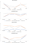
Results: Two patients underwent an Ankle Spacer implantation on the talus. The NRS during walking improved from 6 and 7 preoperatively to 2 and 2 points postoperatively at 2 years, in patient 1 and 2, respectively. The other patient-reported outcome measures also improved substantially. There were no re-operations nor complications. Radiological imaging showed no loosening of the implant and no change of implant position.
Conclusion: The Ankle Spacer showed clinically relevant pain reduction during walking, improvement in clinical outcomes as assessed with PROMs, and no complications or re-operations. This treatment option may evolve as a joint-sparing alternative to invasive end-stage surgeries.
Keywords: Ankle Spacer; End-stage; Hemi-arthroplasty; Osteochondral lesions of the talus; Talar surface implant.
©The Author(s) 2022. Published by Baishideng Publishing Group Inc. All rights reserved.
Conflict of interest statement
Conflict-of-interest statement: The study was sponsored by Arthrex as a post market study with reference number EMEA-17037.
Figures


References
-
- Rikken QGH, Kerkhoffs GMMJ. Osteochondral Lesions of the Talus: An Individualized Treatment Paradigm from the Amsterdam Perspective. Foot Ankle Clin. 2021;26:121–136. - PubMed
-
- Haene R, Qamirani E, Story RA, Pinsker E, Daniels TR. Intermediate outcomes of fresh talar osteochondral allografts for treatment of large osteochondral lesions of the talus. J Bone Joint Surg Am. 2012;94:1105–1110. - PubMed
LinkOut - more resources
Full Text Sources

