

Unusual Location of Pulp Glomus Tumor: A Case Study and Literature Review
- PMID: 35317460
- PMCID: PMC8929525
- DOI: 10.1097/GOX.0000000000004206
Unusual Location of Pulp Glomus Tumor: A Case Study and Literature Review
Abstract
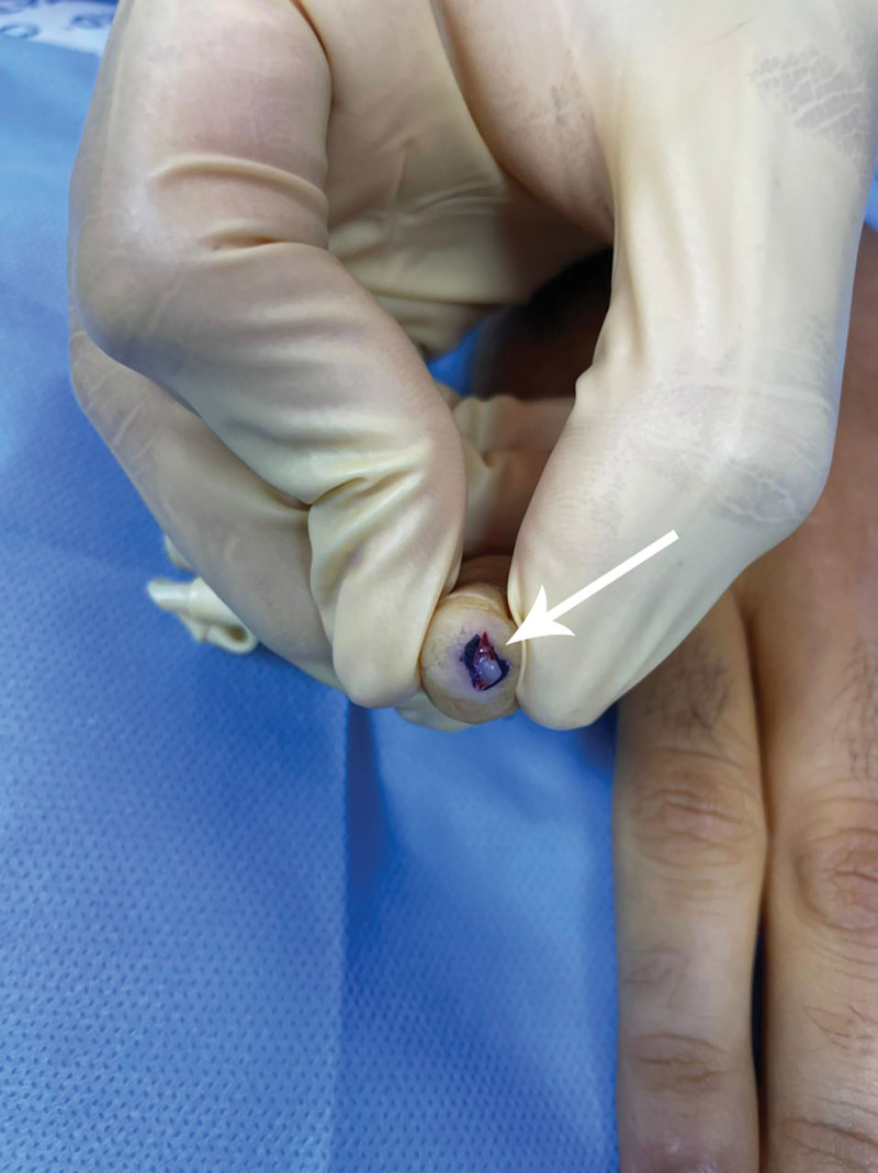
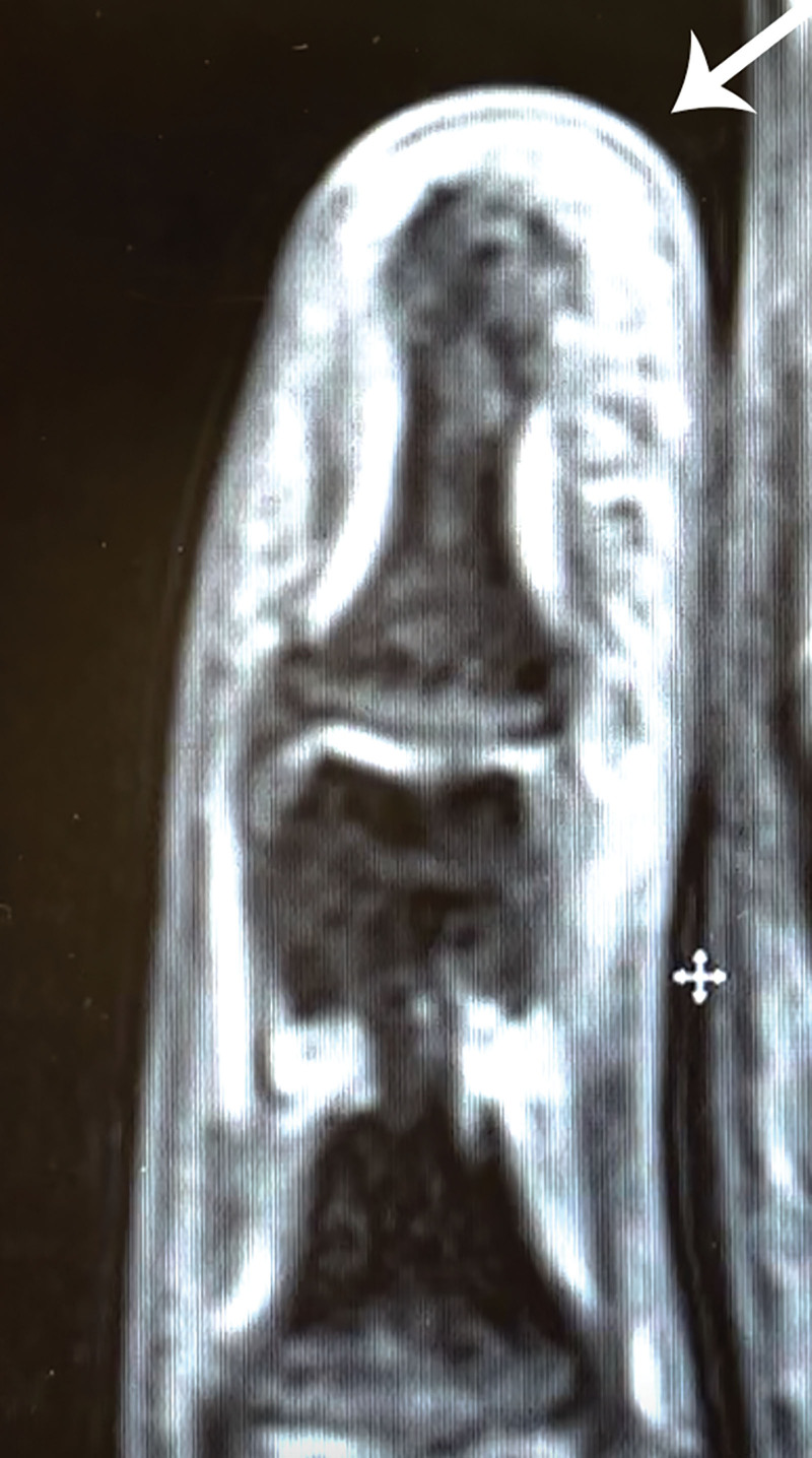
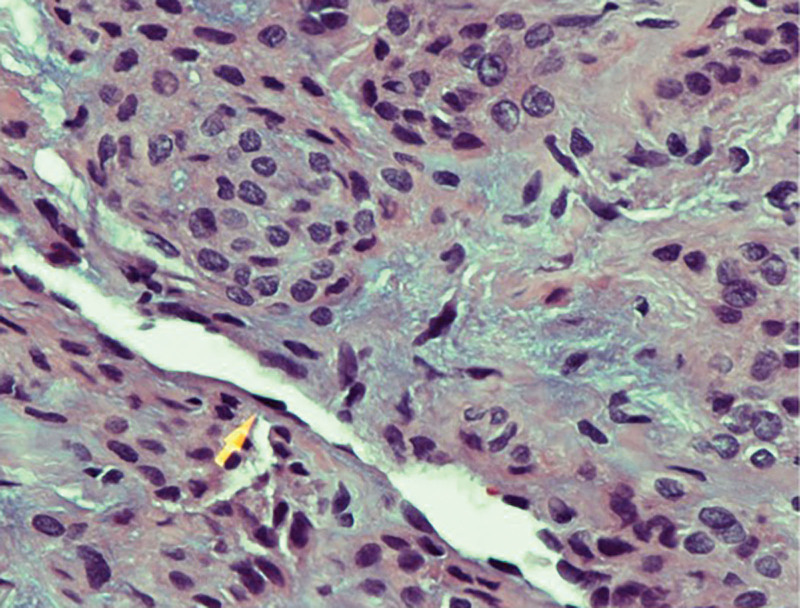
Glomus tumors are painful, benign tumors that develop from the glomus bodies. They account for less than 1% of tumors in hand, and less than 10% present in the pulp of the digits. Cold hypersensitivity, increased pinprick sensitivity, and paroxysmal pain are common glomus tumor symptoms. We describe a 27-year-old man who came with pain in the right little digit, confined to the pulp for 10 years. The tip of the finger was extremely sensitive to touch, and the pain worsened in a cold atmosphere. Upon palpation, no mass was recognized. There was pinpoint tenderness within the distal volar pulp of the little finger. MRI with a contrast of the right little digit showed a 2-mm enhancing lesion in the tip of the little finger. An incision was done over the volar plane of the little finger, removing the tumor bluntly. The tumor was found to be a glomus tumor after histologic evaluation. Glomus tumors of the volar pulp are notoriously hard to detect. Hence, the presence of localized pain in the volar tip for the finger should raise suspicion of the diagnosis of a glomus tumor, and surgical removal should be offered to relieve symptoms and avoid recurrence.
Copyright © 2022 The Authors. Published by Wolters Kluwer Health, Inc. on behalf of The American Society of Plastic Surgeons.
Conflict of interest statement
Disclosure: The authors have no financial interest to declare in relation to the content of this article.
Figures
Similar articles
-
Unusual Volar Pulp Location of Glomus Tumor.Plast Reconstr Surg Glob Open. 2017 Jan 30;5(1):e1215. doi: 10.1097/GOX.0000000000001215. eCollection 2017 Jan. Plast Reconstr Surg Glob Open. 2017. PMID: 28203512 Free PMC article.
-
A painful glomus tumor on the pulp of the distal phalanx.J Korean Neurosurg Soc. 2010 Aug;48(2):185-7. doi: 10.3340/jkns.2010.48.2.185. Epub 2010 Aug 31. J Korean Neurosurg Soc. 2010. PMID: 20856673 Free PMC article.
-
Painful pulp space of a pinky finger: A report of glomus tumor at an unusual site.J Family Med Prim Care. 2020 Aug 25;9(8):4425-4427. doi: 10.4103/jfmpc.jfmpc_602_20. eCollection 2020 Aug. J Family Med Prim Care. 2020. PMID: 33110875 Free PMC article.
-
Glomus tumours of the hand: Review of literature.J Clin Orthop Trauma. 2016 Oct-Dec;7(4):286-291. doi: 10.1016/j.jcot.2016.04.006. Epub 2016 Sep 1. J Clin Orthop Trauma. 2016. PMID: 27857505 Free PMC article. Review.
-
Extradigital Symplastic Glomus Tumor of the Hand: Report of 2 Cases and Literature Review.Am J Dermatopathol. 2015 Jul;37(7):560-2. doi: 10.1097/DAD.0000000000000132. Am J Dermatopathol. 2015. PMID: 25051107 Review.
Cited by
-
Solitary Glomus Tumor on the Base of the Right Thumb: A Rare Case Report and a Literature Review from Saudi Arabia.Int Med Case Rep J. 2024 Apr 23;17:371-380. doi: 10.2147/IMCRJ.S456808. eCollection 2024. Int Med Case Rep J. 2024. PMID: 38681995 Free PMC article.
-
Glomus Tumour of Hand--A Commonly Misdiagnosed Pathology: A Case Series.J West Afr Coll Surg. 2022 Oct-Dec;12(4):39-45. doi: 10.4103/jwas.jwas_171_22. Epub 2022 Nov 23. J West Afr Coll Surg. 2022. PMID: 36590779 Free PMC article.
-
A Glomus Tumor Presenting on the Ventromedial Aspect of the Little Finger Causing Bony Erosion: A Rare Case From India.Cureus. 2024 Feb 14;16(2):e54173. doi: 10.7759/cureus.54173. eCollection 2024 Feb. Cureus. 2024. PMID: 38496094 Free PMC article.
References
-
- Carroll RE, Berman AT. Glomus tumors of the hand: review of the literature and report on twenty-eight cases. J Bone Joint Surg Am. 1972;54:691–703. - PubMed
-
- Takata H, Ikuta Y, Ishida O, et al. . Treatment of subungual glomus tumour. Hand Surg. 2001;6:25–27. - PubMed
-
- Nazerani S, Motamedi MH, Keramati MR. Diagnosis and management of glomus tumors of the hand. Tech Hand Up Extrem Surg. 2010;14:8–13. - PubMed
-
- Hamdi MF. Glomus tumour of fingertip: report of eight cases and literature review. Musculoskelet Surg. 2011;95:237–240. - PubMed
Publication types
LinkOut - more resources
Full Text Sources