. 2022 Apr 29;130(9):1306-1317.
doi: 10.1161/CIRCRESAHA.121.320680. Epub 2022 Mar 23.

Gene Therapy With the N-Terminus of Junctophilin-2 Improves Heart Failure in Mice

Affiliations

Gene Therapy With the N-Terminus of Junctophilin-2 Improves Heart Failure in Mice

Jinxi Wang et al. Circ Res. .

Abstract

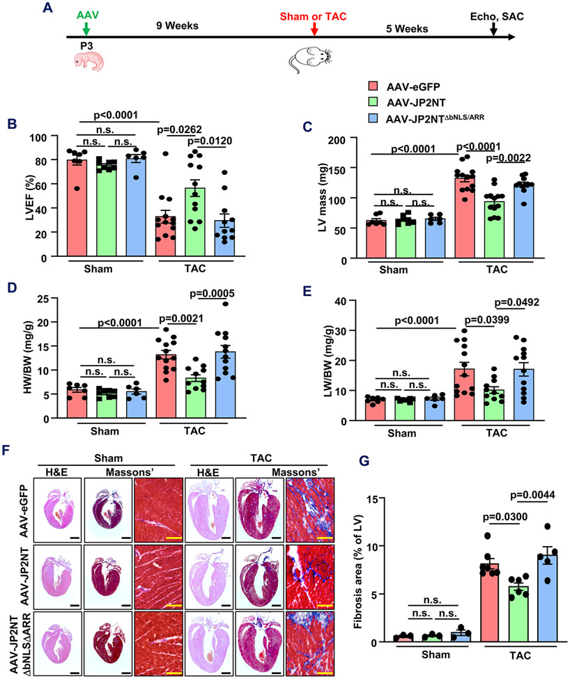
Background: Transcriptional remodeling is known to contribute to heart failure (HF). Targeting stress-dependent gene expression mechanisms may represent a clinically relevant gene therapy option. We recently uncovered a salutary mechanism in the heart whereby JP2 (junctophilin-2), an essential component of the excitation-contraction coupling apparatus, is site-specifically cleaved and releases an N-terminal fragment (JP2NT [N-terminal fragment of JP2]) that translocates into the nucleus and functions as a transcriptional repressor of HF-related genes. This study aims to determine whether JP2NT can be leveraged by gene therapy techniques for attenuating HF progression in a preclinical pressure overload model.

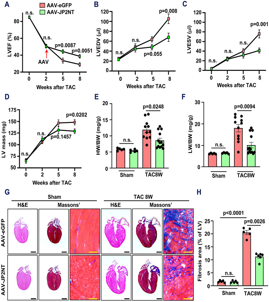
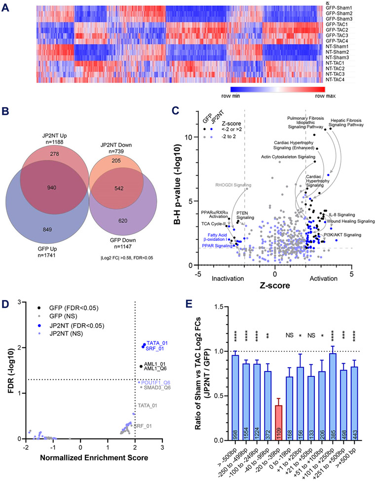
Methods: We intraventricularly injected adeno-associated virus (AAV) (2/9) vectors expressing eGFP (enhanced green fluorescent protein), JP2NT, or DNA-binding deficient JP2NT (JP2NTΔbNLS/ARR) into neonatal mice and induced cardiac stress by transaortic constriction (TAC) 9 weeks later. We also treated mice with established moderate HF from TAC stress with either AAV-JP2NT or AAV-eGFP. RNA-sequencing analysis was used to reveal changes in hypertrophic and HF-related gene transcription by JP2NT gene therapy after TAC. Echocardiography, confocal imaging, and histology were performed to evaluate heart function and pathological myocardial remodeling following stress.

Results: Mice preinjected with AAV-JP2NT exhibited ameliorated cardiac remodeling following TAC. The JP2NT DNA-binding domain is required for cardioprotection as its deletion within the AAV-JP2NT vector prevented improvement in TAC-induced cardiac dysfunction. Functional and histological data suggest that JP2NT gene therapy after the onset of cardiac dysfunction is effective at slowing the progression of HF. RNA-sequencing analysis further revealed a broad reversal of hypertrophic and HF-related gene transcription by JP2NT overexpression after TAC.

Conclusions: Our prevention- and intervention-based approaches here demonstrated that AAV-mediated delivery of JP2NT into the myocardium can attenuate stress-induced transcriptional remodeling and the development of HF when administered either before or after cardiac stress initiation. Our data indicate that JP2NT gene therapy holds great potential as a novel therapeutic for treating hypertrophy and HF.

Keywords: genetic therapy; heart failure; hypertrophy; junctophilin.


Figures

Figure 1.
Figure 1.. In vivo gene therapy with AAV effectively expresses JP2 cargo in cardiomyocytes.
A, Schematic of CMV promoter-driven JP2NT, JP2NTΔbNLS/ARR, JP2, and eGFP adeno-associated viral constructs. B, Western blot analysis of organ lysates from mice 8-weeks post AAV injection for expression of HA-tagged JP2NT or JP2NTΔbNLSΔARR. SKM, skeletal muscle. C, D, Immunohistochemistry micrographs of myocardial sections from AAV-eGFP, AAV-JP2NT, AAV-JP2NTΔbNLS/ARR, and AAV-JP2 injected mice 8 weeks post AAV injection. Scale bar, 20 μm.
Figure 2.
Figure 2.. Neonatal JP2NT gene therapy attenuates TAC-induced dysfunction and fibrosis.
A, Time course of experiments. On the 3rd day after birth, mouse pups were injected intraventricularly with cardiotropic serotype 9 (AAV). TAC or sham surgery was performed 9 weeks later on both male and female mice. Five weeks after surgery, heart function was measured by echocardiography and mice were sacrificed for heart collection. B, C, Echocardiography analysis of LV ejection fraction (LVEF, B) and LV mass (C) on sham and TAC groups (5 weeks post-surgery). n = 7-13 mice per group (sham/AAV-eGFP (7), sham/AAV-JP2NT (8), sham/AAV- JP2NTΔbNLS/ARR (6), TAC/AAV-eGFP (13), TAC/AAV-JP2NT (12), TAC/AAV- JP2NTΔbNLS/ARR (11)). D, Heart weight to body weight ratio (HW/BW) for sham and TAC mice 5 weeks after surgery. E, Lung weight to body weight ratio (LW/BW) for sham and TAC mice 5 weeks after surgery. F, Representative views of hematoxylin & eosin (H&E) and Masson’s trichrome stained hearts isolated from sham or TAC mice 5 weeks after surgery. H&E black scale bars, 1 mm; Masson’s trichrome yellow scale bar, 100 μm. G, Percentage of the fibrotic area in sham and TAC groups (n=3 for each sham group, n= 5-7 for each TAC group). Statistical significance was determined with 2-way ANOVA followed by Tukey post hoc test (B-E) or Kruskal-Wallis test followed by Dunn post hoc test (G). n.s., not significant.
Figure 3.
Figure 3.. JP2NT gene therapy preserves T-tubule integrity after TAC.
A, B, Representative LV T-tubule images stained with lipophilic marker MM4–64 (A) and quantitative T-tubule regularity (TTpower) summary (B) of AAV-eGFP, AAV-JP2NT and JP2NTΔbNLS/ARR injected mice 5 weeks after TAC (n = 3 for each Sham group, n = 5-7 for each TAC group). C, Summarized T-tubule integrity index (TTintegrity) data for GFP-positive AAV-transduced cardiomyocytes and GFP-negative non-transduced cardiomyocytes in AAV-eGFP, AAV-JP2NT, and AAV- JP2NTΔbNLS/ARR injected mice 5 weeks after TAC, n = 30 fields from 3 hearts for each group, 5-10 AAV (eGFP) positive or negative cardiomyocytes on each field. Scale bar, 20 μm. Statistical significance was determined with 2-way ANOVA followed by Tukey post hoc test (B) or Kruskal-Wallis test followed by Dunn post hoc test (C). n.s., not significant.
Figure 4.
Figure 4.. Protocol for investigating AAV-JP2NT gene therapy in hearts with established cardiac dysfunction.
A, Map of CMV promoter-driven JP2NT and eGFP adeno-associated viral constructs. B-C, The experimental workflow (B) and flow chart of sequential inclusion criteria (C) used to obtain a cohort of mice with similar levels of moderate heart failure that were then randomized to receive AAV-eGFP or AAV-JP2NT. The TAC model was performed on 9-10-week-old C57BL/6N male mice. Mice with ejection fractions (EF) ranging from 40%–60% 2-weeks post-TAC were selected for gene therapy administration. Heart function was assessed by echocardiography 3 and 5 weeks after AAV injection. Mice were euthanized for collecting heart tissue after the last echocardiography measurement. D, Western blot analysis of HA-tagged JP2NT organ distribution from mice intravenously injected with AAVs at 9-10 weeks of age and harvested 8-weeks later. SKM, skeletal muscle.
Figure 5.
Figure 5.. JP2NT gene therapy ameliorated heart failure development after pressure-overload induced pathological remodeling.
A-D, Echocardiography analysis of left ventricular ejection fraction (LVEF) (A), left ventricular end-diastolic volume (B), left ventricular end-systolic volume (C), and LV mass (D) in animals at baseline (0) and 2, 5, and 8 weeks post-TAC. Arrow indicates the time of AAV-eGFP or -JP2NT injection at 2 weeks post-TAC. n = 5 mice to each group at baseline (0), n=11-15 mice to each group after TAC (AAV-eGFP: 15, 14 and 11 mice at 2, 5 and 8 weeks, respectively. AAV-JP2NT: 15, 15 and 13 mice at 2, 5 and 8 weeks, respectively). E, Heart weight to body weight ratio (HW/BW) for baseline and TAC mice 8 weeks after surgery. F, Lung weight to body weight ratio (LW/BW) for baseline and TAC mice 8 weeks after surgery. n = 5-13 mice for each group (baseline AAV-eGFP (5), baseline AAV-JP2NT (5), TAC/AAV-eGFP (11), TAC/AAV-JP2NT (13). G, Representative views of hematoxylin & eosin (H&E) and Masson’s trichrome stained hearts isolated from sham or TAC mice 8 weeks after surgery. H&E black scale bars, 1 mm; Masson’s trichrome yellow scale bars, 100 μm. H, Percentage of the fibrotic area in sham and TAC groups (n=5~6 mice for each group). Two sided unpaired t test was used to determine statistical significance between AAV-eGFP and AAV-JP2NT groups over time (A-D). Two-way ANOVA followed by Tukey post hoc test was used to determine statistical significance (E-G). n.s., not significant.
Figure 6.
Figure 6.. RNA-Seq reveals a broad reversal of hypertrophic and HF-related gene transcription through JP2NT transduction after TAC.
A, Heatmap of significant differentially expressed genes (DEGs, 1.5-fold change, FDR (padj) < 0.05,) for all comparison groups as determined by RNA-seq analysis. Surgery was performed in 9–10-week-old male mice and injected with AAV-eGFP or AAV-JP2NT (NT) viruses via tail vein two weeks later and harvested 6 weeks after surgery. B, Venn diagram of upregulated and downregulated DEGs after TAC versus sham surgery in AAV-eGFP and AAV-JP2NT infected ventricles. C, Ingenuity Pathway Analysis results for enriched predicted canonical pathways. A threshold of ±2 Z-score and Benjamini-Hochberg (B-H) multiple test-corrected p<0.05 (dashed lines) was used for determining activation. Arrows indicate viral-dependent changes in the coordinates of example pathways known to be altered during cardiac function or are fibrosis related in other tissues. D, Gene set enrichment analysis (GSEA) for transcription factor binding motifs within the promoters of DEGs. E, TAC-regulated DEGs with canonical proximal promoter TATA box motifs are sensitive to repression by JP2NT. The ratio of log2 fold changes for DEGs (FDR < 0.05) in JP2NT (Sham vs. TAC) versus GFP (Sham vs. TAC) treated groups. Distances of closest TATA box motifs from DEG TSSs were binned as shown. All bins were compared with that of TATA boxes located 20-39 bp upstream of the TSS (red bar, one-way ANOVA). Number of DEGs in each bin is indicated within the bars. FDR, false discovery rate; NS, not significant; *, p<0.5; **, p<0.01; ***, p<0.001; ****, p<0.0001.

Comment in

References

    1. Ziaeian B and Fonarow GC. Epidemiology and aetiology of heart failure. Nature reviews Cardiology. 2016;13:368–78. - PMC - PubMed
    1. Savarese G and Lund LH. Global Public Health Burden of Heart Failure. Cardiac failure review. 2017;3:7–11. - PMC - PubMed
    1. Virani SS, Alonso A, Aparicio HJ, Benjamin EJ, Bittencourt MS, Callaway CW, Carson AP, Chamberlain AM, Cheng S, Delling FN, Elkind MSV, Evenson KR, Ferguson JF, Gupta DK, Khan SS, Kissela BM, Knutson KL, Lee CD, Lewis TT, Liu J, Loop MS, Lutsey PL, Ma J, Mackey J, Martin SS, Matchar DB, Mussolino ME, Navaneethan SD, Perak AM, Roth GA, Samad Z, Satou GM, Schroeder EB, Shah SH, Shay CM, Stokes A, VanWagner LB, Wang NY, Tsao CW, American Heart Association Council on E, Prevention Statistics C and Stroke Statistics S. Heart Disease and Stroke Statistics-2021 Update: A Report From the American Heart Association. Circulation. 2021;143:e254–e743. - PubMed
    1. Ishikawa K, Weber T and Hajjar RJ. Human Cardiac Gene Therapy. Circulation research. 2018;123:601–613. - PMC - PubMed
    1. Lohse MJ, Engelhardt S and Eschenhagen T. What is the role of beta-adrenergic signaling in heart failure? Circulation research. 2003;93:896–906. - PubMed

Publication types

Supplementary concepts