. 2022 Mar 22;22(1):308.
doi: 10.1186/s12885-022-09404-8.

MiR-106b-5p regulates esophageal squamous cell carcinoma progression by binding to HPGD

Affiliations

MiR-106b-5p regulates esophageal squamous cell carcinoma progression by binding to HPGD

Fan Yang et al. BMC Cancer. .

Abstract

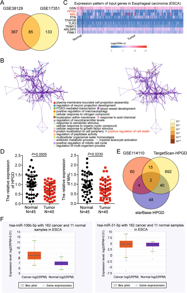
Background: Several studies have documented the key role of microRNAs (miRNAs) in esophageal squamous cell carcinoma (ESCC). Although the expression of the 15-hydroxyprostaglandin dehydrogenase (HPGD) gene and miR-106b-5p are reportedly linked to cancer progression, their underlying mechanisms in ESCC remain unclear.

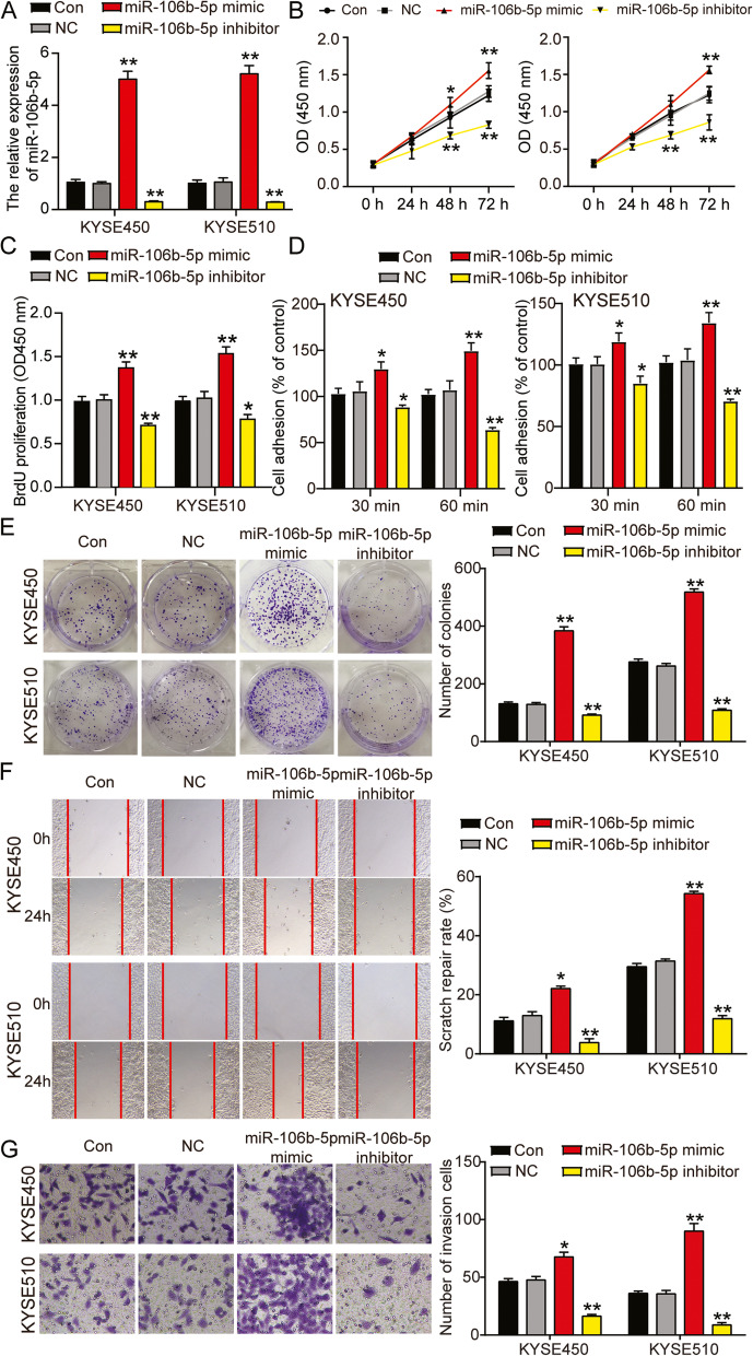
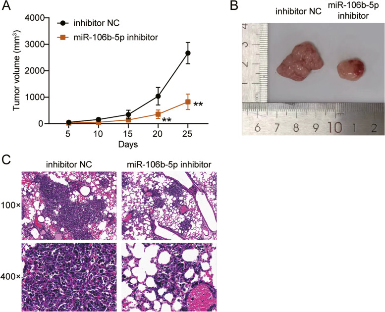
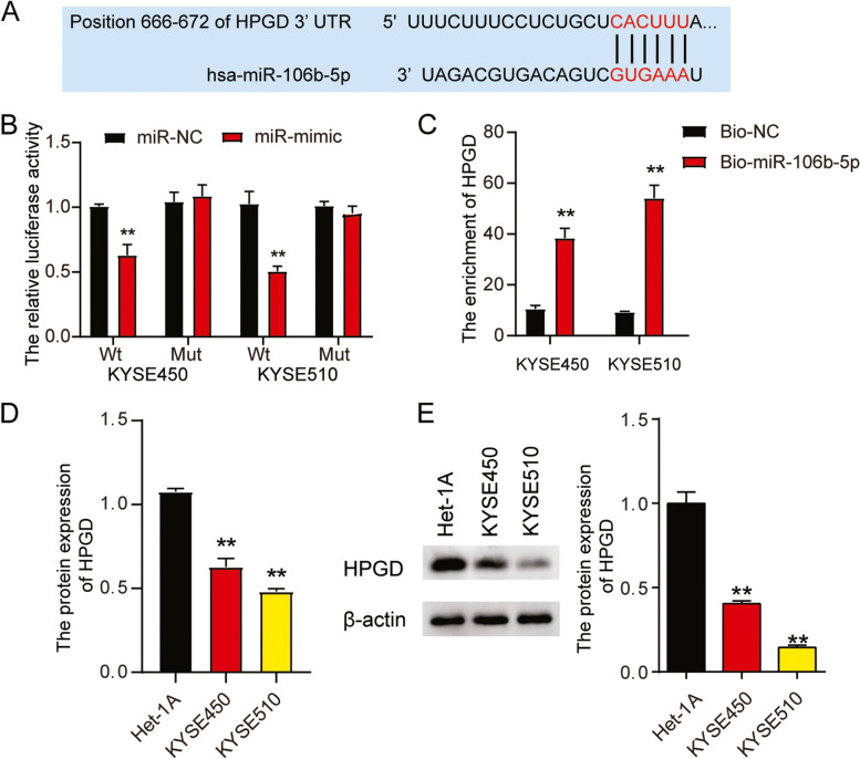
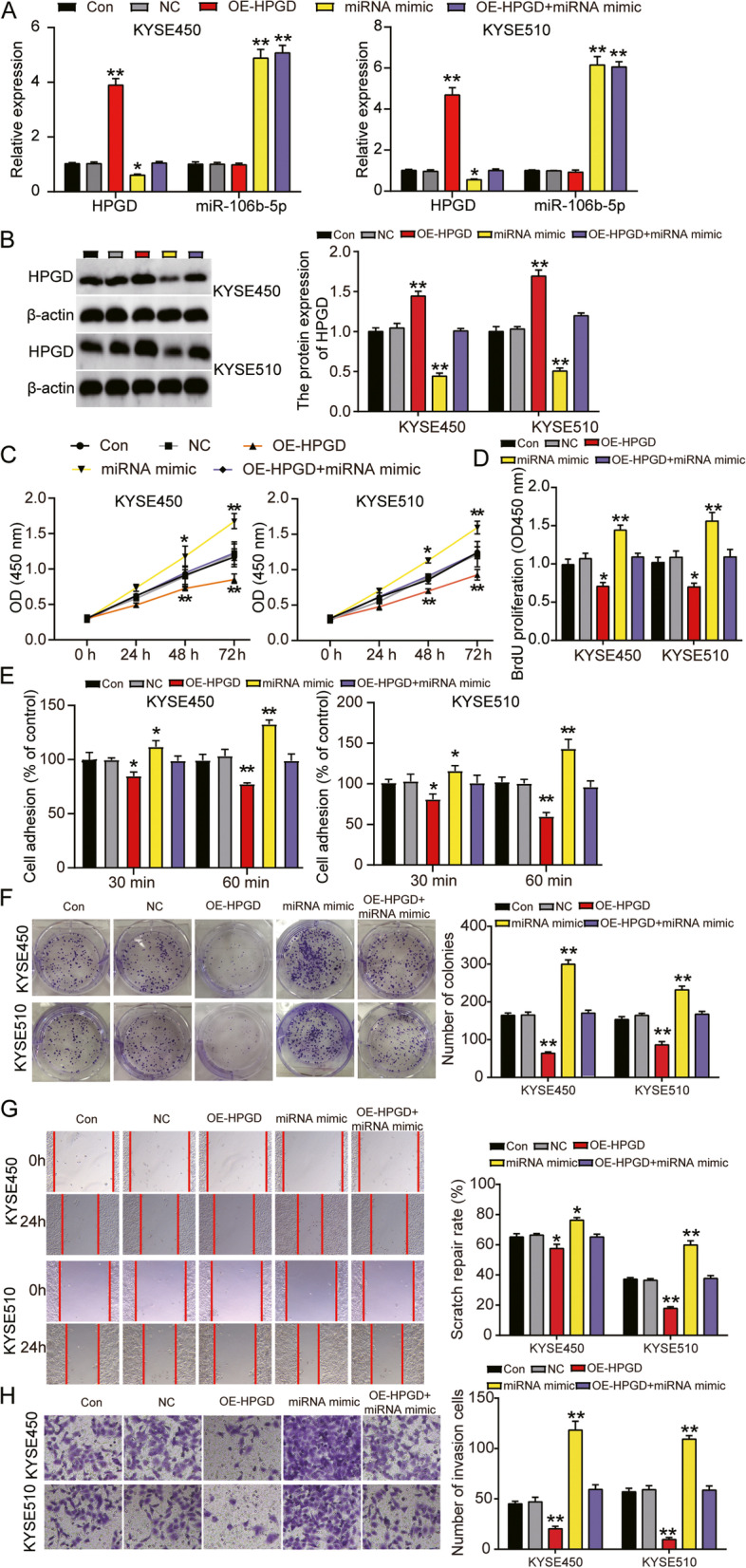
Methods: mRNA and miRNA expression in ESCC tissues and cells were analyzed using RT-qPCR. Luciferase and RNA pull-down assays were used to identify the interaction between miR-106b-5p and HPGD. Xenograft and pulmonary metastasis models were used to assess tumor growth and metastasis. CCK-8, BrdU, colony formation, adhesion, cell wound healing, Transwell, and caspase-3/7 activity assays, and flow cytometry and western blot analyses were used to examine the function of miR-106-5p and HPGD in ESCC cell lines.

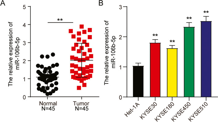
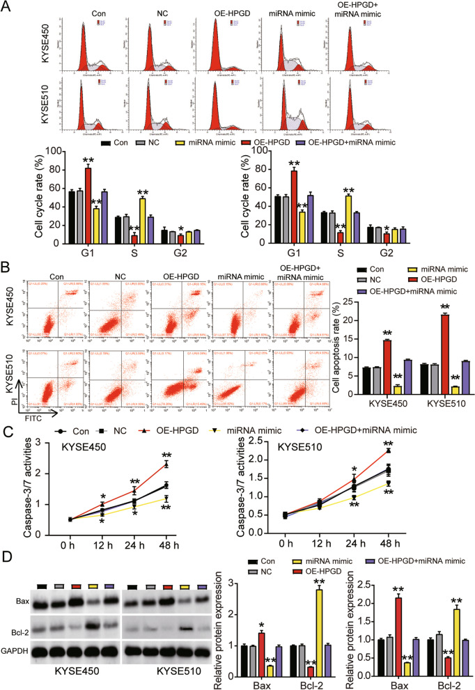
Results: The findings revealed that miR-106b-5p expression was upregulated in ESCC tissues and cell lines. miR-106b-5p augmented cellular proliferation, colony formation, adhesion, migration, invasion, and proportion of cells in the S-phase, but reduced apoptosis and the proportion of cells in G1-phase. Silencing of miR-106-5p inhibited tumor growth in vivo and pulmonary metastasis. Although HPGD overexpression suppressed proliferation, colony formation, adhesion, migration, and invasion of ESCC cells, it promoted apoptosis and caused cell cycle arrest of the ESCC cells. The results also indicated a direct interaction of HPGD with miR-106b-5p in ESCC cells. Furthermore, miR-106b-5p inhibited HPGD expression, thereby suppressing ESCC tumorigenesis.

Conclusion: Our data suggest that miR-106b-5p enhances proliferation, colony formation, adhesion, migration, and invasion, and induces the cycle progression, but represses apoptosis of ESCC cells by targeting HPGD. This suggests that the miR-106b-5p/HPGD axis may serve as a promising target for the diagnosis and treatment of ESCC.

Keywords: 15-hydroxyprostaglandin dehydrogenase; Adhesion; Apoptosis; Cell cycle; Colony formation; Esophageal squamous cell carcinoma; Invasion; MiR-106b-5p; Migration; Proliferation.


Conflict of interest statement

The authors declare that they have no conflict of interests.

Figures

Fig. 1
Fig. 1
HPGD and miR-106b-5p were selected to be further investigate in ESCC by microarray analysis. A 85 DEGs were overlapped from GSE38129 and GSE17351 by Venny 2.1.0. GSE38129 and GSE17351 were the mRNA expression profiles. B The positive regulation of cell death containing 9 DEGs was screen out as the key progress by Metascape analysis. C The expression of HPGO and MAL in ESCA was significant reduced. D The expression of HPGO and MAL was detected in the ESCC tissue samples (n = 45) and normal tissues (n = 45) by qRT-PCR. E miR-106b-5p and miR-31-5p were overlapped from GSE114110, TargetScan, and starBase. GSE114110 was the miRNA expression microarray. TargetScan and starBase were used to predict the miRNAs targeting HPGD. F The expression of miR-106b-5p was significant down-regulated in ESCA by starBase analysis
Fig. 2
Fig. 2
MiR-106b-5p was upregulated in ESCC tissues. A RT-qPCR detection of miR-106b-5p expression in ESCC tissues (n = 45) and normal tissues (n = 45). **, P < 0.001 compared to normal tissues. B Measurement of miR-106b-5p expression in ESCC cells lines (KYSE30, KYSE180, KYSE450, and KYSE510) and normal esophageal epithelial cells (Het-1A). Data are presented as means± SD of at least three independent tests per experiment. **, P < 0.001 compared to Het-1A cells
Fig. 3
Fig. 3
MiR-106b-5p promoted cell proliferation, adhesion, colony formation, migration and invasion in ESCC. A Measurement of miR-106b-5p expression in KYSE450 and KYSE510 cells transfected with NC, miR-106b-5p mimic, and miR-106b-5p inhibitor with RT-qPCR. B Cell viability was detected in KYSE450 and KYSE510 cells transfected with miR-106b-5p mimic, and miR-106b-5p inhibitor by CCK-8 assay. C Cell proliferation was detected in KYSE450 and KYSE510 cells transfected with, miR-106b-5p mimic, and miR-106b-5p inhibitor by BrdU assay. D Cell adhesion was detected in KYSE450 and KYSE510 cells transfected with NC, miR-106b-5p mimic, and miR-106b-5p inhibitor by cell adhesion assay kit. E Cell colony formation was detected in KYSE450 and KYSE510 cells transfected with NC, miR-106b-5p mimic, and miR-106b-5p inhibitor by colony formation assay. F Cell migration rate was detected in KYSE450 and KYSE510 cells transfected with NC, miR-106b-5p mimic, and miR-106b-5p inhibitor by cell wound healing assay. G Cell invasion was detected in KYSE450 and KYSE510 cells transfected with NC, miR-106b-5p mimic, and miR-106b-5p inhibitor by transwell assay. Data are presented as means± SD of at least three independent tests per experiment. *, P < 0.05; **, P < 0.001 compared to CON group. CON, blank control; NC, mimic-NC + inhibitor-NC
Fig. 4
Fig. 4
MiR-106b-5p promoted cell cycle progression and suppressed cell apoptosis in ESCC. A Cell cycle was detected in KYSE450 and KYSE510 cells transfected with NC, miR-106b-5p mimic, and miR-106b-5p inhibitor by flow cytometry assay. B Cell apoptosis rate was detected in KYSE450 and KYSE510 cells transfected with NC, miR-106b-5p mimic, and miR-106b-5p inhibitor by flow cytometry assay. C Cell apoptosis was determined in KYSE450 and KYSE510 cells transfected with NC, miR-106b-5p mimic, and miR-106b-5p inhibitor by caspase-3/7 activity assay kit. D The protein expression of Bax and Bcl-2 were determined in KYSE450 and KYSE510 cells transfected with NC, miR-106b-5p mimic, and miR-106b-5p inhibitor by western blot analysis. Data are presented as means± SD of at least three independent tests per experiment. *, P < 0.05; **, P < 0.001 compared to CON group. CON, blank control; NC, mimic-NC + inhibitor-NC
Fig. 5
Fig. 5
MiR-106b-5p promoted s xenograft and pulmonary metastasis in ESCC. A Tumor growth curves measured after the inoculation of nude mice injected with KYSE510 cells transfected with inhibitor-NC, and miR-106b-5p inhibitor. B Photographs of tumors in nude mice injected with KYSE510 cells transfected with inhibitor-NC, and miR-106b-5p inhibitor. C Representative photographs of H&E stained spontaneous lung metastases in nude mice injected with KYSE510 cells transfected with inhibitor-NC, and miR-106b-5p inhibitor
Fig. 6
Fig. 6
HPGD was a direct target ofmiR-106b-5p. A Bioinformatics analysis showed the predicted binding sequence of HPGD 3’-UTR. B Dual luciferase assay was performed in cells co-transfected with plasmids HPGD-Wt or HPGD-Mut and miR-NC or miR-106b-5p mimic in KYSE450 and KYSE510 cells. **, P < 0.001 compared to co-transfection of HPGD-Wt and miR-NC group. C RNA pull down assay was used to detect the association between HPGD and miR-106b-5p in KYSE450 and KYSE510 cells. **, P < 0.001 compared to Bio-NC group. D RT-qPCR detection of expression of HPGD in Het-1A, KYSE450 and KYSE510 cells. **, P < 0.001 compared to Het-1A cells. E Western blot detection of protein expression of HPGD in Het-1A, KYSE450 and KYSE510 cells. Data are presented as means ± SD of at least three independent tests per experiment. *, P < 0.05; **, P < 0.001 compared to Het-1A cells. Wt, wild-type; Mut, mutant; NC, negative control
Fig. 7
Fig. 7
MiR-106b-5p targeting to HPGD promoted proliferation, adhesion, colony formation, migration and invasion of ESCC cells. A Measurement of HPGD and miR-106b-5p gene expression in KYSE450 and KYSE510 cells transfected with NC, OE-HPGD, miR-106b-5p mimic, and OE-HPGD+miR-106b-5p mimic by RT-qPCR. B Measurement of HPGD protein expression in KYSE450 and KYSE510 cells transfected with NC, OE-HPGD, miR-106b-5p mimic, and OE-HPGD+miR-106b-5p mimic by western blot. C Cell viability was detected in KYSE450 and KYSE510 cells transfected with NC, OE-HPGD, miR-106b-5p mimic, and OE-HPGD+miR-106b-5p mimic by CCK-8 assay. D Cell proliferation was detected in KYSE450 and KYSE510 cells transfected with NC, OE-HPGD, miR-106b-5p mimic, and OE-HPGD+miR-106b-5p mimic by BrdU assay. E Cell adhesion was detected in KYSE450 and KYSE510 cells transfected with NC, OE-HPGD, miR-106b-5p mimic, and OE-HPGD+miR-106b-5p mimic by cell adhesion assay kit. F Cell colony formation was detected in KYSE450 and KYSE510 cells transfected with NC, OE-HPGD, miR-106b-5p mimic, and OE-HPGD+miR-106b-5p mimic by colony formation assay. G Cell migration rate was detected in KYSE450 and KYSE510 cells transfected with NC, OE-HPGD, miR-106b-5p mimic, and OE-HPGD+miR-106b-5p mimic by cell wound healing assay. H Cell invasion was detected in KYSE450 and KYSE510 cells transfected with NC, OE-HPGD, miR-106b-5p mimic, and OE-HPGD+miR-106b-5p mimic by transwell assay. Data are presented as means± SD of at least three independent tests per experiment. *, P < 0.05; **, P < 0.001 compared to CON group. CON, blank control; NC, empty vectors+mimic-NC; OE-HPGD, overexpression-HPGD; OE-HPGD+miR-106b-5p mimic, overexpression-HPGD+ miR-106b-5p mimic
Fig. 8
Fig. 8
MiR-106b-5p targeting to HPGD promoted cell cycle progression and suppressed cell apoptosis of ESCC cells. A Cell cycle was detected in KYSE450 and KYSE510 cells transfected with NC, OE-HPGD, miR-106b-5p mimic, and OE-HPGD+miR-106b-5p mimic by flow cytometry assay. B Cell apoptosis rate was detected in KYSE450 and KYSE510 cells transfected with NC, OE-HPGD, miR-106b-5p mimic, and OE-HPGD+miR-106b-5p mimic by flow cytometry assay. C Cell apoptosis was determined in KYSE450 and KYSE510 cells transfected with NC, OE-HPGD, miR-106b-5p mimic, and OE-HPGD+miR-106b-5p mimic by caspase-3/7 activity assay kit. D The protein expression of Bax and Bcl-2 were determined in KYSE450 and KYSE510 cells transfected with NC, OE-HPGD, miR-106b-5p mimic, and OE-HPGD+miR-106b-5p mimic by western blot analysis. Data are presented as means± SD of at least three independent tests per experiment. *, P < 0.05; **, P < 0.001 compared to CON group. CON, blank control; NC, empty vectors+mimic-NC; OE-HPGD, overexpression-HPGD; OE-HPGD+miR-106b-5p mimic, overexpression-HPGD+ miR-106b-5p mimic

Similar articles

Cited by

References

    1. Bray F, Ferlay J, Soerjomataram I, Siegel RL, Torre LA, Jemal A. Global cancer statistics 2018: GLOBOCAN estimates of incidence and mortality worldwide for 36 cancers in 185 countries. CA Cancer J Clin. 2018;68(6):394–424. doi: 10.3322/caac.21492. - DOI - PubMed
    1. Pennathur A, Gibson MK, Jobe BA, Luketich JD. Oesophageal carcinoma. Lancet. 2013;381(9864):400–412. doi: 10.1016/S0140-6736(12)60643-6. - DOI - PubMed
    1. Song Y, Li L, Ou Y, Gao Z, Li E, Li X, Zhang W, Wang J, Xu L, Zhou Y, et al. Identification of genomic alterations in oesophageal squamous cell cancer. Nature. 2014;509(7498):91–95. doi: 10.1038/nature13176. - DOI - PubMed
    1. Matsushima K, Isomoto H, Yamaguchi N, Inoue N, Machida H, Nakayama T, Hayashi T, Kunizaki M, Hidaka S, Nagayasu T, et al. MiRNA-205 modulates cellular invasion and migration via regulating zinc finger E-box binding homeobox 2 expression in esophageal squamous cell carcinoma cells. J Transl Med. 2011;9:30. doi: 10.1186/1479-5876-9-30. - DOI - PMC - PubMed
    1. Li B, Xu WW, Han L, Chan KT, Tsao SW, Lee NPY, Law S, Xu LY, Li EM, Chan KW, et al. MicroRNA-377 suppresses initiation and progression of esophageal cancer by inhibiting CD133 and VEGF. Oncogene. 2017;36(28):3986–4000. doi: 10.1038/onc.2017.29. - DOI - PMC - PubMed

MeSH terms

Substances