

Aqueous Thunbergia laurifolia leaf extract alleviates paraquat-induced lung injury in rats by inhibiting oxidative stress and inflammation
- PMID: 35317802
- PMCID: PMC8939148
- DOI: 10.1186/s12906-022-03567-4
Aqueous Thunbergia laurifolia leaf extract alleviates paraquat-induced lung injury in rats by inhibiting oxidative stress and inflammation
Abstract
Background: Paraquat (PQ) has been reported to have a high mortality rate. The major target organ of PQ poisoning is the lungs. The pathogenesis of PQ-induced lung injury involves oxidative stress and inflammation. Unfortunately, there is still no effective antidote for PQ poisoning. We hypothesized that aqueous Thunbergia laurifolia (TL) leaf extract is a possible antidote for PQ-induced lung injury.
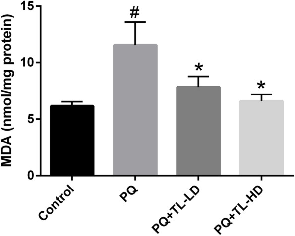
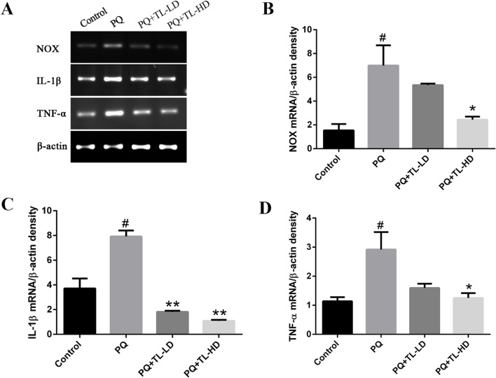
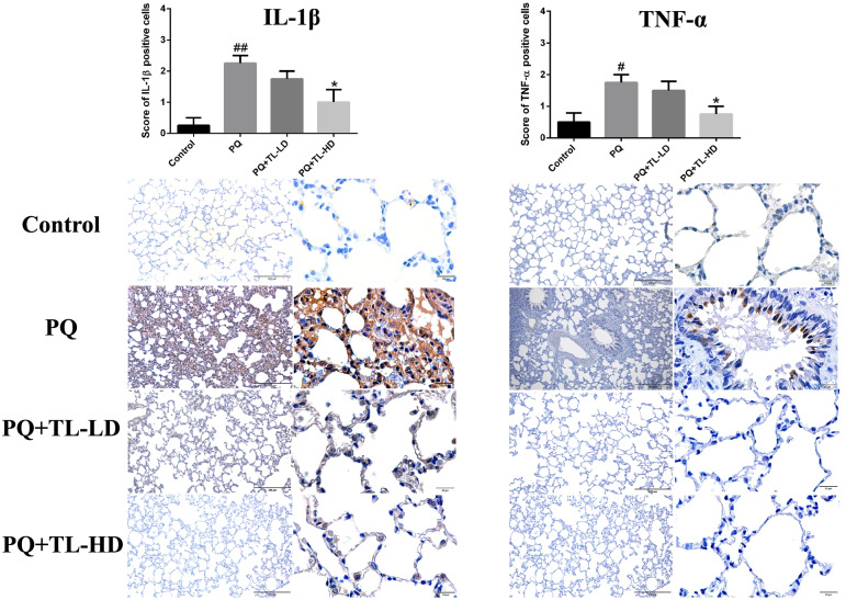
Methods: The total phenolic content and caffeic acid content of an aqueous extract of TL leaves were analyzed. Male Wistar rats were randomly divided into four groups (n = 4 per group): the control group (administered normal saline), the PQ group (administered 18 mg/kg body weight (BW) PQ dichloride subcutaneously), the PQ + TL-low-dose (LD) group (administered PQ dichloride subcutaneously and 100 mg/kg BW aqueous TL leaf extract by oral gavage) and the PQ + TL-high-dose (HD) group (administered PQ dichloride subcutaneously and 200 mg/kg BW aqueous TL leaf extract by oral gavage). Malondialdehyde (MDA) levels and lung histopathology were analyzed. In addition, the mRNA expression of NADPH oxidase (NOX), interleukin 1 beta (IL-1β), and tumor necrosis factor alpha (TNF-α) was assessed using reverse transcription-polymerase chain reaction (RT-PCR), and the protein expression of IL-1β and TNF-α was analyzed using immunohistochemistry.
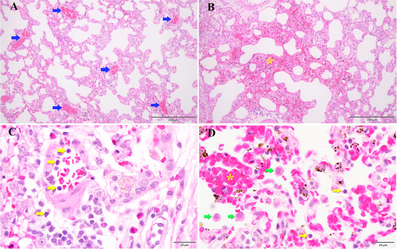
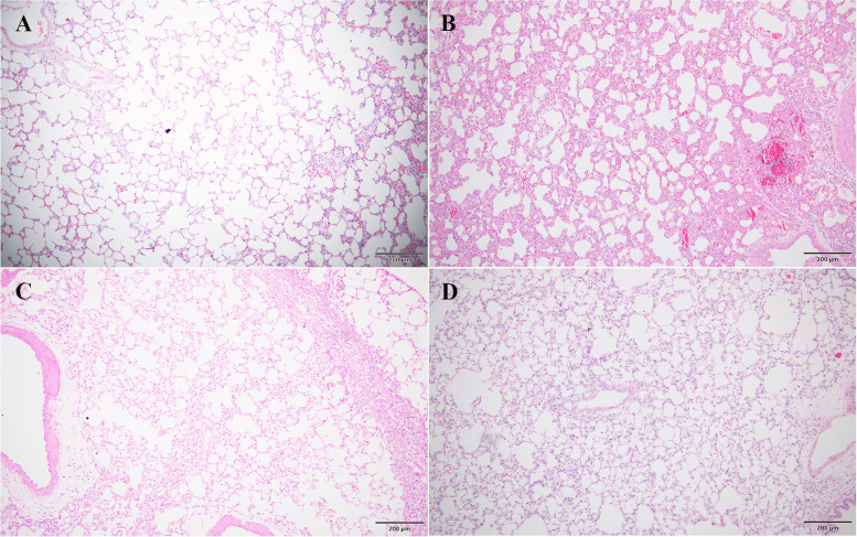
Results: The total phenolic content of the extract was 20.1 ± 0.39 μg gallic acid equivalents (Eq)/mg extract, and the caffeic acid content was 0.31 ± 0.01 μg/mg. The PQ group showed significantly higher MDA levels and NOX, IL-1β and TNF-α mRNA expression than the control group. Significant pathological changes, including alveolar edema, diffuse alveolar collapse, hemorrhage, leukocyte infiltration, alveolar septal thickening and vascular congestion, were observed in the PQ group compared with the control group. However, the aqueous TL leaf extract significantly attenuated the PQ-induced increases in MDA levels and NOX, IL-1β and TNF-α expressions. Moreover, the aqueous TL leaf extract ameliorated PQ-induced lung pathology.
Conclusion: This study indicates that aqueous TL leaf extract can ameliorate PQ-induced lung pathology by modulating oxidative stress through inhibition of NOX and by regulating inflammation through inhibition of IL-1β and TNF-α expressions. We suggest that aqueous TL leaf extract can be used as an antidote for PQ-induced lung injury.
Keywords: Inflammation; Interleukin 1 beta; Lung injury; Malondialdehyde; NADPH oxidase; Oxidative stress; Paraquat; Thunbergia laurifolia; Tumor necrosis factor alpha.
© 2022. The Author(s).
Conflict of interest statement
The authors declare that they have no competing interests.
Figures
Similar articles
-
Thunbergia laurifolia aqueous leaf extract ameliorates paraquat-induced kidney injury by regulating NADPH oxidase in rats.Heliyon. 2022 Apr 1;8(4):e09234. doi: 10.1016/j.heliyon.2022.e09234. eCollection 2022 Apr. Heliyon. 2022. PMID: 35399379 Free PMC article.
-
[Protective effect of thalidomide on ALI induced by paraquat poisoning in rats and its mechanism].Zhonghua Wei Zhong Bing Ji Jiu Yi Xue. 2017 Nov;29(11):977-981. doi: 10.3760/cma.j.issn.2095-4352.2017.11.004. Zhonghua Wei Zhong Bing Ji Jiu Yi Xue. 2017. PMID: 29151411 Chinese.
-
[Effect of pirfenidone on paraquat-induced pulmonary fibrosis in rats].Zhonghua Lao Dong Wei Sheng Zhi Ye Bing Za Zhi. 2023 Feb 20;41(2):104-111. doi: 10.3760/cma.j.cn121094-20211008-00489. Zhonghua Lao Dong Wei Sheng Zhi Ye Bing Za Zhi. 2023. PMID: 36882273 Chinese.
-
[Effects of Nintedanib associated with Shenfu Injection on paraquat-induced lung injury in rats].Zhonghua Lao Dong Wei Sheng Zhi Ye Bing Za Zhi. 2023 Feb 20;41(2):81-86. doi: 10.3760/cma.j.cn121094-20220419-00203. Zhonghua Lao Dong Wei Sheng Zhi Ye Bing Za Zhi. 2023. PMID: 36882270 Chinese.
-
The efficacy of hemodialysis on paraquat poisoning mortality: A systematic review and meta-analysis.J Res Med Sci. 2022 Sep 27;27:74. doi: 10.4103/jrms.jrms_235_21. eCollection 2022. J Res Med Sci. 2022. PMID: 36353345 Free PMC article. Review.
Cited by
-
Unprecedented Approach for Using Misoprostol Alongside Low-Dose Gamma Radiation to Alleviate Paraquat-Induced Pulmonary Injury in Rats.Dose Response. 2025 Mar 25;23(1):15593258251326707. doi: 10.1177/15593258251326707. eCollection 2025 Jan-Mar. Dose Response. 2025. PMID: 40144808 Free PMC article.
-
Andrographolide alleviates paraquat-induced acute lung injury by activating the Nrf2/HO-1 pathway.Iran J Basic Med Sci. 2023;26(6):653-661. doi: 10.22038/IJBMS.2023.68827.15000. Iran J Basic Med Sci. 2023. PMID: 37275765 Free PMC article.
References
-
- Shashibhushan J, Venugopal K, Lingaraja M, Patanjali C, Suresh C, Huggi V. Paraquat: a fatal poison. Med J DY Patil Univ. 2015;8(3):370–374. doi: 10.4103/0975-2870.157090. - DOI
MeSH terms
Substances
LinkOut - more resources
Full Text Sources
Research Materials
