

Endothelial dysfunction and altered endothelial biomarkers in patients with post-COVID-19 syndrome and chronic fatigue syndrome (ME/CFS)
- PMID: 35317812
- PMCID: PMC8938726
- DOI: 10.1186/s12967-022-03346-2
Endothelial dysfunction and altered endothelial biomarkers in patients with post-COVID-19 syndrome and chronic fatigue syndrome (ME/CFS)
Abstract
Background: Fatigue, exertion intolerance and post-exertional malaise are among the most frequent symptoms of Post-COVID Syndrome (PCS), with a subset of patients fulfilling criteria for Myalgic Encephalomyelitis/Chronic Fatigue Syndrome (ME/CFS). As SARS-CoV-2 infects endothelial cells, causing endotheliitis and damaging the endothelium, we investigated endothelial dysfunction (ED) and endothelial biomarkers in patients with PCS.
Methods: We studied the endothelial function in 30 PCS patients with persistent fatigue and exertion intolerance as well as in 15 age- and sex matched seronegative healthy controls (HCs). 14 patients fulfilled the diagnostic criteria for ME/CFS. The other patients were considered to have PCS. Peripheral endothelial function was assessed by the reactive hyperaemia index (RHI) using peripheral arterial tonometry (PAT) in patients and HCs. In a larger cohort of patients and HCs, including post-COVID reconvalescents (PCHCs), Endothelin-1 (ET-1), Angiopoietin-2 (Ang-2), Endocan (ESM-1), IL-8, Angiotensin-Converting Enzyme (ACE) and ACE2 were analysed as endothelial biomarkers.
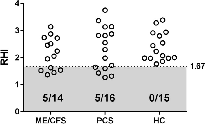
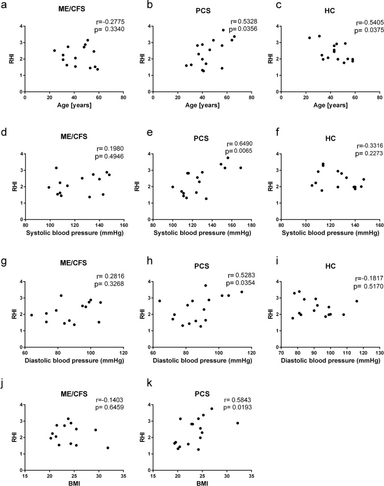
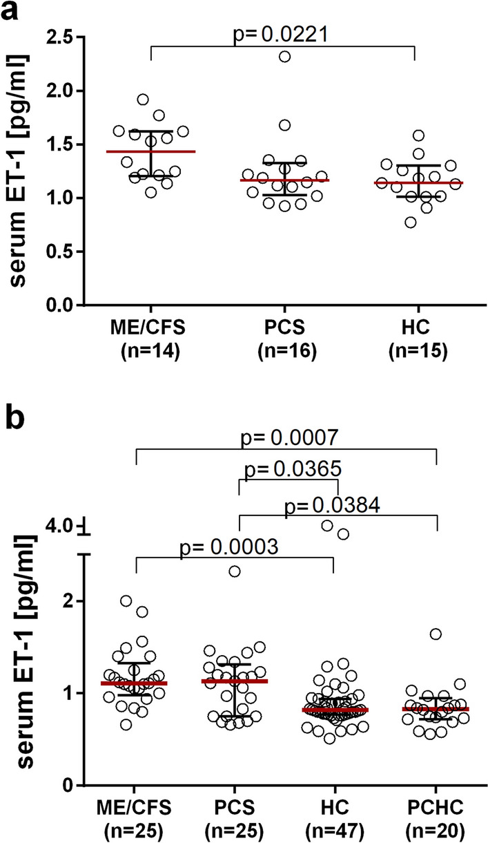
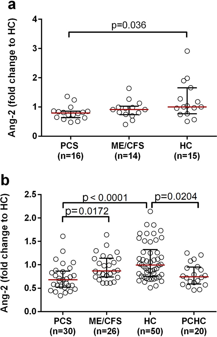
Results: Five of the 14 post-COVID ME/CFS patients and five of the 16 PCS patients showed ED defined by a diminished RHI (< 1.67), but none of HCs exhibited this finding. A paradoxical positive correlation of RHI with age, blood pressure and BMI was found in PCS but not ME/CFS patients. The ET-1 concentration was significantly elevated in both ME/CFS and PCS patients compared to HCs and PCHCs. The serum Ang-2 concentration was lower in both PCS patients and PCHCs compared to HCs.
Conclusion: A subset of PCS patients display evidence for ED shown by a diminished RHI and altered endothelial biomarkers. Different associations of the RHI with clinical parameters as well as varying biomarker profiles may suggest distinct pathomechanisms among patient subgroups.
Keywords: Endothelial dysfunction; Endothelin-1; Myalgic encephalomyelitis/chronic fatigue syndrome; Post-COVID syndrome; Reactive hyperaemia index.
© 2022. The Author(s).
Conflict of interest statement
The authors declare no competing interests.
Figures
Similar articles
-
Serum of Post-COVID-19 Syndrome Patients with or without ME/CFS Differentially Affects Endothelial Cell Function In Vitro.Cells. 2022 Aug 2;11(15):2376. doi: 10.3390/cells11152376. Cells. 2022. PMID: 35954219 Free PMC article.
-
Association of circulating biomarkers with illness severity measures differentiates myalgic encephalomyelitis/chronic fatigue syndrome and post-COVID-19 condition: a prospective pilot cohort study.J Transl Med. 2024 Apr 10;22(1):343. doi: 10.1186/s12967-024-05148-0. J Transl Med. 2024. PMID: 38600563 Free PMC article.
-
Long-term symptom severity and clinical biomarkers in post-COVID-19/chronic fatigue syndrome: results from a prospective observational cohort.EClinicalMedicine. 2023 Aug 19;63:102146. doi: 10.1016/j.eclinm.2023.102146. eCollection 2023 Sep. EClinicalMedicine. 2023. PMID: 37662515 Free PMC article.
-
Key Pathophysiological Role of Skeletal Muscle Disturbance in Post COVID and Myalgic Encephalomyelitis/Chronic Fatigue Syndrome (ME/CFS): Accumulated Evidence.J Cachexia Sarcopenia Muscle. 2025 Feb;16(1):e13669. doi: 10.1002/jcsm.13669. J Cachexia Sarcopenia Muscle. 2025. PMID: 39727052 Free PMC article. Review.
-
The persistence of myalgic encephalomyelitis/chronic fatigue syndrome (ME/CFS) after SARS-CoV-2 infection: A systematic review and meta-analysis.J Infect. 2024 Dec;89(6):106297. doi: 10.1016/j.jinf.2024.106297. Epub 2024 Sep 29. J Infect. 2024. PMID: 39353473
Cited by
-
Post-COVID exercise intolerance is associated with capillary alterations and immune dysregulations in skeletal muscles.Acta Neuropathol Commun. 2023 Dec 8;11(1):193. doi: 10.1186/s40478-023-01662-2. Acta Neuropathol Commun. 2023. PMID: 38066589 Free PMC article.
-
Alterations in plasma proteome during acute COVID-19 and recovery.Mol Med. 2024 Aug 25;30(1):131. doi: 10.1186/s10020-024-00898-5. Mol Med. 2024. PMID: 39183264 Free PMC article.
-
Serum of Post-COVID-19 Syndrome Patients with or without ME/CFS Differentially Affects Endothelial Cell Function In Vitro.Cells. 2022 Aug 2;11(15):2376. doi: 10.3390/cells11152376. Cells. 2022. PMID: 35954219 Free PMC article.
-
Understanding Endothelial Dysfunction and Its Role in Ischemic Stroke After the Outbreak of Recanalization Therapies.Int J Mol Sci. 2024 Oct 29;25(21):11631. doi: 10.3390/ijms252111631. Int J Mol Sci. 2024. PMID: 39519182 Free PMC article. Review.
-
Blood virome research in myalgic encephalomyelitis/chronic fatigue syndrome: challenges and opportunities.Curr Opin Virol. 2024 Sep-Dec;68-69:101437. doi: 10.1016/j.coviro.2024.101437. Epub 2024 Nov 12. Curr Opin Virol. 2024. PMID: 39537445 Free PMC article. Review.
References
-
- Kedor C, Freitag H, Meyer-Arndt L, Wittke K, Zoller T, Steinbeis F, et al. Chronic COVID-19 Syndrome and Chronic Fatigue Syndrome (ME/CFS) following the first pandemic wave in Germany—a first analysis of a prospective observational study. medRxiv. 2021 doi: 10.1101/2021.02.06.21249256. - DOI
-
- Sotzny F, Blanco J, Capelli E, Castro-Marrero J, Steiner S, Murovska M, et al. Myalgic encephalomyelitis/chronic fatigue syndrome—evidence for an autoimmune disease. Autoimmun Rev. 2018;17(6):601–609. - PubMed
Publication types
MeSH terms
Substances
LinkOut - more resources
Full Text Sources
Medical
Research Materials
Miscellaneous
