

CircESRP1 enhances metastasis and epithelial-mesenchymal transition in endometrial cancer via the miR-874-3p/CPEB4 axis
- PMID: 35317822
- PMCID: PMC8939068
- DOI: 10.1186/s12967-022-03334-6
CircESRP1 enhances metastasis and epithelial-mesenchymal transition in endometrial cancer via the miR-874-3p/CPEB4 axis
Abstract
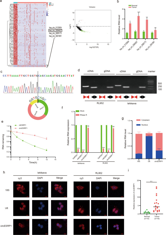
Background: Metastasis is critical for endometrial cancer (EC) progression and prognosis. Accumulating evidence suggests that circular RNAs (circRNAs) can operate as independent functional entities. However, the functional regulatory mechanisms of circRNAs in EC remain unclear.
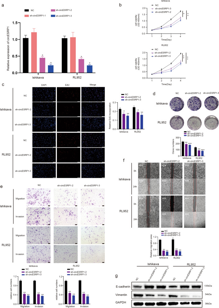
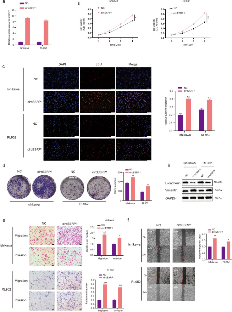
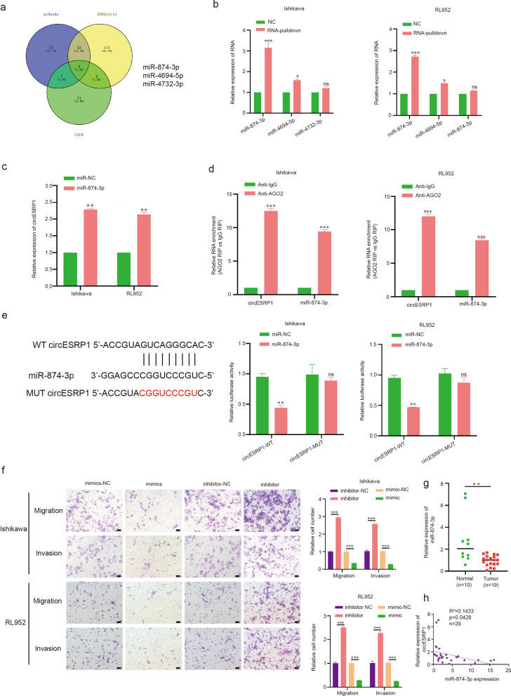
Methods: The levels of circESRP1, miR-874-3p, and CPEB4 mRNA in EC tissues and cells were determined by qRT-PCR. Sanger sequencing, PCR with divergent primers, an actinomycin D assay, and RNase R treatment were applied to verify the circular properties. Fluorescence in situ hybridization (FISH) and nuclear-cytoplasmic fractionation were used to determine the localization of circESRP1. CCK-8, EdU incorporation, colony formation, Transwell, and wound healing assays were applied to assess the effects of circESRP1 on cell proliferation, migration, and invasion. The mutual regulatory mechanism of ceRNAs was investigated using dual-luciferase reporter, RNA pulldown, RNA immunoprecipitation (RIP), and Western blot assays. The biological effects were further validated in vivo in nude mouse xenograft models.
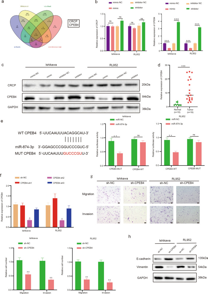
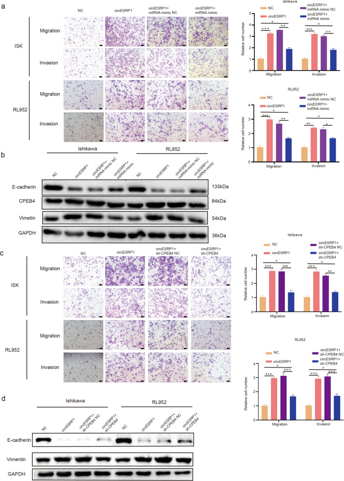
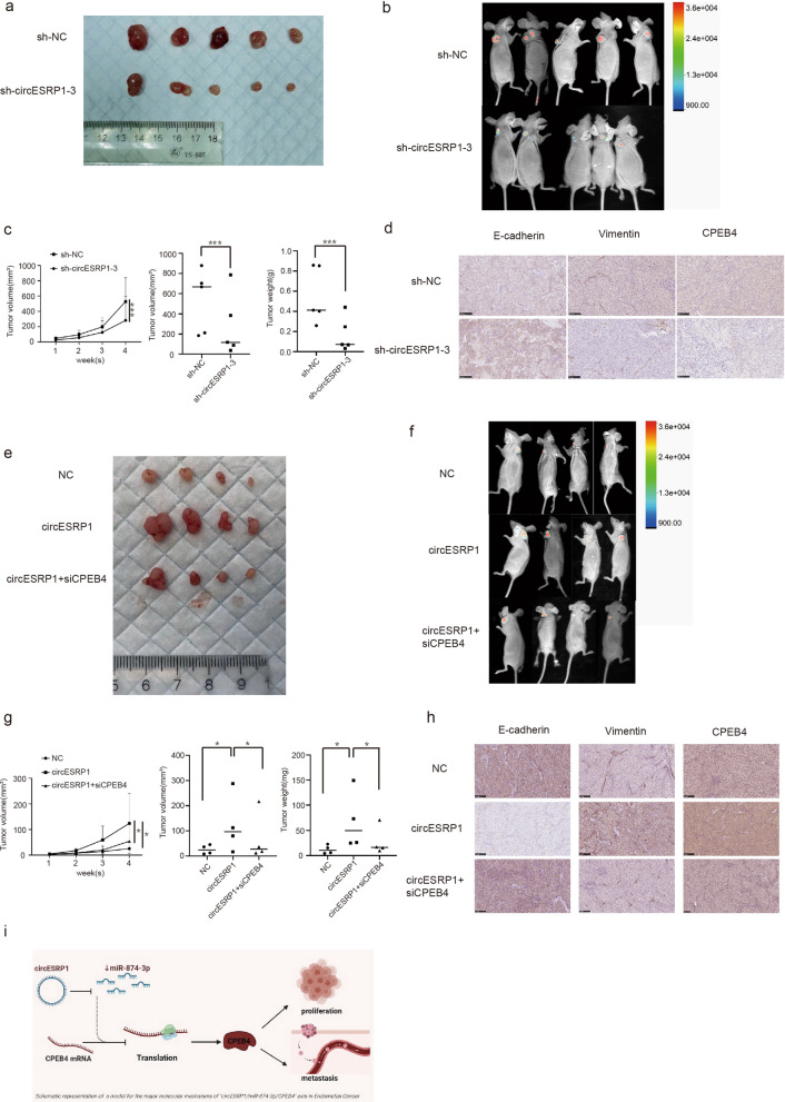
Results: circESRP1 was highly expressed in EC tissues and cells and was mainly localized in the cytoplasm. Silencing circESRP1 inhibited the proliferation, migration, and invasion of EC cells in vitro and in vivo; however, overexpression of circESRP1 had the opposite effects. Mechanistically, circESRP1 sponged miR-874-3p to upregulate CPEB4 expression and ultimately contribute to EC cell proliferation and metastasis. Furthermore, circESRP1 regulated tumour growth in xenograft models.
Conclusions: CircESRP1 can interact with miR-874-3p to regulate EMT in endometrial cancer via the miR-874-3p/CPEB4 axis. CircESRP1 may serve as a promising therapeutic target for endometrial cancer.
Keywords: CPEB4; CircESRP1; EMT; Endometrial cancer; Proliferation; miR-874-3p.
© 2022. The Author(s).
Conflict of interest statement
The authors declare no competing interests.
Figures
Similar articles
-
Long non-coding RNA VPS9D1-AS1 enhances proliferation, invasion, and epithelial-mesenchymal transition in endometrial cancer via miR-377-3p/SGK1.Kaohsiung J Med Sci. 2022 Nov;38(11):1048-1059. doi: 10.1002/kjm2.12606. Epub 2022 Oct 17. Kaohsiung J Med Sci. 2022. PMID: 36245426 Free PMC article.
-
Silencing of hsa_circ_0101145 reverses the epithelial-mesenchymal transition in hepatocellular carcinoma via regulation of the miR-548c-3p/LAMC2 axis.Aging (Albany NY). 2020 Jun 18;12(12):11623-11635. doi: 10.18632/aging.103324. Epub 2020 Jun 18. Aging (Albany NY). 2020. PMID: 32554866 Free PMC article.
-
Circular RNA circNTRK2 facilitates the progression of esophageal squamous cell carcinoma through up-regulating NRIP1 expression via miR-140-3p.J Exp Clin Cancer Res. 2020 Jul 11;39(1):133. doi: 10.1186/s13046-020-01640-9. J Exp Clin Cancer Res. 2020. PMID: 32653032 Free PMC article.
-
MiR-1236: Key controller of tumor development and progression: Focus on the biological functions and molecular mechanisms.Pathol Res Pract. 2023 Aug;248:154671. doi: 10.1016/j.prp.2023.154671. Epub 2023 Jul 3. Pathol Res Pract. 2023. PMID: 37418995 Review.
-
RNA-binding proteins and cancer metastasis.Semin Cancer Biol. 2022 Nov;86(Pt 2):748-768. doi: 10.1016/j.semcancer.2022.03.018. Epub 2022 Mar 23. Semin Cancer Biol. 2022. PMID: 35339667 Review.
Cited by
-
circRNAs in Endometrial Cancer-A Promising Biomarker: State of the Art.Int J Mol Sci. 2024 Jun 9;25(12):6387. doi: 10.3390/ijms25126387. Int J Mol Sci. 2024. PMID: 38928094 Free PMC article. Review.
-
MicroRNAs, long non-coding RNAs, and circular RNAs and gynecological cancers: focus on metastasis.Front Oncol. 2023 Oct 3;13:1215194. doi: 10.3389/fonc.2023.1215194. eCollection 2023. Front Oncol. 2023. PMID: 37854681 Free PMC article. Review.
-
CircUSP10 promotes liver cancer progression by regulating miR-211-5p/TCF12/EMT signaling pathway.Heliyon. 2023 Oct 4;9(10):e20649. doi: 10.1016/j.heliyon.2023.e20649. eCollection 2023 Oct. Heliyon. 2023. PMID: 37829805 Free PMC article.
-
Advances in MiRNAs Involved in Endometrial Carcinoma.Comb Chem High Throughput Screen. 2025;28(1):3-11. doi: 10.2174/0113862073299444240308145725. Comb Chem High Throughput Screen. 2025. PMID: 38504572 Review.
-
Hsa_circ_0000069 Accelerates Cervical Cancer Progression by Sponging miR-1270 to Facilitate CPEB4 Expression.Biochem Genet. 2024 Jun;62(3):1638-1656. doi: 10.1007/s10528-023-10494-7. Epub 2023 Sep 4. Biochem Genet. 2024. PMID: 37667097
References
-
- Brooks RA, Fleming GF, Lastra RR, Lee NK, Moroney JW, et al. Current recommendations and recent progress in endometrial cancer. CA Cancer J Clin. 2019;69:258–279. - PubMed
Publication types
MeSH terms
Substances
LinkOut - more resources
Full Text Sources
Miscellaneous
