. 2022 Jun:159:116393.
doi: 10.1016/j.bone.2022.116393. Epub 2022 Mar 19.

Histone deacetylase 5 is a phosphorylation substrate of protein kinase D in osteoclasts

Affiliations

Histone deacetylase 5 is a phosphorylation substrate of protein kinase D in osteoclasts

Carina Mello Guimaraes Meyers et al. Bone. 2022 Jun.

Abstract

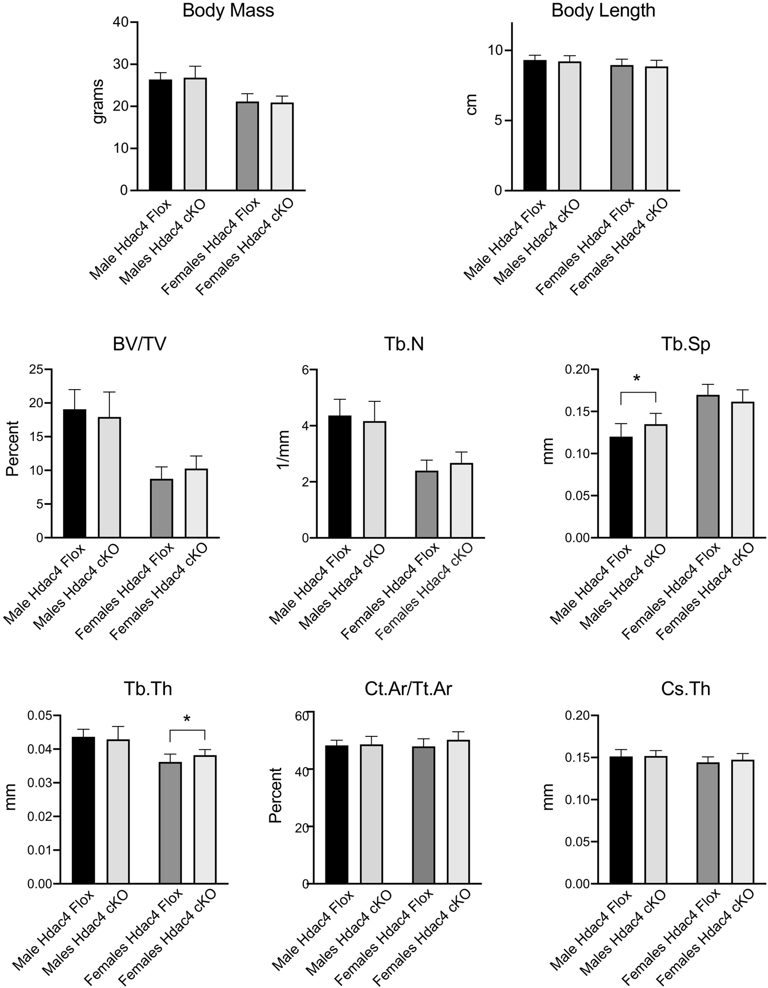
Protein kinase D (PRKD) family kinases are required for formation and function of osteoclasts. However, the substrates of PRKD in osteoclasts are unknown. To identify PRKD-dependent protein phosphorylation in osteoclasts, we performed a quantitative LC-MS/MS phosphoproteomics screen for proteins showing differential phosphorylation in osteoclasts after treatment with the PRKD inhibitor CRT0066101. We identified 757 phosphopeptides showing significant changes following PRKD inhibition. Among the changes, we found a group of 13 proteins showing decreased phosphorylation at PRKD consensus phosphorylation motifs. This group includes histone deacetylase 5 (HDAC5), which is a previously validated PRKD target. Considering this known interaction, work suggesting HDACs may be important regulators of osteoclasts, and studies suggesting potential functional redundancy between HDACs, we further investigated the relationship between PRKD and class IIa HDACs in osteoclasts. We confirmed that CRT0066101 inhibits phosphorylation of endogenous HDAC5 and to a lesser extent HDAC4, whereas HDAC7 phosphorylation was not affected. Osteoclast cultures from Hdac5 global knockout mice displayed impaired differentiation and reduced ability to resorb bone, while conditional knockout of Hdac4 in osteoclasts showed no phenotype in vitro or in vivo. The inhibitory effect of CRT0066101 was reduced in Hdac5 KO osteoclasts. Together these data indicate that the PRKD/HDAC5 axis contributes to osteoclast formation in vitro and suggest that this pathway may contribute to regulation of skeletal dynamics in vivo.

Keywords: Bone remodeling; Histone deacetylases; Osteoclast differentiation; Protein kinase D; Protein phosphorylation.


Conflict of interest statement

Conflicts of Interest: none

Figures

Figure 1
Figure 1
Phosphoproteomics analysis of CRT0066101-responsive protein phosphorylation (A) Schematic of workflow. Osteoclast cultures on day 3 of RANKL stimulation were treated with 200 nM CRT0066101 for 5 hours, lysed and subjected to quantitative phosphoproteomics screening. (B) Concentric Venn diagram illustrating total peptides (grey), phosphopeptides (green), and significantly changed phosphopeptides (innermost circle), divided into increased abundance (blue) and decreased abundance (red) areas. (C) Volcano plot of the 15,269 phosphopeptides that were quantitatively identified comparing Log2 fold change versus adjusted p-value. Significantly increased phosphopeptides are in blue, decreased abundance in red. (D) Top 15 functional annotation clusters from the 757 significantly altered phosphopeptides, analyzed using the DAVID gene ontology tool. (E) Significantly decreased phosphopeptides mapping to PRKD consensus motifs.
Figure 2
Figure 2
Protein kinase D regulates HDAC phosphorylation (A) mean abundance of HDAC5 phosphopeptide (amino acids 648–659, phosphoserine 650) in CRT0066101 and untreated osteoclast cultures. (B) Schematic of class IIa HDACs and conserved regulatory phosphoserine residues. Amino acid numbering is based on mouse HDAC proteins. Position of conserved phosphorylated residues and the sites targeted by anti-P-HDAC antibodies #3424 and 3443 are indicated above. CtBP and MEF2 interaction domains, Nuclear Localization Sequence (NLS) and Nuclear Export Sequences (NES) and the deacetylase catalytic functional domains are also illustrated (C-D) Western blotting against phosphorylated PRKD, P-HDACs and additional phospho-proteins following treatment of day 3 osteoclasts with 200 nM CRT0066101 for the indicated times. α-tubulin was blotted as a loading control (E) Osteoclasts treated with 200 nM CRT0066101 for 60 minutes were lysed and immunoprecipitated (IP) with antibodies against HDAC4, HDAC5, HDAC7 or normal rabbit IgG (control IP). Western blots were blotted with the indicated P-HDAC and total HDAC antibodies. (F) HEK293T cells were transfected with FLAG-tagged HDAC5, PRKD2 and PRKD3 proteins as indicated, lysed and immunoblotted for the FLAG and P-HDAC. (G) Western blotting against Prkd3 control and Prkd3 cKO osteoclast cultures. ** p< 0.005.
Figure 3
Figure 3
In vitro culture of Hdac5 KO osteoclasts (A) real-time RT-PCR (left) and Western blotting (right) against HDAC5 expression in wild-type and Hdac5 KO osteoclast cultures (B) Proliferation/ survival curves showing the number of nuclei per field in cultures of WT cells (dark bars) and Hdac5 KO (light bars) from Day -1 (the day prior to RANKL), Day 0 (the day of RANKL addition), and Days 1–3 of osteoclast differentiation. Data are graphed as mean number of nuclei per field relative to WT Day -1. (C) TRAP staining (top row), rhodamine-phalloidin (middle row) and resorption pits on bone slices visualized by hematoxylin staining (bottom row) of wild type control and Hdac5 KO osteoclast cultures. (D) Quantitation of mature osteoclasts comparing mean number of osteoclasts per field, mean number of nuclei per osteoclast, largest number of nuclei per osteoclast determined from cells double stained for TRAP and DAPI, and total resorbed area fraction from resorption assay on bone slices. (E) Western blotting for HDAC5, phospho-SRC Y416. Total SRC and ACTIN were visualized as loading controls P-SRC to total SRC ratios are indicated below the blots. (F) Real-time RT-PCR from wild-type and Hdac5 KO osteoclast cultures. Graphs show the mean + SD from three independent experiments * p<0.05, ** p<0.005.
Figure 4
Figure 4
CRT0066101 treatment of wild-type and Hdac5 knockout osteoclast cultures. Cells were treated with CRT0066101 at 0, 20nM or 100 nM beginning at the time of RANKL stimulation and stained for TRAP (top rows) or rhodamine-phalloidin (red) and DAPI (blue), middle rows. Bottom rows show resorption pit staining on bone slices. Hdac5 KO or wild-type osteoclasts were cultured on bone slices for 6 days, swabbed to remove the cells and visualized with hematoxylin staining. Graphs at the right present comparisons in mean and maximum nuclei per osteoclast and total resorbed area for wild-type (dark bars) and Hdac5 KO (lighter grey bars). Quantitative data for CRT0066101-treated cells are presented as change relative to the corresponding untreated WT or KO cells. Actin morphology was manually scored for each multinucleated osteoclast and the population distribution is graphed for peripheral actin belts, smaller internal actin rings and disordered/other. * p<0.05, ** p<0.005.
Figure 5
Figure 5
In vitro culture of Hdac4 cKO osteoclasts (A) Western blotting against HDAC4 or β-actin as a loading control from osteoclast cultures during differentiation. The blots from Hdac4 flox and cKO cells shown are taken from the same photo of a single western blot. (B) Proliferation/ survival curves graphing number of nuclei per field in cultures of WT cells (dark bars) and Hdac4 cKO (light bars) in osteoclast cultures from Day -1 (the day prior to RANKL), Day 0 (the day of RANKL addition), and Days 1–3 of osteoclast differentiation. Data are graphed as mean number of nuclei per field relative to WT Day -1. (C) TRAP staining (left), rhodamine-phalloidin (middle column, red) and resorption pits on bone slices (right column, wheat germ agglutinin-HRP staining) of wild type control and Hdac4 cKO osteoclast cultures. (D) Quantitation of mature osteoclasts, comparing number of osteoclasts per field, number of nuclei per osteoclast, largest number of nuclei per osteoclast determined from cells double stained for TRAP and DAPI, and total resorbed area fraction from resorption assay on bone slices.
Figure 6
Figure 6
Analysis of skeletal parameters of Hdac4 cKO male and female mice at 12 weeks age. Mass and body length were measured at sacrifice. μCT analysis of trabecular bone was performed at the distal femur; cortical measures were obtained at the mid-diaphysis. Differences between Hdac4flox and Hdac4 cKO of the same sex did not reach statistical significance unless specifically indicated. * p<0.05.
Figure 7
Figure 7
Model of regulation of osteoclasts by PRKD and HDAC5. Under normal conditions (upper schematic), PRKD activates factors that promote osteoclast formation, regulates actin cytoskeleton and phosphorylates HDAC5 to give a moderate level of inhibitory factors, ultimately giving balanced level of osteoclastogenesis. In Hdac5 KO osteoclasts (middle schematic), loss of HDAC5 dysregulates expression of osteoclast inhibitory factors thereby reducing osteoclasts. Treatment with PRKD inhibitors (bottom panel) reduces HDAC5 phosphorylation thus reducing expression of HDAC5-responsive inhibitory factors, but loss of PRKD-responsive stimulatory factors results in diminished osteoclast formation.

Similar articles

References

    1. Briggs AM, Cross MJ, Hoy DG, Sànchez-Riera L, Blyth FM, Woolf AD, March L, Musculoskeletal Health Conditions Represent a Global Threat to Healthy Aging: A Report for the 2015 World Health Organization World Report on Ageing and Health, The Gerontologist 56(Suppl_2) (2016) S243–S255. - PubMed
    1. Boyce BF, Advances in the regulation of osteoclasts and osteoclast functions, J Dent Res 92(10) (2013) 860–7. - PMC - PubMed
    1. Brown JP, Antiresorptives: Safety Concerns-Clinical Perspective, Toxicol Pathol 45(7) (2017) 859–863. - PubMed
    1. Ukon Y, Makino T, Kodama J, Tsukazaki H, Tateiwa D, Yoshikawa H, Kaito T, Molecular-Based Treatment Strategies for Osteoporosis: A Literature Review, Int J Mol Sci 20(10) (2019). - PMC - PubMed
    1. Leightner AC, Mello Guimaraes Meyers C, Evans MD, Mansky KC, Gopalakrishnan R, Jensen ED, Regulation of Osteoclast Differentiation at Multiple Stages by Protein Kinase D Family Kinases, Int J Mol Sci 21(3) (2020). - PMC - PubMed

Publication types