Review
. 2022 Dec 22;109(2):88-95.
doi: 10.1136/heartjnl-2021-320718.

Invasive coronary physiology in patients with angina and non-obstructive coronary artery disease: a consensus document from the coronary microvascular dysfunction workstream of the British Heart Foundation/National Institute for Health Research Partnership

Collaborators, Affiliations
Review

Invasive coronary physiology in patients with angina and non-obstructive coronary artery disease: a consensus document from the coronary microvascular dysfunction workstream of the British Heart Foundation/National Institute for Health Research Partnership

Divaka Perera et al. Heart. .

Abstract

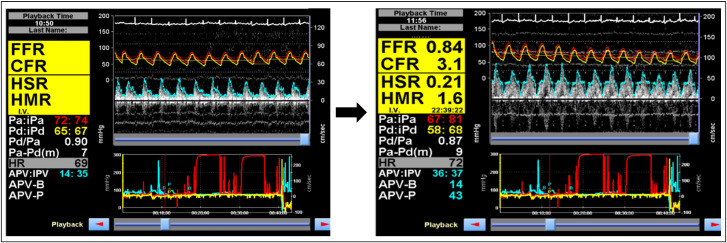
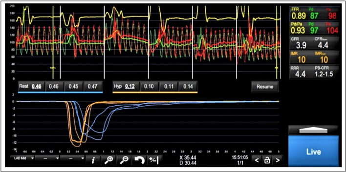
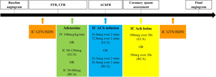
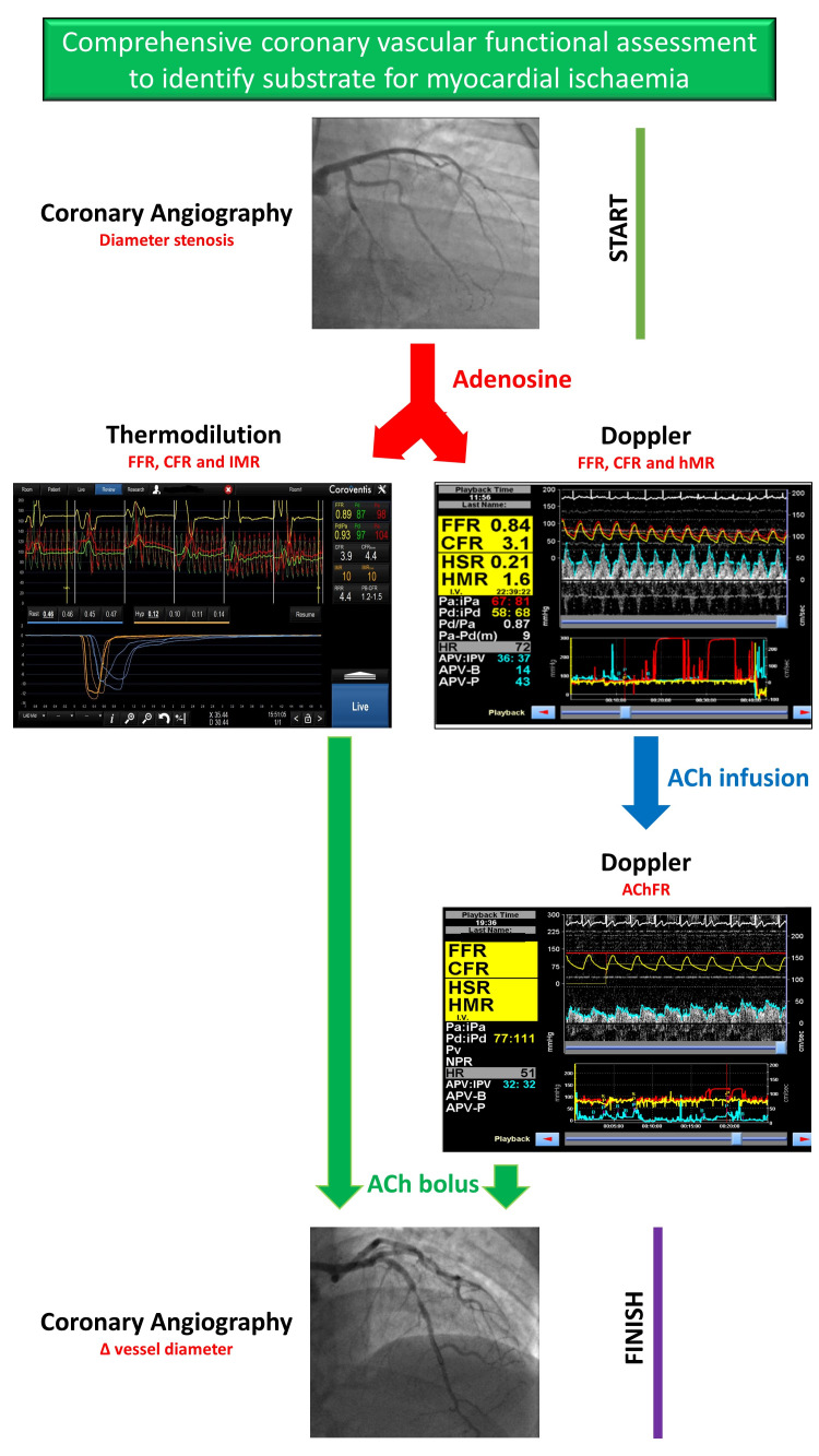
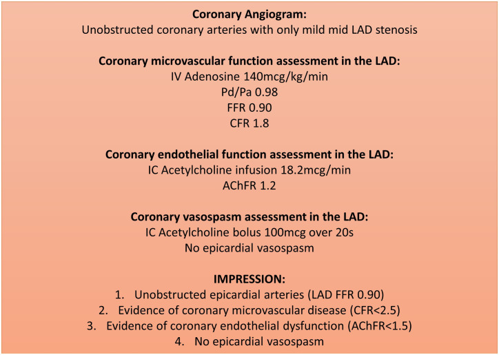
Nearly half of all patients with angina have non-obstructive coronary artery disease (ANOCA); this is an umbrella term comprising heterogeneous vascular disorders, each with disparate pathophysiology and prognosis. Approximately two-thirds of patients with ANOCA have coronary microvascular disease (CMD). CMD can be secondary to architectural changes within the microcirculation or secondary to vasomotor dysfunction. An inability of the coronary vasculature to augment blood flow in response to heightened myocardial demand is defined as an impaired coronary flow reserve (CFR), which can be measured non-invasively, using imaging, or invasively during cardiac catheterisation. Impaired CFR is associated with myocardial ischaemia and adverse cardiovascular outcomes.The CMD workstream is part of the cardiovascular partnership between the British Heart Foundation and The National Institute for Health Research in the UK and comprises specialist cardiac centres with expertise in coronary physiology assessment. This document outlines the two main modalities (thermodilution and Doppler techniques) for estimation of coronary flow, vasomotor testing using acetylcholine, and outlines a standard operating procedure that could be considered for adoption by national networks. Accurate and timely disease characterisation of patients with ANOCA will enable clinicians to tailor therapy according to their patients' coronary physiology. This has been shown to improve patients' quality of life and may lead to improved cardiovascular outcomes in the long term.

Keywords: Angina Pectoris; Chest Pain; Coronary Angiography; Microvascular Angina.


Conflict of interest statement

Competing interests: DP has received support for a research grant from Abbott Vascular and Philips Volcano in the past. CB is employed by The University of Glasgow, which holds research and consultancy agreements for work done in the course of his employment with the following companies: AstraZeneca, Abbott Vascular, GSK, Heartflow, Menarini, Novartis, Philips and Siemens Healthcare. PDM has received speaker’s honoraria from Abbott Vascular. RP has received speaker’s honoraria from Philips Volcano.

Figures

Figure 1
Figure 1
Coronary vascular physiology assessment, using Doppler wire, demonstrating normal epicardial coronary artery (FFR=0.84) and endothelium-independent coronary microvascular (CFR=3.1) physiology. The aortic (red) and distal coronary (yellow) pressures are used to calculate Pd/Pa and FFR. The Doppler flow signals are used to derive the instantaneous peak velocity (IPV) and averaged peak velocity (APV), which are used as a surrogate for coronary blood flow. The pressure and flow traces are both ECG-gated. CFR=APV-P/APV-B; hMR=Pd/APV-P; hSR=(Pa-Pd)/APV. APV-B, basal averaged peak velocity; APV-P, hyperaemic averaged peak velocity; CFR, coronary flow reserve; FFR, fractional flow reserve; HMR, hyperaemic microvascular resistance; HSR, hyperaemic stenosis resistance.
Figure 2
Figure 2
Coronary vascular physiology assessment demonstrating normal epicardial coronary artery (FFR=0.89) and endothelium-independent coronary microvascular (CFR=3.9) physiology. The red (aortic) and green (distal coronary) pressure traces at the top half of the figure are used to calculate Pd/Pa and FFR. The blue traces at the bottom of the figure represent resting transit times (0.46 s, 0.45 s and 0.47 s) and the yellow traces represent hyperaemic transit times (0.10 s, 0.11 s and 0.14 s). CFR=resting mean transit time/hyperaemic mean transit time; IMR=Pd * hyperaemic mean transit time. CFR, coronary flow reserve; FFR, fractional flow reserve.
Figure 3
Figure 3
Coronary angiography images of the left coronary artery at baseline (left hand side) and after acetylcholine bolus (right hand side), in a patient with epicardial coronary artery vasospasm. The gold arrows demonstrate the diffuse segments of spasm in the left anterior descending artery.
Figure 4
Figure 4
The sequence of vasodilator infusions utilised to assess coronary vascular physiology in the catheter laboratory. AChFR, acetylcholine flow reserve; CFR, coronary flow reserve; FFR, fractional flow reserve; GTN, glyceryl trinitrate; IC, intracoronary; ISDN, isosorbide dinitrate; LCA, left coronary artery; RCA, right coronary artery.
Figure 5
Figure 5
This figure represents our proposed algorithm for any patient coming to the catheter laboratory with symptoms suggestive of myocardial ischaemia who is found to have unobstructed epicardial arteries. AchFR, acetylcholine flow reserve; CFR, coronary flow reserve; FFR, fractional flow reserve; hMR, hyperaemic microvascular resistance; HSR, hyperaemic stenosis resistance.
Figure 6
Figure 6
An example of a standardised report for a patient with coronary vascular physiology assessment. We recommend recording the following data on every report: (1) epicardial artery indices (FFR/NHPR); (2) coronary endothelium-independent microvascular function (CFR); (3) coronary endothelial function (AChFR) (if measured) and (4) evidence of coronary vasospasm (if measured), including maximum induced diameter stenosis, symptoms and ECG changes; (5) systemic haemodynamics: aortic blood pressure, left ventricular end-diastolic pressure and rate pressure product. AChFR, acetylcholine flow reserve; CFR, coronary flow reserve; FFR, fractional flow reserve; LAD, left anterior descending.

Similar articles

Cited by

References

    1. Knuuti J, Wijns W, Saraste A, et al. . 2019 ESC guidelines for the diagnosis and management of chronic coronary syndromes. Eur Heart J 2020;41:407–77. 10.1093/eurheartj/ehz425 - DOI - PubMed
    1. Sara JD, Widmer RJ, Matsuzawa Y, et al. . Prevalence of coronary microvascular dysfunction among patients with chest pain and nonobstructive coronary artery disease. JACC Cardiovasc Interv 2015;8:1445–53. 10.1016/j.jcin.2015.06.017 - DOI - PubMed
    1. Ford TJ, Stanley B, Good R, et al. . Stratified medical therapy using invasive coronary function testing in angina: the CorMicA trial. J Am Coll Cardiol 2018;;72:2841–55. 10.1016/j.jacc.2018.09.006 - DOI - PubMed
    1. Radico F, Zimarino M, Fulgenzi F, et al. . Determinants of long-term clinical outcomes in patients with angina but without obstructive coronary artery disease: a systematic review and meta-analysis. Eur Heart J 2018;39:2135–46. 10.1093/eurheartj/ehy185 - DOI - PubMed
    1. Rahman H, Ryan M, Lumley M, et al. . Coronary microvascular dysfunction is associated with myocardial ischemia and abnormal coronary perfusion during exercise. Circulation 2019;140:1805–16. 10.1161/CIRCULATIONAHA.119.041595 - DOI - PMC - PubMed

Publication types