. 2022 Mar 22;12(1):4903.
doi: 10.1038/s41598-022-08865-y.

Subretinal fluid disturbs the retinal venous blood flow in central serous chorioretinopathy

Affiliations

Subretinal fluid disturbs the retinal venous blood flow in central serous chorioretinopathy

Joon Seo Lim et al. Sci Rep. .

Abstract

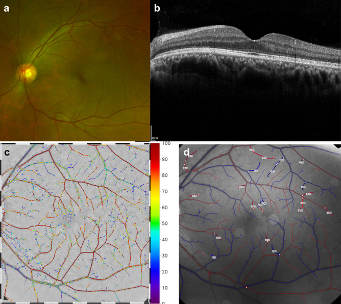
The significance of subretinal fluid in the retinal blood flow is unclear. Here, we evaluated the association between subretinal fluid (SRF) and retinal blood flow in eyes with central serous chorioretinopathy (CSC) using a retinal functional imager (RFI) and optical coherence tomography angiography (OCTA). In this retrospective case-control study involving 26 eyes from 18 CSC patients and 25 eyes from 21 age- and sex-matched controls, we found that the CSC group showed significant differences from the control group in terms of the retinal venule blood flow velocity (3.60 ± 0.43 vs 3.96 ± 0.56 mm/s; p = 0.030), retinal venule blood flow rate (8.75 ± 2.67 vs 12.51 ± 7.12 nl/s; p = 0.040), and the diameter of retinal venules (118.26 ± 14.25 vs 126.92 ± 35.31 μm; p = 0.045). Linear regression analysis showed that SRF thickness accounted for a 36.9% reduction in venous BFR (p = 0.013). The difference in the O2 saturation between retinal arteries and veins was greater in the CSC group. There was no correlation between SRF thickness and capillary densities in OCTA. Our findings suggest that disturbance in venous return and the associated altered oxygen may be significant changes in the retinal blood flow dynamics in eyes with SRF.


Conflict of interest statement

The authors declare no competing interests.

Figures

Figure 1
Figure 1
Relationship between subretinal fluid (SRF) thickness and arterial blood flow velocity (a), venous blood flow velocity (b), arterial blood flow rate (c), and venous blood flow rate (d) in central serous chorioretinopathy. Pearson’s correlation. BFR blood flow rate, BFV blood flow velocity, SRF subretinal fluid.
Figure 2
Figure 2
Relationship between subretinal fluid (SRF) thickness and OCTA parameters: FCP density (a), SCP density (b), DCP density (c), and FAZ area (d) in central serous chorioretinopathy. Pearson’s correlation. SRF subretinal fluid, DCP deep capillary plexus, FAZ foveal avascular zone, FCP full capillary plexus, SCP superficial capillary flexus, SRF subretinal fluid.
Figure 3
Figure 3
Retinal angiographic images from age-matched control group using fundus photography (a), optical coherence tomography (b), and retinal functional imager (RFI) (c,d). (c) O2 saturation image and (d) retinal vessel velocity image.
Figure 4
Figure 4
Retinal angiographic images from a case of central serous chorioretinopathy using fundus photography (a), optical coherence tomography (b), and retinal functional imager (RFI) (c,d). (c) O2 saturation image and (d) retinal vessel velocity image.

References

    1. Daruich A, et al. Mechanisms of macular edema: Beyond the surface. Prog. Retin. Eye Res. 2018;63:20–68. doi: 10.1016/j.preteyeres.2017.10.006. - DOI - PubMed
    1. Sharma S, et al. Macular morphology and visual acuity in the second year of the comparison of age-related macular degeneration treatments trials. Ophthalmology. 2016;123:865–875. doi: 10.1016/j.ophtha.2015.12.002. - DOI - PMC - PubMed
    1. Zarbin, M.A., Hill, L., Maunz, A., Gliem, M. & Stoilov, I. Anti-VEGF-resistant subretinal fluid is associated with better vision and reduced risk of macular atrophy. Br J. Ophthalmol. (2021). - PMC - PubMed
    1. Cheung CMG, et al. Pachychoroid disease. Eye (Lond) 2019;33:14–33. doi: 10.1038/s41433-018-0158-4. - DOI - PMC - PubMed
    1. Beutelspacher SC, et al. Central serous chorioretinopathy shows reduced retinal flow circulation in retinal function imaging (RFI) Acta Ophthalmol. 2011;89:e479–482. doi: 10.1111/j.1755-3768.2011.02136.x. - DOI - PubMed

Publication types

MeSH terms