. 2022 Jul;127(1):56-68.
doi: 10.1038/s41416-022-01778-7. Epub 2022 Mar 22.

VIVA1: a more invasive subclone of MDA-MB-134VI invasive lobular carcinoma cells with increased metastatic potential in xenograft models

Affiliations

VIVA1: a more invasive subclone of MDA-MB-134VI invasive lobular carcinoma cells with increased metastatic potential in xenograft models

Victoria Allen et al. Br J Cancer. 2022 Jul.

Abstract

Background: Invasive lobular carcinoma (ILC) is the second most common type of breast cancer. As few tools exist to study ILC metastasis, we isolated ILC cells with increased invasive properties to establish a spontaneously metastasising xenograft model.

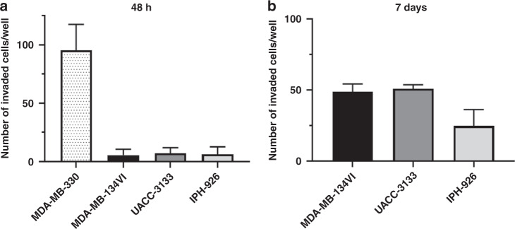
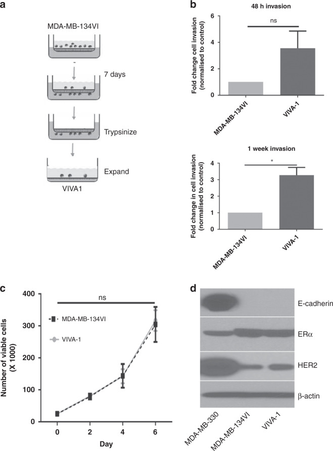
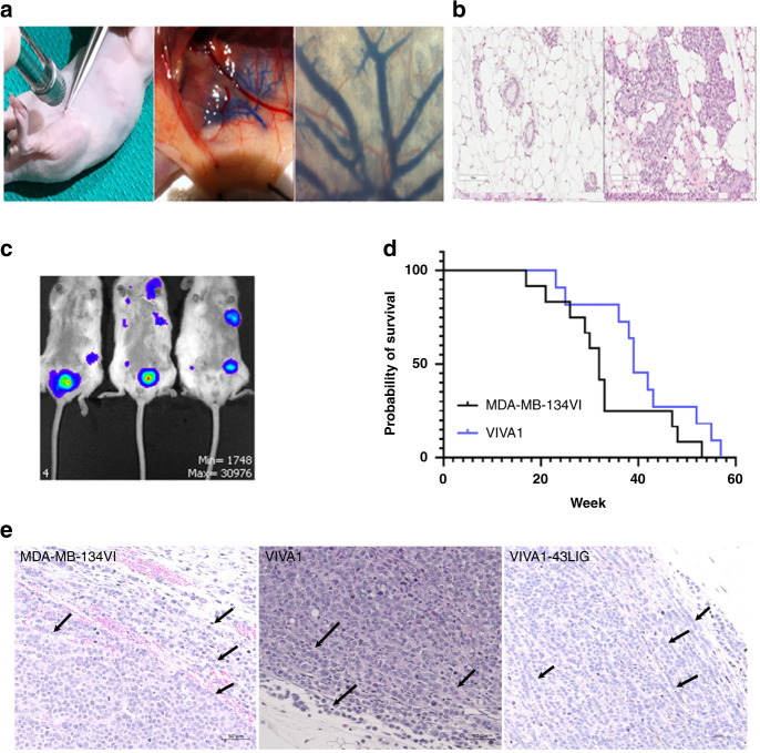
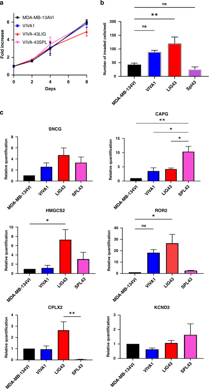
Methods: MDA-MB-134VI ILC cells were placed in transwells for 7 days. Migrated cells were isolated and expanded to create the VIVA1 cell line. VIVA1 cells were compared to parental MDA-MB-134VI cells in vitro for ILC marker expression and relative proliferative and invasive ability. An intraductally injected orthotopic xenograft model was used to assess primary and metastatic tumour growth in vivo.

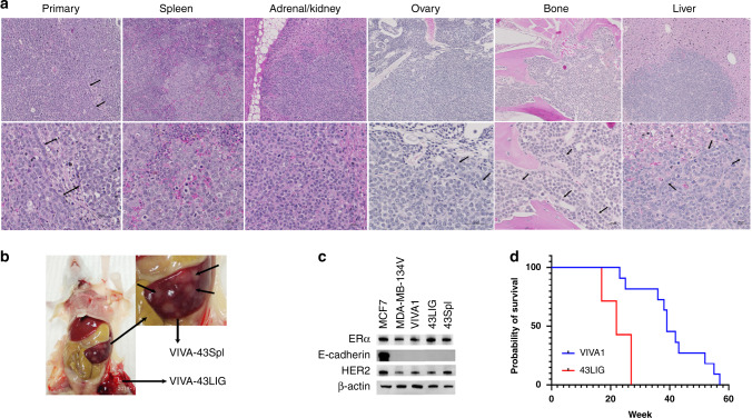
Results: Similar to MDA-MB-134VI, VIVA1 cells retained expression of oestrogen receptor (ER) and lacked expression of E-cadherin, however showed increased invasion in vitro. Following intraductal injection, VIVA1 and MDA-MB-134VI cells had similar primary tumour growth and survival kinetics. However, macrometastases were apparent in 7/10 VIVA1-injected animals. Cells from a primary orthotopic tumour (VIVA-LIG43) were isolated and showed similar proliferative rates but were also more invasive than parental cells. Upon re-injection intraductally, VIVA-LIG43 cells had more rapid tumour growth with similar metastatic incidence and location.

Conclusions: We generated a new orthotopic spontaneously metastasising xenograft model for ER+ ILC amenable for the study of ILC metastasis.


Conflict of interest statement

The authors declare no competing interests.

Figures

Fig. 1
Fig. 1. ILC cell lines are generally minimally invasive.
The ILC cell lines, MDA-MB-330, MDA-MB-134VI, UACC-3133 and IPH926 were plated on the surface of Matrigel-coated transwell invasion chambers in their respective growth media with reduced FBS (5%) and were allowed to migrate towards increased FBS concentrations (20%) for (a) 48h or (b) 7 days. Invading cells were stained with crystal violet, counted from five representative images and plotted with the SEM. We considered MDA-MB-330 as an invasive ILC cell line as they were able to invade within 48 h of plating in an invasion chamber, while all other lines tested were considered minimally invasive (n = 3 biological replicates, each with duplicate technical replicates).
Fig. 2
Fig. 2. Isolation and characterisation of a more invasive subclone from MDA-MB-134VI.
a MDA-MB-134VI cells were seeded into Matrigel-coated transwell invasion chambers and allowed to migrate for 7 days, at which time migrated cells were isolated from membranes by trypsinization and expanded to generate the VIVA1 subclone. b Invasion of VIVA1 subclone was compared to parental MDA-MB-134VI cells using Matrigel-coated transwell invasion chambers and incubated for 48 h (top panels) or 7 days (bottom panels), at which time cells were counted in five fields of view in duplicate chambers. Graphs represent mean and SEM of migrated cells for n = 3 biological replicates. * represents P value ≤0.05 using unpaired t test with Welch’s correction. c Cell growth and viability over time were measured for VIVA1 cells compared to control parental MDA-MB-134VI cells using trypan blue exclusion and counting of cells on the ViaXR cell counter. Graphs represent the mean number of viable cells per well with associated SEM in duplicate wells for n = 3 biological replicates. Significance was tested using a two-way ANOVA with Sidak’s multiple comparison testing (ns represents P value >0.05). d Protein lysates from plated cells were generated and 50µg of protein was subjected to western blot analysis for the proteins indicated. Western blot detection of β-actin was used as the loading control.
Fig. 3
Fig. 3. RNA-seq analysis shows the altered gene expression in VIVA1 cells associated with increased cell growth and invasion.
RNA was isolated from cell lines using the mRNeasy kit and subjected to next-generation RNA-seq using the Illumina NextSeq as described in 'Materials and methods'. Following sequence analysis and determination of differential gene expression, targets which showed statistically significant log fold changes >0.5 or <−0.5 were submitted to the DAVID online pathway analysis to generate the GoTerm list illustrated in (a) with P values colour-coded as indicated and those with FDR <0.05 indicated with *. b Significantly altered gene lists were also subjected to KEGG pathway analysis and graphically represented with P values colour-coded as indicated and those with FDR <0.05 indicated with *. c Log2 differential expression was plotted against the associated inverse log 10 P value to generate volcano plots. Red dots represent those gene targets with significantly different expression (i.e. P value <0.05) between VIVA1 and MDA-MB-134VI. Blue dots are also significantly differentially expressed genes but are highlighted for attention.
Fig. 4
Fig. 4. VIVA1 subclone and parental MDA-MB-134VI grow in immunocompromised animals following intraductal injection of tumour cells.
a ILC Tumour cells were injected intraductally in immunocompromised mice via injection through the nipple, which resulted in the dissemination of injected material throughout the mammary ductal tree as evidenced by the distribution of Evan’s blue dye. b Intraductal injection of MDA-MB-134VI cells in CD-1 nude mice resulted in growth of tumours within the mammary ductal lumen by 24 weeks post injection as evident in H&E-stained tissue sections (right panel) compared to the appearance of normal ductal structures observed in uninjected mammary glands (left panel). c Growth of luciferase-expressing VIVA1 cells is evident in injected mammary glands by ~24–26 weeks post injection as detected by IVIS. d MDA-MB-134VI (12 animals), and VIVA1 (11 animals) were injected intraductally into the inguinal and abdominal mammary glands of NSG mice, and animals were euthanized upon reaching a clinical endpoint, with survival time recorded. Overall survival of mice was plotted against time. e Tumours arising following intraductal injection of MDA-MB-134VI parental cells, VIVA1 cells or the VIVA1-LIG43 primary tumour-derived cells were stained with H&E to visualise tumour cell histology and tumour growth patterns. Single-file growth patterns were observed along invasive edges of tumours for all three cell lines.
Fig. 5
Fig. 5. Intraductally injected VIVA1 cells generate tumours with features of ILC and spontaneously metastasize from the orthotopic site.
At clinical endpoint, animals injected with ILC tumour cell lines were autopsied and portions of tumours retrieved for H&E histological assessment, while other portions were used to generate single-cell suspensions which were then cultured and expanded in vitro. a H&E staining of VIVA1 induced primary tumour and metastasis to spleen, adrenal gland, ovary, bone and liver were also observed with single-file growth patterns often observed at invasive edges or in the presence of additional stroma (indicated by arrows in each labelled panel). b Cells isolated from both primary tumour (43LIG) and the splenic metastasis (43Spl) of the same animal were expanded and protein lysates generated. c Western blot analysis of ILC markers showed the primary and metastases derived cell lines retained expression of ILC markers. MCF7 ductal carcinoma cells were included as a positive control for E-cadherin expression. β-actin was included as a loading control. d Cells isolated from a VIVA1 induced primary tumour (43LIG) were reinjected intraductally into additional animals (n = 6). Overall survival was plotted against the original survival of animals intraductally injected with VIVA1 cells previously shown in Fig. 4 for comparison.
Fig. 6
Fig. 6. Cells isolated from VIVA1-derived primary or metastatic tumours retain increased invasive properties compared to parental MDA-MB-134VI cells.
Cells were isolated from primary tumour or splenic metastatic sites from animals intraductally injected with VIVA1 cells and expanded in vitro. a Cell growth over time was measured in 2D in vitro culture following viable cell counting with AlamarBlue as described in 'Methods'. All cell lines tested showed similar growth kinetics with the graph representing the mean and standard error of eight technical replicates in each of three biological replicates. b Cell invasion was tested using Matrigel-coated invasion chambers and incubation for 7 days, at which time membranes were isolated, stained and invaded cells counted as described in 'Methods'. Graph is the mean and standard error of duplicate wells in each of three independent biological replicates. VIVA1 (P = 0.051) and primary tumour-derived VIVA1-LIG43 cells retained increased invasive abilities compared to MDA-MB-134VI parental cells. **P value <0.001. c RNA isolated from the various cell lines was subjected to qRT-PCR for highly differentially expressed genes identified in previous RNA-seq experiments. Graphs represent mean and standard error for relative levels of expression of various targets as indicated following normalisation to levels of β-actin as a control from three technical replicates for each of three independent biological replicates. *P value < 0.05; **P value <0.001.

Similar articles

References

    1. Iorfida M, Maiorano E, Orvieto E, Maisonneuve P, Bottiglieri L, Rotmensz N, et al. Invasive lobular breast cancer: subtypes and outcome. Breast Cancer Res Treat. 2012;133:713–23. - PubMed
    1. Nielsen TO, Parker JS, Leung S, Voduc D, Ebbert M, Vickery T, et al. A comparison of PAM50 intrinsic subtyping with immunohistochemistry and clinical prognostic factors in tamoxifen-treated estrogen receptor-positive breast cancer. Clin Cancer Res. 2010;16:5222–32. - PMC - PubMed
    1. Hugh J, Hanson J, Cheang MC, Nielsen TO, Perou CM, Dumontet C, et al. Breast cancer subtypes and response to docetaxel in node-positive breast cancer: use of an immunohistochemical definition in the BCIRG 001 trial. J Clin Oncol. 2009;27:1168–76. - PMC - PubMed
    1. Cheang MC, Chia SK, Voduc D, Gao D, Leung S, Snider J, et al. Ki67 index, HER2 status, and prognosis of patients with luminal B breast cancer. J Natl Cancer Inst. 2009;101:736–50. - PMC - PubMed
    1. Weigelt B, Geyer FC, Natrajan R, Lopez-Garcia MA, Ahmad AS, Savage K, et al. The molecular underpinning of lobular histological growth pattern: a genome-wide transcriptomic analysis of invasive lobular carcinomas and grade- and molecular subtype-matched invasive ductal carcinomas of no special type. J Pathol. 2010;220:45–57. - PubMed

Publication types

Substances