. 2022 Mar 22;5(1):252.
doi: 10.1038/s42003-022-03205-2.

Porcine intraepithelial lymphocytes undergo migration and produce an antiviral response following intestinal virus infection

Affiliations

Porcine intraepithelial lymphocytes undergo migration and produce an antiviral response following intestinal virus infection

Yuchen Li et al. Commun Biol. .

Abstract

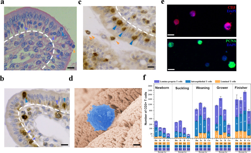
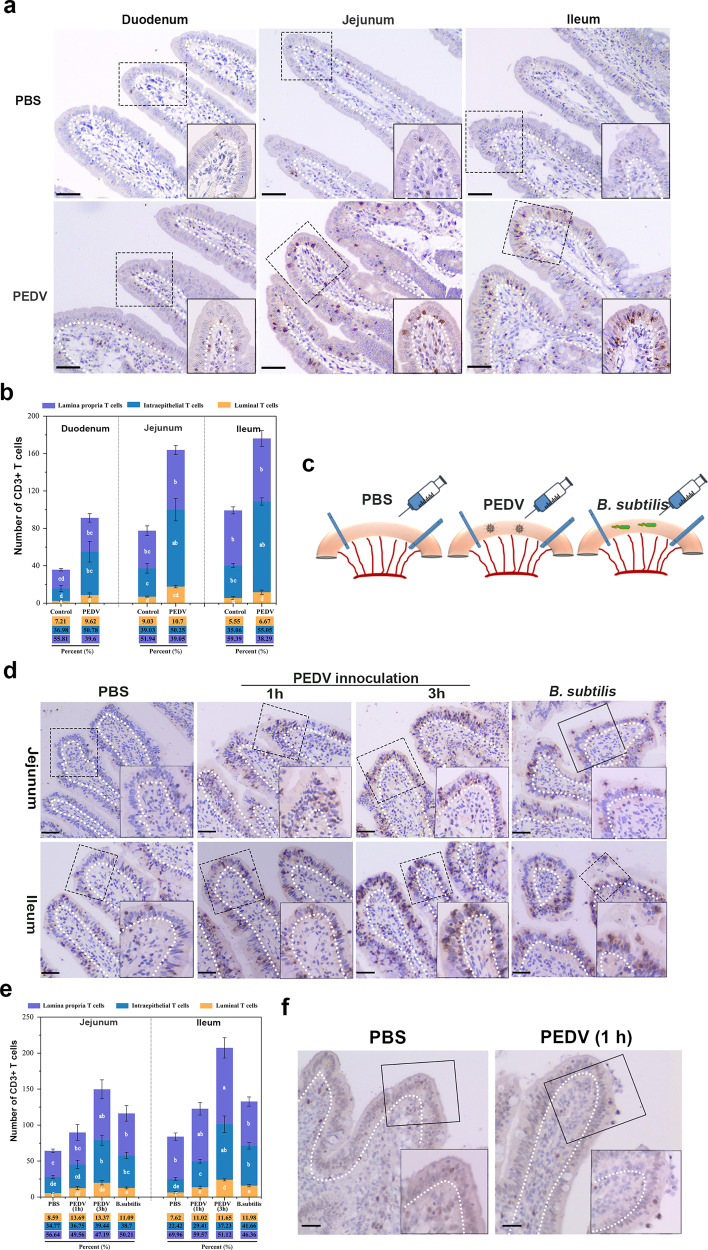
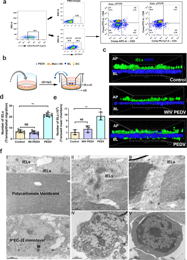
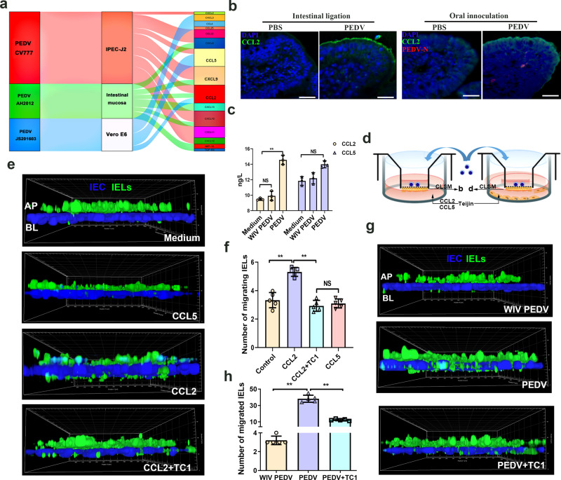
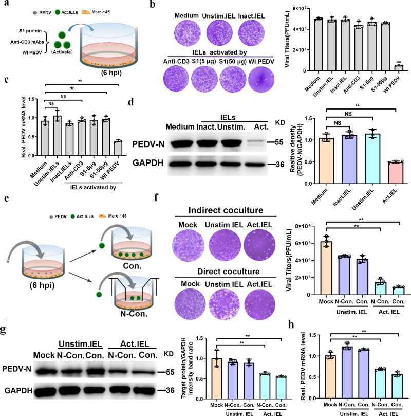
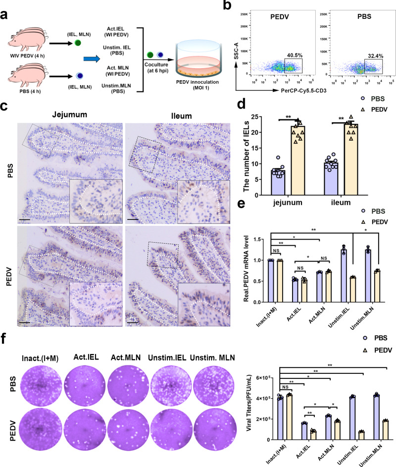
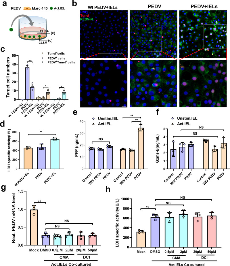
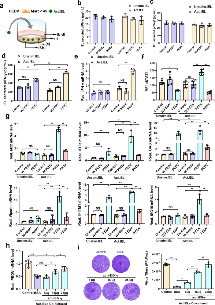
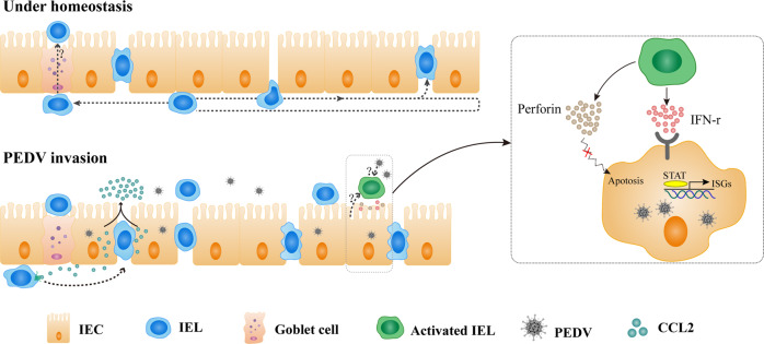
The location of intraepithelial lymphocytes (IELs) between epithelial cells provide a first line of immune defense against enteric infection. It is assumed that IELs migrate only along the basement membrane or into the lateral intercellular space (LIS) between epithelial cells. Here, we identify a unique transepithelial migration of porcine IELs as they move to the free surface of the intestinal epithelia. The major causative agent of neonatal diarrhea in piglets, porcine epidemic diarrhea virus (PEDV), increases the number of IELs entering the LIS and free surface of the intestinal epithelia, driven by chemokine CCL2 secreted from virus-infected intestinal epithelial cells. Remarkably, only virus pre-activated IELs inhibits PEDV infection and their antiviral activity depends on the further activation by virus-infected cells. Although high levels of perforin is detected in the co-culture system, the antiviral function of activated IELs is mainly mediated by IFN-γ secretion inducing robust antiviral response in virus-infected cells. Our results uncover a unique migratory behavior of porcine IELs as well as their protective role in the defense against intestinal infection.


Conflict of interest statement

The authors declare no competing interests.

Figures

Fig. 1
Fig. 1. The distribution of porcine intestinal intraepithelial lymphocytes (IELs).
a Hematoxylin and eosin (HE) staining showing the distribution of IELs in the basal surface (white arrowheads), lateral intercellular space (LIS) (black arrowheads), or free-surface (red arrowheads) of epithelial layer in jejunum tissues (45-day-old piglets). Scale bars, 10 μm. b Representative images of the CD3-positive IELs in the basal (white arrowheads) or intraepithelial of jejunal epithelia (black arrowheads) (45-day-old piglets). Scale bars, 10 μm. c Representative images of the CD3-positive IELs located in LIS (black arrowheads) or free-surface (red arrowheads) of jejunal epithelia (45-day-old piglets). Scale bars, 10 μm. d Representative SEM image showed the luminally located IELs (blue) in jejunum tissue (45-day-old piglets). Scale bars, 5 μm. e IELs were isolated from ileal contents and stained for CD3 and PCNA protein. The scale bar represents 10 μm. f Quantitative analysis of IELs within various intestinal segments collected from pigs (n = 3) in different growth stages. The numbers of CD3-positive cells in different sites of the intestinal epithelial layer were counted in 20 random intestinal villi. The frequency of total T cells in each area (%) was presented in the bar chart. All data shown are the mean results ± SD from three independent experiments. Statistical significance was shown using one-way ANOVA. NS: no significance; *P < 0.05, **P < 0.01. The differences are indicated by different letters. Letters above the graphs indicate statistical significance in which treatments with a letter in common are not significantly different from each other.
Fig. 2
Fig. 2. Influence of porcine epidemic diarrhea virus (PEDV) infection on porcine intestinal intraepithelial lymphocytes (IELs).
a Pigs (n = 3) from mock or PEDV challenge groups were euthanatized at 48 hpi, and small intestinal tissues were fixed and subjected to IHC analysis. Representative image of IELs distribution in the small intestine of PBS-inoculated or PEDV-inoculated weaned piglets (one-month-old). Scale bars, 20 μm. b Quantitative analysis of IELs showed in (a) was performed, in which 20 random intestinal villi were counted. The frequency of total T cells in each area (%) were also presented. c Schematic of the experimental setting of the intestinal ligated loop model. Anesthetized piglets (one-month-old, n = 3) were subjected to intestinal ligation followed by injection with PBS, PEDV, and Bacillus subtilis, and were euthanatized at 3-hours post-treatment. d Immunohistochemistry (IHC) analysis demonstrated the distribution pattern of IELs influenced by PEDV or B. subtilis in the ligated loop of the terminal jejunum. Scale bars, 20 μm. e Quantification of IELs (d) in 20 random intestinal villi. The frequency of total T cells in each area (%) was also calculated. f The proliferative activity of intraepithelial and transepithelial IELs in the ligated loop was detected via proliferating cell nuclear antigen (PCNA) staining. The scale bar represents 50 μm. All data shown are the mean results ± SD from three independent experiments. Statistical significance was obtained using one-way ANOVA. NS no significance, *P < 0.05, **P < 0.01. The differences are indicated by different letters. Letters above the graphs indicate statistical significance in which treatments with a letter in common are not significantly different from each other.
Fig. 3
Fig. 3. Porcine epidemic diarrhea virus (PEDV) infection promotes the transepithelial migration of intestinal intraepithelial lymphocytes (IELs) in an in vitro co-culture system.
a Flow cytometry analysis revealed population and subpopulations of isolated porcine intestinal IELs. b Schematic of the experimental setting to study the migration of IELs in the co-culture system. Mac-145 cells (70% confluent) were inoculated with PEDV at a multiplicity of infection (MOI) of 0.1 for 24 h at 37 °C. Next, the transwell insert containing the IELs/IPEC-J2 co-culture system was cultured with PEDV-infected Marc 145 cells for 2 h. c The migration of IELs in the co-culture system was determined using immunofluorescence. The filters in the coculture system were processed and viewed using confocal laser scanning microscopy (CLSM; AP to BL). CFSE labeled IELs (green) were detected in the apical or basal side of the monolayer intestinal epithelial cells (DAPI, blue). d Quantification of basolateral IELs from fluorescence images in (c); each value was calculated from the averaged values of three individual filters. e The number of CFSE-labeled IELs in lower chambers of co-culture system were counted by FACS. f The filters from the co-culture system were processed for TEM. The representative image shows the cellular composition of the co-culture system (panel I), and the migrating IELs (panels II and III). Moreover, the IELs in apical side (mock-infected group; panel IV) or basal surface (PEDV infected group; panel V) of IPEC-J2 in the co-culture system were also collected separately and sent for TEM observation. The scale bar represents 3 μm (I); and 2 μm (II to V). All data shown are the mean results ± SD from three independent experiments. Statistical significance was obtained using one-way ANOVA. NS no significance, *P < 0.05, **P < 0.01.
Fig. 4
Fig. 4. Role of CCL2 in the intraepithelial and transepithelial migration of intestinal intraepithelial lymphocytes (IELs) induced by porcine epidemic diarrhea virus (PEDV) infection.
a Sankey diagram depicts the chemokine secretion in the host cells and the intestinal mucosa infected with variant and classical PEDV strains. b The protein expression of CCL2 in the ligated terminal jejunum injected with PEDV and in the jejunum from piglets orally inoculated with PEDV was detected via immunofluorescence analysis. CCL2 and PEDV were immunolabeled with anti-CCL2 mAb (green) and anti-PEDV polyclonal antibody (Red), respectively; the cell nuclei were stained with DAPI (blue). Scale bars, 50 μm. c The protein expression of CCL2 and CCL5 in the medium of Marc-145 cells was detected using ELISA kits. d Schematic of experimental setting used to study the migration of IELs influenced by chemokines induced by PEDV infection. e Chemokines CCL2, CCL5, and the CCL2 inhibitor TC1 were added into the medium of the basolateral side. Filters from the co-culture system were determined via CLSM, and the intraepithelial and transepithelial (DAPI, blue) migration of IELs (CFSE, green) in response to different treatments was shown by a three-dimensional (3D) rendering of representative fields. f Quantitative analysis of transepithelial IELs was performed. The number of transepithelial IELs was counted from five random fields of view at a unit area (0.078 mm2) for each of the three individual filters. g The IELs/IPEC-J2 co-culture model was pretreated with an CCL2 inhibitor TC1 for 2 h (DMSO as a negative control) by upper compartment inoculation, followed by a culture with PEDV-infected or whole inactivated PEDV-treated Marc-145 cells. The intraepithelial and transepithelial (DAPI, blue) migration of IELs (CFSE, green) was detected via CLSM. h Quantitative analysis of transepithelial IELs in each experimental group. All data are the mean ± SD, and comparisons were performed using one-way ANOVA. *P < 0.05, **P < 0.01. The results are from at least three different experiments.
Fig. 5
Fig. 5. Preactivated intestinal intraepithelial lymphocytes (IELs) protect epithelial cells from porcine epidemic diarrhea virus (PEDV) infection in a TCR-independent manner.
Experimental setting to study the antiviral effect of intestinal IELs. a The scheme depicts that intestinal IELs were pretreated with CD3 antibody, whole inactivated PEDV, and the PEDV spike protein; thereafter, they were co-cultured with PEDV-infected epithelial cells for 24 h. b The viral titers in the culture supernatant were detected via a plaque assay and summarized in a histogram. c The intracellular viral RNA levels were quantitated via RT-qPCR. d The expression of PEDV-N protein in epithelial cells (Marc-145) was detected using western blot. e The model used to study the protective role of intestinal IELs against PEDV infection. The scheme depicts the whole inactivated PEDV pre-activated or unstimulated IELs that were co-cultured with epithelial cells (Marc-145) using two methods (contact and noncontact co-culture). f Viral titers in the supernatant of the coculture system were measured via a plaque assay after a co-culture of 24 h. Meanwhile, the expression of the viral protein (g) and mRNA e (h) in epithelial cells (Marc-145) was also evaluated. All data are the mean ± SD, and comparisons were performed using one-way ANOVA. *P < 0.05, **P < 0.01. The results are from at least three different experiments.
Fig. 6
Fig. 6. Oral inoculation with porcine epidemic diarrhea virus (PEDV) pre-activated intestinal intraepithelial lymphocytes (IELs) and increased their antiviral activity against virus infection.
a For in vivo IELs activation experiment, piglets were sacrificed 4 h after PEDV inoculation, n = 3 piglets per group. The scheme depicts that intestinal IELs isolated from piglets orally inoculated with PEDV (PBS inoculation was used as a negative control) were pretreated with whole inactivated PEDV for pre-activation (DMEM medium was used as a negative control) and then co-cultured with PEDV-infected epithelial cells for 24 h. b The percentage of CD3-positive IELs in the jejunum of piglets were analyzed using fluorescence-activated cell sorting (FACS). c Immunohistochemistry (IHC) analysis displayed the distribution pattern of IELs influenced by PEDV inoculation. Scale bars, 10 μm. d The numbers of IELs shown in (c) were quantified and displayed on a histogram. e The PEDV RNA expression in epithelial cells from the co-culture system in (a) was quantitated using RT-qPCR. f Moreover, the infectious viral particles in the culture medium of the coculture system in (a) were detected by plaque formation. The histogram summarizes the plaque assay results. All data are the mean ± SD, and comparisons were performed using one-way ANOVA. *P < 0.05, **P < 0.01. The results are from at least three different experiments.
Fig. 7
Fig. 7. The antiviral activity of IELs was independent from their cytotoxic effects.
a The design for detecting the cytotoxicity of IELs. The virus pre-activated IELs were co-cultured with porcine epidemic diarrhea virus (PEDV)-infected epithelial cells for 24 h. b TUNEL staining was used to determine the apoptosis rate of PEDV-infected or whole inactivated PEDV-treated epithelial cells. c The number of TUNEL+, PEDV+, and TUNEL+PEDV+ cells in each group was calculated and shown in a bar graph, which was produced from five random fields of view for each of three individual sections. d The released lactate dehydrogenase (LDH) activity assay was also used to evaluate the cytotoxicity of IELs in different groups. e, f Levels of perforin (e) and granzyme B (f) release in the culture supernatants of different groups were measured via ELISA. g, h The pre-reactivated IELs were treated with CMA at two concentrations (1 and 0.5 μM) or DCI at two concentrations (20 and 50 μM) for 3 h, and their antiviral activity were further detected by co-culturing with virus-infected epithelial cells. Both of the inhibitors were also maintained for the whole duration of the co-culturing process. The intracellular viral RNA expression (g) and extracellular LDH activity (h) in each experimental group were determined. All data are the mean ± SD and comparisons were performed using one-way ANOVA. *P < 0.05, **P < 0.01. The results are from at least three different experiments.
Fig. 8
Fig. 8. Pre-activated intestinal intraepithelial lymphocytes (IELs) produced IFN-γ and induced antiviral pathways in virus-infected epithelial cells.
a The scheme depicts that mock or whole inactivated virus (WIV) porcine epidemic diarrhea virus (PEDV) pre-activated IELs were co-cultured with PEDV-infected epithelial cells for 24 h. bd The production of type I (b, c), II (d), and III IFNs (e) by IELs in the co-culture system was detected using ELISA kits. e The mRNA expression of IFN-γ in IELs was also measured via RT-qPCR. f Phosphorylation of STAT1 in the epithelial cells was analyzed by flow cytometry. g The transcription of a set of IFN stimulated genes (ISGs) in PEDV-infected epithelial cells were detected via RT-qPCR. h A similar experiment was performed as in (a), but blocking antibodies against IFN-γ were added to the medium with a certain concentration gradient. i The viral titers in the supernatant, as well as the production of viral RNA in epithelial cells from different groups were detected. All data are the mean ± SD, comparisons performed using one-way ANOVA. *P < 0.05, **P < 0.01. The results are from at least three different experiments.
Fig. 9
Fig. 9. Schematic of the proposed mechanism of intestinal intraepithelial lymphocytes (IELs) functions in response to intestinal porcine epidemic diarrhea virus (PEDV) infection.
Under homeostatic conditions, porcine intestinal IELs not only exhibit a flossing behavior in which the cells move into the LIS between two adjacent enterocytes, but also move to the free end of the intestinal epithelia to perform their daily immune surveillance behavior. During PEDV infection, IELs were recruited by the polarity distribution of CCL2 secreted from virus-infected intestinal epithelial cells (IECs). The IELs’ enhanced intraepithelial and transepithelial migration may benefit their interaction upon encountering viral antigens. Intestinal IELs could exert potent innate antiviral resistance by viral pre-activation and the signaling of virus-infected IECs. However, the integral mechanisms involved in this process are not well elucidated. Although the secretion of perforin from IELs was significantly increased, the antiviral function of activated IELs was mainly mediated by IFN-γ secretion that induced robust antiviral response in virus-infected cells.

Similar articles

Cited by

References

    1. Perez-Lopez A, Behnsen J, Nuccio S-P, Raffatellu M. Mucosal immunity to pathogenic intestinal bacteria. Nat. Rev. Immunol. 2016;16:135. doi: 10.1038/nri.2015.17. - DOI - PubMed
    1. Olivares-Villagómez D, Van Kaer L. Intestinal intraepithelial lymphocytes: sentinels of the mucosal barrier. Trends Immunol. 2018;39:264–275. doi: 10.1016/j.it.2017.11.003. - DOI - PMC - PubMed
    1. Cheroutre H. Starting at the beginning: new perspectives on the biology of mucosal T cells. Annu Rev. Immunol. 2004;22:217–246. doi: 10.1146/annurev.immunol.22.012703.104522. - DOI - PubMed
    1. Ma H, Qiu Y, Yang H. Intestinal intraepithelial lymphocytes: Maintainers of intestinal immune tolerance and regulators of intestinal immunity. J. Leukoc. Biol. 2021;109:339–347. doi: 10.1002/JLB.3RU0220-111. - DOI - PMC - PubMed
    1. Nielsen MM, Witherden DA, Havran WL. γδ T cells in homeostasis and host defence of epithelial barrier tissues. Nat. Rev. Immunol. 2017;17:733–745. doi: 10.1038/nri.2017.101. - DOI - PMC - PubMed

Publication types

Substances