. 2022 Sep;59(9):e14051.
doi: 10.1111/psyp.14051. Epub 2022 Mar 23.

Suppression of alpha-band power underlies exogenous attention to emotional distractors

Affiliations

Suppression of alpha-band power underlies exogenous attention to emotional distractors

Lydia Arana et al. Psychophysiology. 2022 Sep.

Abstract

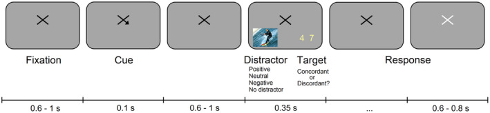
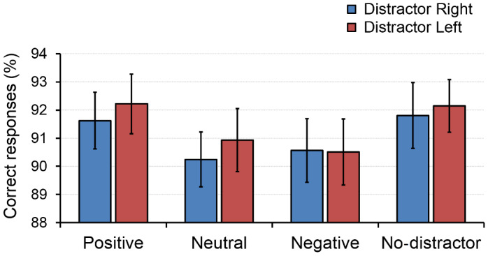
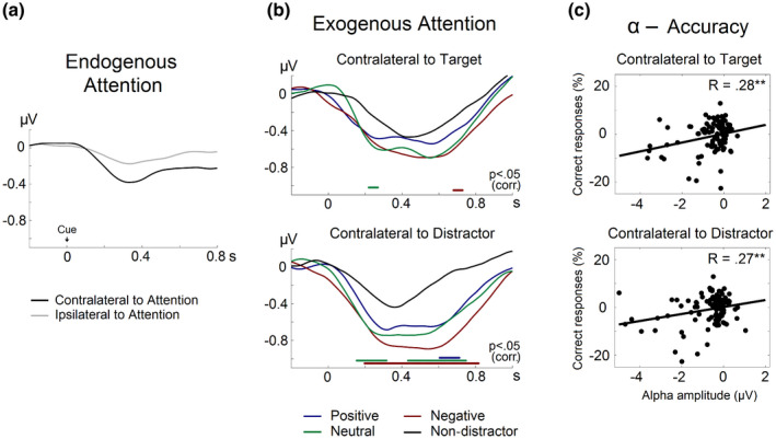
Alpha-band oscillations (8-14 Hz) are essential for attention and perception processes by facilitating the selection of relevant information. Directing visuospatial endogenous (voluntary) attention to a given location consistently results in a power suppression of alpha activity over occipito-parietal areas contralateral to the attended visual field. In contrast, the neural oscillatory dynamics underlying the involuntary capture of attention, or exogenous attention, are currently under debate. By exploiting the inherent capacity of emotionally salient visual stimuli to capture attention, we aimed to investigate whether exogenous attention is characterized by either a reduction or an increase in alpha-band activity. Electroencephalographic activity was recorded while participants completed a Posner visuospatial cueing task, in which a lateralized image with either positive, negative, or neutral emotional content competed with a target stimulus presented in the opposite hemifield. Compared with trials with no distractors, alpha power was reduced over occipital regions contralateral to distracting images. This reduction of alpha activity turned out to be functionally relevant, as it correlated with impaired behavioral performance on the ongoing task and was enhanced for distractors with negative valence. Taken together, our results demonstrate that visuospatial exogenous attention is characterized by a suppression of alpha-band activity contralateral to distractor location, similar to the oscillatory underpinnings of endogenous attention. Further, these results highlight the key role of exogenous attention as an adaptive mechanism for the efficient detection of biologically salient stimuli.

Our findings reveal that alpha‐band oscillations do not only index the locus of top‐down visuospatial attention, as it is well known, but also the degree of exogenous attentional capture. Thus, the suppression of alpha‐band power might be taken to indicate the amount of attentional resources diverted to distractor stimuli and, importantly, to describe the advantage of emotional over neutral stimuli.

Keywords: EEG; alpha oscillations; emotion; exogenous attention; time-frequency analyses.


Figures

FIGURE 1
FIGURE 1
Experimental task. A spatial cue indicated with a probability of 100% the location of an upcoming target stimulus (i.e., a pair a numbers). Participants performed a digit‐parity task while an emotional distractor appeared in the uncued hemifield. No distractors were presented in the control block
FIGURE 2
FIGURE 2
Behavioral results. Percentage of correct responses in the digit‐parity task under the presence of positive, neutral, and negative images, as well as in the absence of distraction. Blue bars indicate distractors presented on the right; red bars indicate left. Error bars represent the standard error of the mean
FIGURE 3
FIGURE 3
TF results: Alpha‐band power suppression time‐locked to cue and target/distractor. In response to the cue, alpha power decreased over contralateral posterior sites, whereas in response to target/distractor, alpha‐band exhibited a bilateral power reduction. The topography shows the modulation of alpha activity between 9 and 13 Hz and from 0.3 to 0.7 s after target/distractor onset. Occipital electrodes with maximal power suppression are highlighted in white
FIGURE 4
FIGURE 4
Alpha‐band TSE and correlation with accuracy. (a) TSE of alpha‐band amplitude time‐locked to cue onset. The black line represents alpha‐TSE in the hemisphere contralateral to the focus of endogenous attention; the gray line represents the ipsilateral hemisphere. (b) TSE of alpha‐band amplitude time‐locked to target/distractor onset. Alpha‐TSE contralateral to target and distractor location are shown separately. The figure shows a more pronounced alpha‐band desynchronization contralateral to the presentation of distractor images (positive, neutral, and negative) compared with the absence of distraction. Significant differences between distractor and non‐distractor conditions are indicated by colored horizontal lines (p < .05 corrected for multiple comparisons). (c) Correlation between alpha‐band amplitude and accuracy in the digit‐parity task in the hemisphere contralateral to target and distractor. **p < .01

References

    1. Adolphs, R. (2004). Emotional vision. Nature Neuroscience, 7(11), 1167–1168. 10.1038/nn1104-1167 - DOI - PubMed
    1. Alamia, A. , & VanRullen, R. (2019). Alpha oscillations and traveling waves: Signatures of predictive coding? PLoS Biology, 17(10), e3000487. 10.1371/journal.pbio.3000487 - DOI - PMC - PubMed
    1. Babiloni, C. , Vecchio, F. , Bultrini, A. , Romani, G. L. , & Rossini, P. M. (2006). Pre‐ and poststimulus alpha rhythms are related to conscious visual perception: A high‐resolution EEG study. Cerebral Cortex, 16(12), 1690–1700. 10.1093/cercor/bhj104 - DOI - PubMed
    1. Bacigalupo, F. , & Luck, S. J. (2019). Lateralized suppression of alpha‐band EEG activity as a mechanism of target processing. Journal of Neuroscience, 39(5), 900–917. 10.1523/JNEUROSCI.0183-18.2018 - DOI - PMC - PubMed
    1. Bagherzadeh, Y. , Baldauf, D. , Pantazis, D. , & Desimone, R. (2020). Alpha synchrony and the neurofeedback control of spatial attention. Neuron, 105(3), 577–587.e5. 10.1016/j.neuron.2019.11.001 - DOI - PubMed

Publication types