Publication guidelines and recommendations for pupillary measurement in psychophysiological studies
- PMID: 35318693
- PMCID: PMC9272460
- DOI: 10.1111/psyp.14035
Publication guidelines and recommendations for pupillary measurement in psychophysiological studies
Abstract
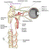
A variety of psychological and physical phenomena elicit variations in the diameter of pupil of the eye. Changes in pupil size are mediated by the relative activation of the sphincter pupillae muscle (decrease pupil diameter) and the dilator pupillae muscle (increase pupil diameter), innervated by the parasympathetic and sympathetic branches, respectively, of the autonomic nervous system. The current guidelines are intended to inform and guide psychophysiological research involving pupil measurement by (1) summarizing important aspects concerning the physiology of the pupil, (2) providing methodological and data-analytic guidelines and recommendations, and (3) briefly reviewing psychological phenomena that modulate pupillary reactivity. Because of the increased ease and tractability of pupil measurement, the goal of these guidelines is to promote accurate recording, analysis, and reporting of pupillary data in psychophysiological research.
Keywords: dilation; parasympathetic; pupil constriction; pupillography; pupillometry; sympathetic.
© 2022 Society for Psychophysiological Research.
Figures


Similar articles
-
Pupillometry measures of autonomic nervous system regulation with advancing age in a healthy pediatric cohort.Clin Auton Res. 2020 Feb;30(1):43-51. doi: 10.1007/s10286-019-00639-3. Epub 2019 Sep 25. Clin Auton Res. 2020. PMID: 31555934
-
Sympathetic nervous system does not mediate reflex pupillary dilation during desflurane anesthesia.Anesthesiology. 1996 Oct;85(4):748-54. doi: 10.1097/00000542-199610000-00009. Anesthesiology. 1996. PMID: 8873544
-
Emotion lies in the eye of the listener: Emotional arousal to novel sounds is reflected in the sympathetic contribution to the pupil dilation response and the P3.Biol Psychol. 2018 Mar;133:10-17. doi: 10.1016/j.biopsycho.2018.01.010. Epub 2018 Feb 4. Biol Psychol. 2018. PMID: 29378283
-
[Evaluation of autonomic nervous function by pupil dynamics recording].Nihon Rinsho. 1992 Apr;50(4):708-16. Nihon Rinsho. 1992. PMID: 1619750 Review. Japanese.
-
Disorders of the pupil.Handb Clin Neurol. 2011;102:427-66. doi: 10.1016/B978-0-444-52903-9.00022-4. Handb Clin Neurol. 2011. PMID: 21601076 Review.
Cited by
-
The Acute Effects of a Fast-Food Meal Versus a Mediterranean Food Meal on the Autonomic Nervous System, Lung Function, and Airway Inflammation: A Randomized Crossover Trial.Nutrients. 2025 Feb 8;17(4):614. doi: 10.3390/nu17040614. Nutrients. 2025. PMID: 40004945 Free PMC article. Clinical Trial.
-
Seeing a Talker's Mouth Reduces the Effort of Perceiving Speech and Repairing Perceptual Mistakes for Listeners With Cochlear Implants.Ear Hear. 2025 Jun 16:10.1097/AUD.0000000000001683. doi: 10.1097/AUD.0000000000001683. Online ahead of print. Ear Hear. 2025. PMID: 40518561
-
Assessment of Speech Processing and Listening Effort Associated With Speech-on-Speech Masking Using the Visual World Paradigm and Pupillometry.Trends Hear. 2025 Jan-Dec;29:23312165241306091. doi: 10.1177/23312165241306091. Trends Hear. 2025. PMID: 39800920 Free PMC article.
-
Intra-individual comparison of appetitive trace and delay conditioning in humans across acquisition and extinction.Sci Rep. 2025 Jun 20;15(1):20156. doi: 10.1038/s41598-025-05350-0. Sci Rep. 2025. PMID: 40542024 Free PMC article.
-
A novel method for modeling tonic and phasic pupil dynamics in humans.Behav Res Methods. 2025 Jul 25;57(9):233. doi: 10.3758/s13428-025-02755-7. Behav Res Methods. 2025. PMID: 40715885 Free PMC article.
References
-
- Alexandridis E (1985). The Pupil (Die Pupille, translated by T. Telger). Springer.
-
- American Conference of Governmental Industrial Hygienists (2010). Nonionizing radiation and fields. In Documentation of the threshold limit values and biological exposure indices (pp. 135–147). American Conference of Governmental Industrial Hygienists.
Publication types
MeSH terms
Grants and funding
LinkOut - more resources
Full Text Sources
Other Literature Sources

