

Micromanipulation in Paramecium: From non-mendelian inheritance to the outlook for versatile micromachines
- PMID: 35318763
- PMCID: PMC9543784
- DOI: 10.1111/jeu.12909
Micromanipulation in Paramecium: From non-mendelian inheritance to the outlook for versatile micromachines
Abstract
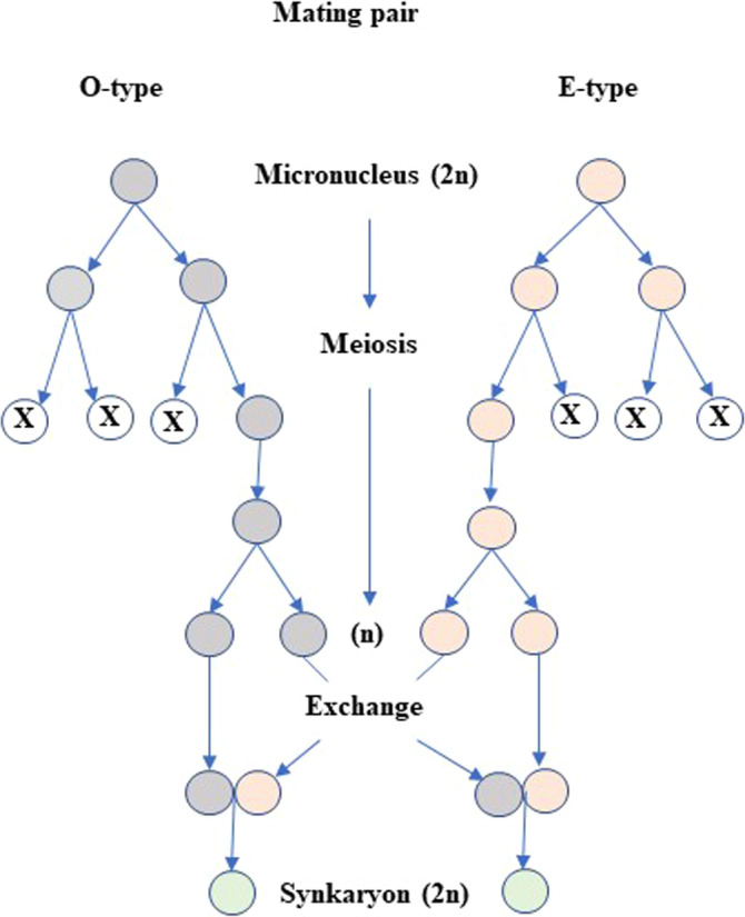
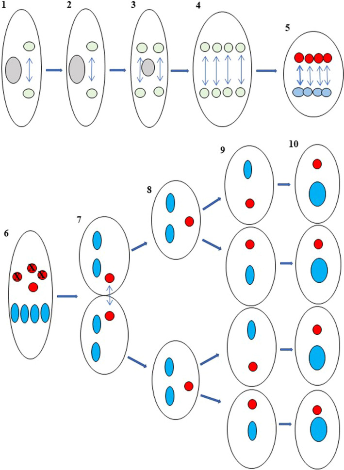
This review addresses nine areas of knowledge revealed by micromanipulations performed with Paramecium. Microinjection has shown that sexual maturation and senescence of Paramecium caudatum is a programmed process conducted by a specific gene and its product protein. In Paramecium tetraurelia, autogamy was revealed to depend on the number of DNA syntheses rather than the number of cell divisions in clonal aging. The cytoplasmic complementarity test established that microinjection of wild-type cytoplasm can correct genetic defects of mutants. The concept of complementarity together with protein chemistry revealed compounds that control membrane excitability. In non-Mendelian inheritance, noncoding small RNAs made from the parental micronucleus regulate the rearrangement of the progeny's macronuclear DNA. The macronucleus has the potential to be used as a factory for genetic engineering. The development and differentiation of progeny's nuclei in mating pairs are controlled by the parental macronucleus. The chemical reaction processes associated with exocytosis have been revealed by microinjection of various enzymes and antibodies. Using the fusion gene of histone H2B and yellow-fluorescence protein, it was revealed that the fusion gene-mRNA is transferred between cells during mating. Experiments with endosymbiotic bacteria and the host shed light on the conditions needed to establish sustainable symbiotic relationships.
Keywords: behavioral mutants; complementarity test; conjugation; endosymbiosis; exocytosis; life history; membrane excitation; nucleus differentiation; scnRNA.
© 2022 The Authors. Journal of Eukaryotic Microbiology published by Wiley Periodicals LLC on behalf of International Society of Protistologists.
Figures
Similar articles
-
Non-Mendelian inheritance of macronuclear mutations is gene specific in Paramecium tetraurelia.Mol Cell Biol. 1994 Apr;14(4):2479-84. doi: 10.1128/mcb.14.4.2479-2484.1994. Mol Cell Biol. 1994. PMID: 8139550 Free PMC article.
-
Elucidation of nucleus-cytoplasm interaction: change in ability of the nucleus to express sexuality according to clonal age in Paramecium.J Cell Sci. 1995 Dec;108 ( Pt 12):3671-6. doi: 10.1242/jcs.108.12.3671. J Cell Sci. 1995. PMID: 8719873
-
A Mendelian mutation affecting mating-type determination also affects developmental genomic rearrangements in Paramecium tetraurelia.Genetics. 1996 May;143(1):191-202. doi: 10.1093/genetics/143.1.191. Genetics. 1996. PMID: 8722774 Free PMC article.
-
Non-Mendelian inheritance and homology-dependent effects in ciliates.Adv Genet. 2002;46:305-37. doi: 10.1016/s0065-2660(02)46011-7. Adv Genet. 2002. PMID: 11931229 Review.
-
Paramecium epigenetics in development and proliferation.J Eukaryot Microbiol. 2022 Sep;69(5):e12914. doi: 10.1111/jeu.12914. Epub 2022 May 4. J Eukaryot Microbiol. 2022. PMID: 35363910 Review.
Cited by
-
Application of RNA interference and protein localization to investigate housekeeping and developmentally regulated genes in the emerging model protozoan Paramecium caudatum.Commun Biol. 2024 Feb 19;7(1):204. doi: 10.1038/s42003-024-05906-2. Commun Biol. 2024. PMID: 38374195 Free PMC article.
-
A Review for the Special Issue on Paramecium as a Modern Model Organism.Microorganisms. 2023 Apr 3;11(4):937. doi: 10.3390/microorganisms11040937. Microorganisms. 2023. PMID: 37110360 Free PMC article. Review.
References
-
- Aufderheide, K. (1984) Clonal aging in Paramecium tetraurelia. Absence evidence for a cytoplasmic factor. Mechanisms of Ageing and Development, 28, 57–66. - PubMed
-
- Barber, M. (1904) A new method of isolating microorganisms. The Journal of the Kansas Medical Society, 4, 489–494.
Publication types
MeSH terms
LinkOut - more resources
Full Text Sources
