Review
. 2022 Sep;18(36):e2107305.
doi: 10.1002/smll.202107305. Epub 2022 Mar 23.

Reciprocity of Cell Mechanics with Extracellular Stimuli: Emerging Opportunities for Translational Medicine

Affiliations
Review

Reciprocity of Cell Mechanics with Extracellular Stimuli: Emerging Opportunities for Translational Medicine

Yiwei Li et al. Small. 2022 Sep.

Abstract

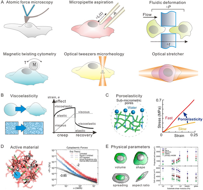
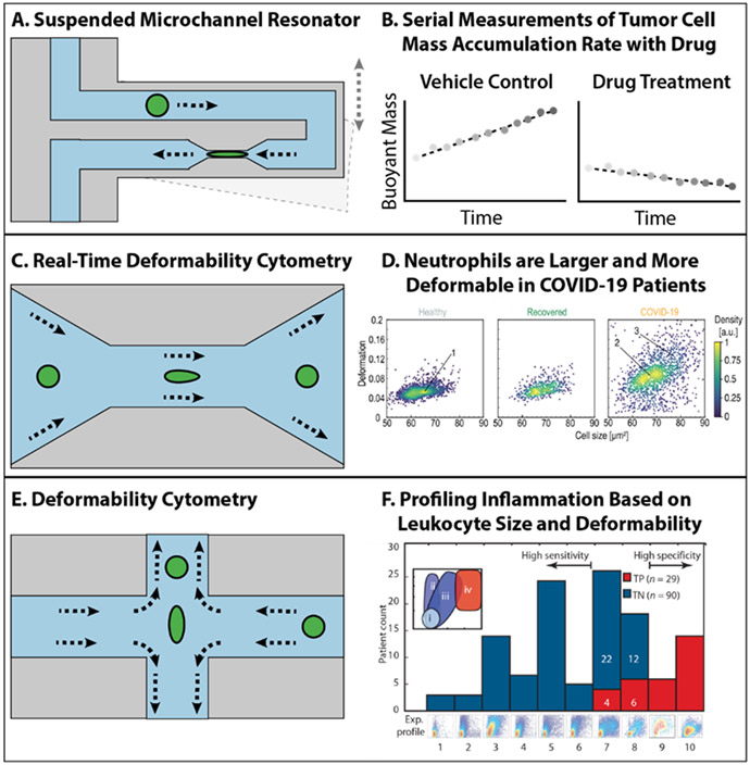
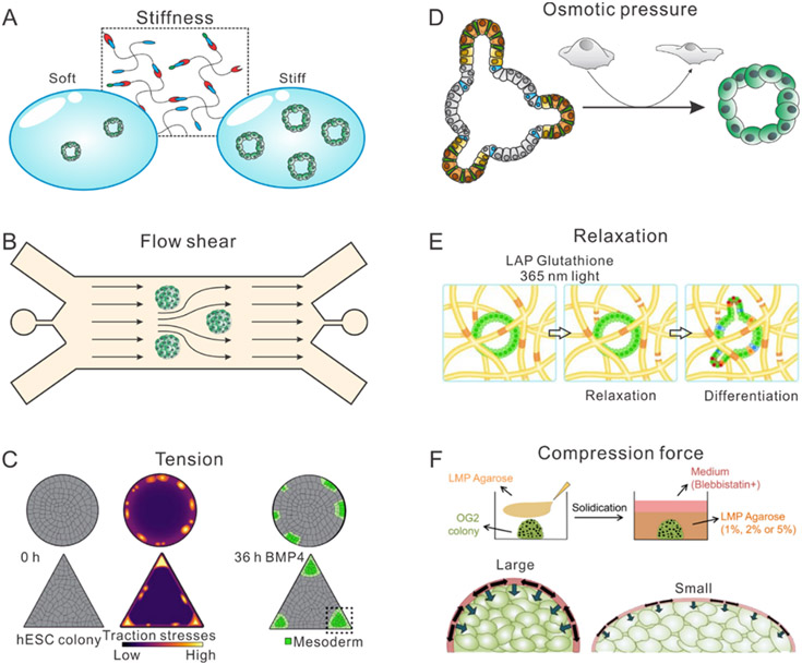
Human cells encounter dynamic mechanical cues in healthy and diseased tissues, which regulate their molecular and biophysical phenotype, including intracellular mechanics as well as force generation. Recent developments in bio/nanomaterials and microfluidics permit exquisitely sensitive measurements of cell mechanics, as well as spatiotemporal control over external mechanical stimuli to regulate cell behavior. In this review, the mechanobiology of cells interacting bidirectionally with their surrounding microenvironment, and the potential relevance for translational medicine are considered. Key fundamental concepts underlying the mechanics of living cells as well as the extracelluar matrix are first introduced. Then the authors consider case studies based on 1) microfluidic measurements of nonadherent cell deformability, 2) cell migration on micro/nano-topographies, 3) traction measurements of cells in three-dimensional (3D) matrix, 4) mechanical programming of organoid morphogenesis, as well as 5) active mechanical stimuli for potential therapeutics. These examples highlight the promise of disease diagnosis using mechanical measurements, a systems-level understanding linking molecular with biophysical phenotype, as well as therapies based on mechanical perturbations. This review concludes with a critical discussion of these emerging technologies and future directions at the interface of engineering, biology, and medicine.

Keywords: biomaterials; cell mechanics; deformability cytometry; directed migration; extracellular matrix; mechanobiology; microfluidics; traction force microscopy.


Figures

Figure 1.
Figure 1.. Schematic illustration of different aspects of cell mechanics and tools for measurement of cell mechanics.
(A) Schematic illustration of tools for measurements of cell mechanics, including atomic force microscopy, micropipette aspiration, fluidic deformation, magnetic twisting cytometry, optical tweezer microrheology, and optical stretcher. (B) Schematic illustration of viscoelasticity of cells. (C) Schematic illustration of poroelasticity of cells. Reproduced with permission.[50] Copyright 2018, Elsevier. (D) Schematic illustration of cells as active materials. Reproduced with permission.[26] Copyright 2014, Elsevier. (E) Schematic illustration of physical parameters of cells, including but not limited to volume, shape, spreading, and aspect ratio. Reproduced with permission.[51] Copyright 2014, American Chemical Society.
Figure 2.
Figure 2.. Schematic illustration of high-throughput measurement of nonadherent single cell mechanics.
(A) Schematic illustration of suspended microchannel resonator. (B) Serial measurements of tumor cell mass accumulation rate with drug using suspended microchannel resonator. (C) Schematic illustration of real-time deformability cytometry. (D) Testing deformability of neutrophils in health people and COVID-19 patients using real-time deformability cytometry. Reproduced with permission.[71] Copyright 2021, Elsevier. (E) Schematic illustration of deformability cytometry. (F) Profilling inflammation based on leukocytes and deformability using deformability cytometry. Reproduced with permission.[74] Copyright 2013, The American Association for the Advancement of Science.
Figure 3.
Figure 3.. Schematic illustration of extracellular mechanical stimuli including
(A) solid stress, Reproduced with permission.[92] Copyright 2014, Ivyspring International Publisher, (B) fluid stress, Reproduced with permission.[92] Copyright 2014, Ivyspring International Publisher, (C) microarchitecture, Reproduced with permission.[93] Copyright 2019, WILEY-VCH, and (D) mechanics, Reproduced with permission.[94] Copyright 2017, Intechopen.
Figure 4.
Figure 4.. Schematic illustration of Interrogating Cell Migration Dynamics for Diagnostics and Therapeutics.
(A) Schematic illustration showed that glioma cell migration is sensitive to PDGF on nanogrooved substrates. (B) Schematic illustration showed that T-cell directional migration is regulated by contractility and microtubule (MT) instability. (C) Schematic illustration showed that metastatic potential of breast cancer cells can be indicated by invasion of branched channels. (D) Schematic illustration showed that active neutrophils from septic patients exhibit spontaneous, sporadic migration.
Figure 5.
Figure 5.. Traction force microscopy in 3D matrix.
(A) i. Representative image of a MDA-MB-231 cell (blue) in a 3D collagen network (green). (Scale bar, 10 μm.) (ii–iv) Scheme of the force–displacement measurement with laser tweezers and the relation between matrix stiffening (blue potential wells) and the cell-generated stress field in the cell contraction direction. Reproduced with permission.[101] Copyright 2018. (B) i. Experimental setup for high-throughput imaging to measure cell-induced matrix deformations. ii. Multicellular clusters were grown inside a silk–collagen matrix with embedded 1-μm red fluorescent tracer particles in a 96-well-plate setup. To achieve high-throughput imaging, clusters were imaged by using a spinning-disk confocal microscopy with a low-NA air objective. iii. The 3D cell-induced matrix deformations recovered by directly tracking tracer particles. Reproduced with permission.[164] Copyright 2020. (C) Optical coherence microscopy of multicellular spheroids of mammary epithelial cells alone (MCF10at1) and in co-culture with wild type adipose stem cells (WT-ASC) or obese adipose stem cells (ob/ob ASC). Reproduced with permission.[167] Copyright 2020, Wiley-VCH.
Figure 6.
Figure 6.. Schematic illustration of mechanical programming of organoid morphogenesis.
(A) Schematic illustration of synthetic hydrogel for organoid culturing with varying stiffness. (B) Schematic illustration of microfluidic device for inducing flow shearing force to cultured organoid. (C) Schematic illustration of micropatterning technology for inducing local tension and contractility for cultured organoids and embryos. Reproduced with permission.[185] Copyright 2020, Elsevier. (D) Schematic illustration of osmotic compression to mimic local fluid pressure in native tissue for organoid culturing. Reproduced with permission.[15] Copyright 2021, Elsevier. (E) Schematic illustration of photo-degradable hydrogel for mimicking tissue stress relaxation for organoid culturing. Reproduced with permission.[186] Copyright 2020, Wiley-VCH. (F) Schematic illustration of hydrogel confinement for mimicking compression force for organoid and embryo. Reproduced with permission[187]. Copyright 2019, Elsevier.
Figure 7.
Figure 7.. Development of new technologies for inducing active mechanical stimuli.
(A) Schematic illustration of soft robot for inducing cyclic mechanical compression. Reproduced with permission.[216] Copyright 2018, Nature Publish Group. (B) Schematic illustration of compressive cranial window for mimicking solid stress generated by brain tumor. Reproduced with permission.[84] Copyright 2020, Nature Publish Group. (C) Schematic illustration of stimuli responsive polymer for inducing local force for cells on substrate. Reproduced with permission.[217] Copyright 2017, Nature Publish Group. (D) Schematic illustration of microfluidic devices for inducing flow shearing force for cells. Reproduced with permission.[218] Copyright 2018, Wiley-VCH. (E) Schematic illustration of organ-on-a-chip for inducing stretching to cultured cells. Reproduced with permission.[219] Copyright 2012, Royal Society of Chemistry. (F) Schematic illustration of magnetic nanomaterials for inducing local forces in mice. Reproduced with permission.[220] Copyright 2015, Nature Publish Group.
Scheme 1.
Scheme 1.
Schematic illustration of reciprocity of cell mechanics with extracellular stimuli for various biomedical regenerative applications. Cell mechanics contains four characteristics including viscoelasticity, poroelasticity, physical properties, and active material behaviors. Extracellular stimuli contains four formulas of stresses, including solid stress, fluid pressure, ECM architecture, and ECM mechanics. The cell mechanics interplay with extracellular stimuli. The study of the interplays benefits fundamental mechanobiology, biophysical disease diagnosis, and therapeutic mechanical stimuli. Reproduced with permission.[11,24-31] Viscoelasticity: Copyright 2020, The American Society for Cell Biology; Poroelasticity: Copyright 2013, Nature Publishing Group; Physical Properties: Copyright 2019, Royal Society of Chemistry; Active materials: Copyright 2014, Cell Press; Extracellular stimuli: Copyright 2019, Nature Publishing Group; Solid stress: Copyright 2020, Elsevier; Fluid pressure: Copyright 1998, Elsevier. Architecture: Copyright 2020, National Academy of Sciences; Mechanics: Copyright 2020, Royal Society of Chemistry; Cell Mechanics: Copyright 2020, Cell Press.

Similar articles

Cited by

References

    1. Engler AJ, Humbert PO, Wehrle-Haller B, Weaver VM, Science 2009, 324, 208. - PMC - PubMed
    1. Jacobs CR, Huang H, Kwon RY, Introduction to cell mechanics and mechanobiology, Garland Science, 2012.
    1. D’Arcy WT, Dover, New York, 1992.
    1. Kechagia JZ, Ivaska J, Roca-Cusachs P, Nature Reviews Molecular Cell Biology 2019, 20, 457. - PubMed
    1. Fletcher DA, Mullins RD, Nature 2010, 463, 485. - PMC - PubMed

Publication types

LinkOut - more resources