

PET/CT imaging of spinal inflammation and microcalcification in patients with low back pain: A pilot study on the quantification by artificial intelligence-based segmentation
- PMID: 35319166
- PMCID: PMC9322590
- DOI: 10.1111/cpf.12751
PET/CT imaging of spinal inflammation and microcalcification in patients with low back pain: A pilot study on the quantification by artificial intelligence-based segmentation
Abstract
Background: Current imaging modalities are often incapable of identifying nociceptive sources of low back pain (LBP). We aimed to characterize these by means of positron emission tomography/computed tomography (PET/CT) of the lumbar spine region applying tracers 18 F-fluorodeoxyglucose (FDG) and 18 F-sodium fluoride (NaF) targeting inflammation and active microcalcification, respectively.
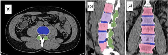
Methods: Using artificial intelligence (AI)-based quantification, we compared PET findings in two sex- and age-matched groups, a case group of seven males and five females, mean age 45 ± 14 years, with ongoing LBP and a similar control group of 12 pain-free individuals. PET/CT scans were segmented into three distinct volumes of interest (VOIs): lumbar vertebral bodies, facet joints and intervertebral discs. Maximum, mean and total standardized uptake values (SUVmax, SUVmean and SUVtotal) for FDG and NaF uptake in the 3 VOIs were measured and compared between groups. Holm-Bonferroni correction was applied to adjust for multiple testing.
Results: FDG uptake was slightly higher in most locations of the LBP group including higher SUVmean in the intervertebral discs (0.96 ± 0.34 vs. 0.69 ± 0.15). All NaF uptake values were higher in cases, including higher SUVmax in the intervertebral discs (11.63 ± 3.29 vs. 9.45 ± 1.32) and facet joints (14.98 ± 6.55 vs. 10.60 ± 2.97).
Conclusion: Observed intergroup differences suggest acute inflammation and microcalcification as possible nociceptive causes of LBP. AI-based quantification of relevant lumbar VOIs in PET/CT scans of LBP patients and controls appears to be feasible. These promising, early findings warrant further investigation and confirmation.
Keywords: fluorodeoxyglucose; low back pain; lumbar vertebrae; positron emission tomography; sodium fluoride.
© 2022 The Authors. Clinical Physiology and Functional Imaging published by John Wiley & Sons Ltd on behalf of Scandinavian Society of Clinical Physiology and Nuclear Medicine.
Conflict of interest statement
The authors declare no conflicts of interest.
Figures
References
-
- Barnsley, L. , Lord, S.M. , Wallis, B.J. & Bogduk, N. (1994) Lack of effect of intraarticular corticosteroids for chronic pain in the cervical zygapophyseal joints. New England Journal of Medicine, 330, 1047–1050. - PubMed
-
- Beattie, P.F. & Meyers, S.P. (1998) Magnetic resonance imaging in low back pain: general principles and clinical issues. Physical Therapy, 78, 738–753. - PubMed
-
- Belal, S.L. , Sadik, M. , Kaboteh, R. , Enqvist, O. , Ulén, J. & Poulsen, M.H. et al. (2019) Deep learning for segmentation of 49 selected bones in CT scans: first step in automated PET/CT‐based 3D quantification of skeletal metastases. European Journal of Radiology, 113, 89–95. - PubMed
-
- Blau, M. , Ganatra, R. & Bender, M.A. (1972) 18F‐fluoride for bone imaging. Seminars in Nuclear Medicine, 2, 31–37. - PubMed
-
- Blomberg, B.A. , Thomassen, A. , Takx, R.A.P. , Hildebrandt, M.G. , Simonsen, J.A. , Buch‐Olsen, K.M. et al. (2014) Delayed 18F‐fluorodeoxyglucose PET/CT imaging improves quantitation of atherosclerotic plaque inflammation: results from the CAMONA study. Journal of Nuclear Cardiology, 21, 588–597. - PubMed
MeSH terms
Substances
Grants and funding
LinkOut - more resources
Full Text Sources
Miscellaneous
