. 2022 Apr 13;96(7):e0005722.
doi: 10.1128/jvi.00057-22. Epub 2022 Mar 23.

Heparanase Blockade as a Novel Dual-Targeting Therapy for COVID-19

Affiliations

Heparanase Blockade as a Novel Dual-Targeting Therapy for COVID-19

Jingyu Xiang et al. J Virol. .

Abstract

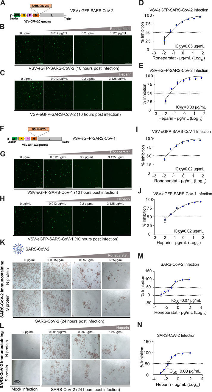
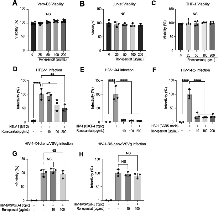
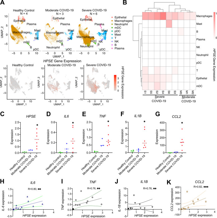
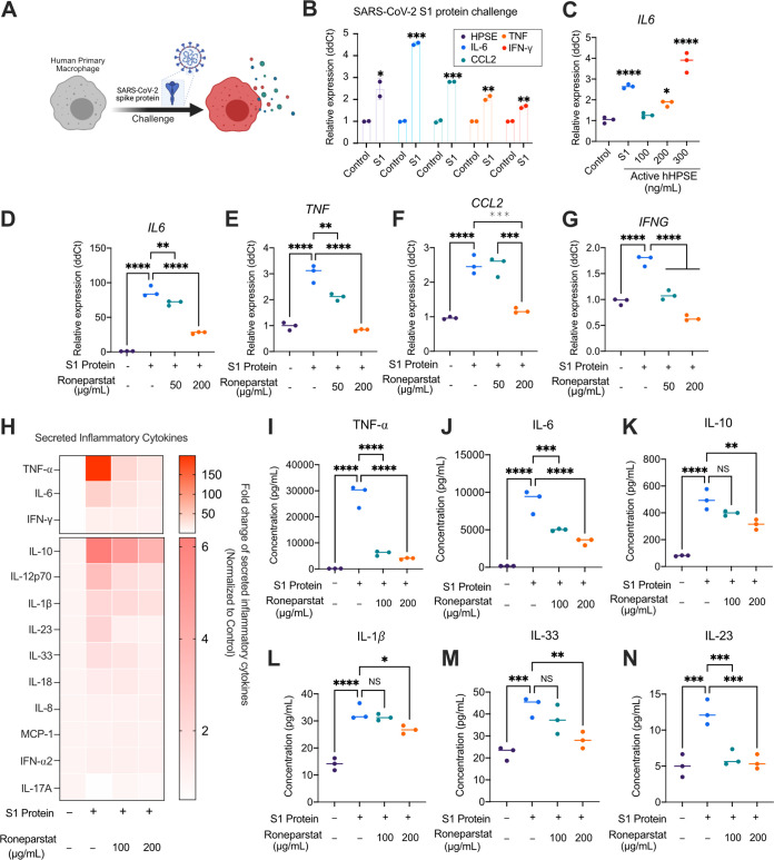
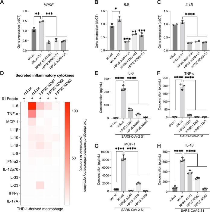
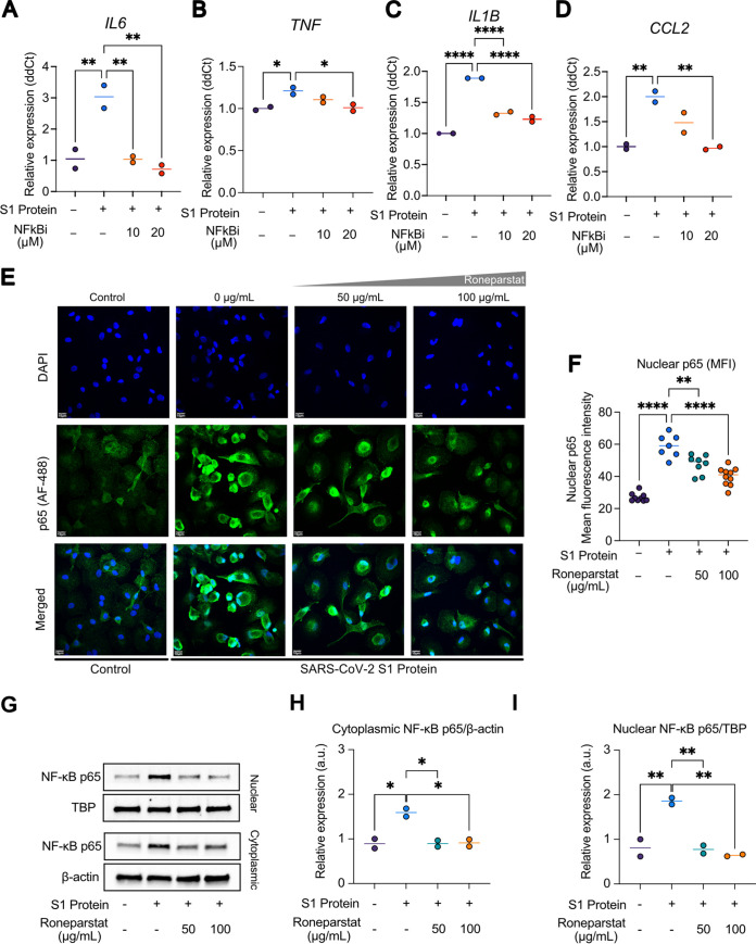
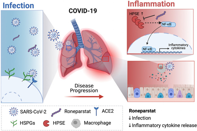
The coronavirus disease 2019 (COVID-19) pandemic, caused by severe acute respiratory syndrome coronavirus 2 (SARS-CoV-2), has caused over 5 million deaths worldwide. Pneumonia and systemic inflammation contribute to its high mortality. Many viruses use heparan sulfate proteoglycans as coreceptors for viral entry, and heparanase (HPSE) is a known regulator of both viral entry and inflammatory cytokines. We evaluated the heparanase inhibitor Roneparstat, a modified heparin with minimum anticoagulant activity, in pathophysiology and therapy for COVID-19. We found that Roneparstat significantly decreased the infectivity of SARS-CoV-2, SARS-CoV-1, and retroviruses (human T-lymphotropic virus 1 [HTLV-1] and HIV-1) in vitro. Single-cell RNA sequencing (scRNA-seq) analysis of cells from the bronchoalveolar lavage fluid of COVID-19 patients revealed a marked increase in HPSE gene expression in CD68+ macrophages compared to healthy controls. Elevated levels of HPSE expression in macrophages correlated with the severity of COVID-19 and the expression of inflammatory cytokine genes, including IL6, TNF, IL1B, and CCL2. In line with this finding, we found a marked induction of HPSE and numerous inflammatory cytokines in human macrophages challenged with SARS-CoV-2 S1 protein. Treatment with Roneparstat significantly attenuated SARS-CoV-2 S1 protein-mediated inflammatory cytokine release from human macrophages, through disruption of NF-κB signaling. HPSE knockdown in a macrophage cell line also showed diminished inflammatory cytokine production during S1 protein challenge. Taken together, this study provides a proof of concept that heparanase is a target for SARS-CoV-2-mediated pathogenesis and that Roneparstat may serve as a dual-targeted therapy to reduce viral infection and inflammation in COVID-19. IMPORTANCE The complex pathogenesis of COVID-19 consists of two major pathological phases: an initial infection phase elicited by SARS-CoV-2 entry and replication and an inflammation phase that could lead to tissue damage, which can evolve into acute respiratory failure or even death. While the development and deployment of vaccines are ongoing, effective therapy for COVID-19 is still urgently needed. In this study, we explored HPSE blockade with Roneparstat, a phase I clinically tested HPSE inhibitor, in the context of COVID-19 pathogenesis. Treatment with Roneparstat showed wide-spectrum anti-infection activities against SARS-CoV-2, HTLV-1, and HIV-1 in vitro. In addition, HPSE blockade with Roneparstat significantly attenuated SARS-CoV-2 S1 protein-induced inflammatory cytokine release from human macrophages through disruption of NF-κB signaling. Together, this study provides a proof of principle for the use of Roneparstat as a dual-targeting therapy for COVID-19 to decrease viral infection and dampen the proinflammatory immune response mediated by macrophages.

Keywords: COVID-19; SARS-CoV-2; heparanase; inflammatory cytokine release; macrophage.


Conflict of interest statement

The authors declare a conflict of interest. Roneparstat (SST0001) is a proprietary drug of Leadiant Biosciences S.p.A. Alessandro Noseda is the employee of Leadiant Biosciences S.p.A. No potential conflicts of interest were disclosed by the other authors.

Figures

FIG 1
FIG 1
The HPSE inhibitor Roneparstat suppresses coronavirus infection. (A) Schematic representation of the genome of VSV-eGFP-SARS-CoV-2. VSV G was deleted from the VSV genome, and the SARS-CoV-2 S gene was inserted between the M and L genes in the VSV genome, which resulted in the construction of replication-competent VSV-eGFP-SARS-CoV-2. Monolayers of Vero-E6 cells were infected with VSV-eGFP-SARS-CoV-2 at an MOI of 0.1 in the presence or absence of various doses of Roneparstat or heparin for 1 h. At 10 h postinfection, infected cells (GFP+) were quantified by a Cytation 5 microscope. (B and C) Representative fluorescence images of Vero-E6 cells infected with VSV-eGFP-SARS-CoV-2 in the presence of 0, 0.012, 0.2, and 3.125 μg/mL of Roneparstat or heparin. (D and E) Logistic inhibition curve of Roneparstat or heparin treatment during VSV-eGFP-SARS-CoV-2 infection. (F) Schematic representation of the genome of VSV-eGFP-SARS-CoV-1. VSV G was deleted from the VSV genome, and the SARS-CoV-1 S gene was inserted between the M and L genes in the VSV genome, which resulted in the construction of replication-competent VSV-eGFP-SARS-CoV-1. Monolayers of Vero-E6 cells were infected with VSV-eGFP-SARS-CoV-1 at an MOI of 0.1 in the presence or absence of various doses of Roneparstat or heparin for 1 h. At 10 h postinfection, infected cells (GFP+) were quantified by a Cytation 5 microscope. (G and H) Representative fluorescence images of Vero-E6 cells infected with VSV-eGFP-SARS-CoV-1 in the presence of 0, 0.012, 0.2, and 3.125 μg/mL of Roneparstat or heparin. (I and J) Logistic inhibition curve of Roneparstat or heparin treatment during VSV-eGFP-SARS-CoV-1 infection. (K and L) Representative images of immunostaining from SARS-CoV-2 (strain USA-WA1/2020) (400 PFU)-infected monolayers of Vero-E6 cells. Antibodies specific to the SARS-CoV-2 spike (S) or nuclear (N) protein were used to identify SARS-CoV-2-infected cells under various Roneparstat or heparin treatment conditions (0, 0.0015, 0.097, and 6.25 μg/mL). (M and N) Logistic inhibition curve of Roneparstat or heparin treatment during SARS-CoV-2 infection. Error bars represent the SEM. *, P < 0.05; **, P < 0.01; ***, P < 0.001 (2-tailed distribution, homoscedastic Student’s t test for 2 groups or 1-way ANOVA for multiple comparisons).
FIG 2
FIG 2
Roneparstat-mediated blockade of infection is dependent on HSPGs. (A to C) The Vero-E6 (A), Jurkat (B), or THP-1 (C) cell line was treated with various doses of Roneparstat (0, 25, 50, 100, and 200 μg/mL) for 24 h. Subsequently, cell viability was examined using a CellTiter-Blue viability assay. (D) Jurkat-LTR-Luc reporter cells cocultured with irradiated HTLV-1-producing MT-2 cells in the presence or absence of various doses (10, 100, and 200 μg/mL) of Roneparstat for 48 h. Infectivity (percent) was quantified via luciferase assays. (E and F) HIV-1-LTR-Luc reporter cells were pretreated overnight with Roneparstat (10, 100, and 200 μg/mL), followed by HIV-1 (CXCR4- or CCR5-tropic strain) infection for 24 h. Infectivity (percent) was quantified via luciferase assays. (G and H) U87/X4 or U87/R5 target cells were infected with the VSVg-pseudotyped virus VSVg-HIV-1Δenv-luc for 48 h in the presence or absence of Roneparstat (10 and 100 μg/mL). Infectivity (percent) was quantified via luciferase assays. Error bars represent the SEM. *, P < 0.05; **, P < 0.01; ***, P < 0.001; NS, not significant (2-tailed distribution, homoscedastic Student’s t test for 2 groups or 1-way ANOVA for multiple comparisons).
FIG 3
FIG 3
The HPSE gene is upregulated in the BALF of COVID-19 patients and correlates with the expression of inflammatory cytokine genes in macrophages. (A, top) Alignment of major BALF cell clusters by UMAP across control (n = 4), moderate COVID-19 (n = 3), and severe COVID-19 (n = 6) samples. (Bottom) UMAP presentation showing the HPSE gene expression levels across control, moderate COVID-19, and severe COVID-19 samples. (B) Heat map of differential HPSE gene expression of different cell types by each COVID-19 sample. Cell markers (cell types are identified by signature genes) for each cell type are defined as follows: CD68 for macrophages, TPSB2 for mast cells, MS4A1 for B cells, IGHG4 for plasma cells, KLRD1 for natural killer (NK) cells, FCGR3B for neutrophils, LILRA4 for plasmacytoid dendritic cells (pDC), CD3D for T cells, TPPP3 and KRT18 for epithelial cells, and CD1C and CLEC9A for myeloid dendritic cells (mDC). (C to G) Expression levels of HPSE, IL6, TNF, IL1B, and CCL2 by BALF macrophages across healthy control, moderate COVID-19, and severe COVID-19 samples. (H to K) Correlations between HPSE expression and the expression of IL6, TNF, IL1B, and CCL2. Error bars represent the SEM. *, P < 0.05; **, P < 0.01; ***, P < 0.001 (2-tailed distribution, homoscedastic Student’s t test for 2 groups or 1-way ANOVA for multiple comparisons; a Spearman correlation coefficient test was adopted to determine statistically significant correlations between two groups).
FIG 4
FIG 4
Inhibition of HPSE by Roneparstat decreases inflammatory cytokine release induced by the SARS-CoV-2 spike protein. (A) Experimental design of human primary monocyte-derived macrophages challenged with SARS-CoV-2 spike (S1) protein. (B) qPCR detection of the transcription of the HPSE, IL6, TNF, CCL2, and IFNG genes in macrophages challenged with S1 protein (0.5 μg/mL) for 12 h. (C) qPCR detection of the transcription of IL-6 in macrophages challenged with S1 protein (0.5 μg/mL) or various doses (100, 200, and 300 ng/mL) of recombinant active heparanase (rHPSE) for 12 h. (D to G) qPCR detection of the transcription of the IL6, TNF, CCL2, and IFNG genes in macrophages cotreated with S1 protein (0.5 μg/mL) and various doses of Roneparstat (50 and 200 μg/mL) for 12 h. (H) Conditioned medium was collected from the macrophages cotreated with S1 protein (0.5 μg/mL) and various doses of Roneparstat (100 and 200 μg/mL) for 24 h. Inflammatory cytokines were quantified using a flow cytometry-based multiplex cytokine array. Fold changes of the secreted inflammatory cytokines were normalized to the values for the untreated control and are presented in a heat map. (I to N) Absolute concentrations of secreted TNF-α, IL-6, IL-10, IL-1β, IL-33, and IL-23 were calculated based on standard curve fitting. Error bars represent the SEM. *, P < 0.05; **, P < 0.01; ***, P < 0.001 (2-tailed distribution, homoscedastic Student’s t test for 2 groups or 1-way ANOVA for multiple comparisons).
FIG 5
FIG 5
SARS-CoV-2 S1-induced inflammatory cytokine release depends on HPSE expression. Control (shLuc) or HPSE knockdown (KD) THP-1-derived macrophages were challenged with S1 protein (0.5 μg/mL) for 12 h. (A to C) Transcription levels of HPSE (A), IL6 (B), and IL1B (C) were examined by qPCR. (D) Conditioned medium was collected from the THP-1 macrophages treated with S1 protein (0.5 μg/mL) for 24 h. Inflammatory cytokines were quantified using a flow cytometry-based multiplex cytokine array. Fold changes of the secreted inflammatory cytokines were normalized to the values for the untreated control and are presented in a heat map. (E to H) Absolute concentrations of secreted IL-6 (E), TNF-α (F), MCP-1 (G), and IL-1β (H) were calculated based on standard curve fitting. Error bars represent the SEM. *, P < 0.05; **, P < 0.01; ***, P < 0.001 (2-tailed distribution, homoscedastic Student’s t test for 2 groups or 1-way ANOVA for multiple comparisons).
FIG 6
FIG 6
Roneparstat decreases SARS-CoV-2 spike protein-induced inflammatory cytokine release via NF-κB signaling. Human primary macrophages were pretreated with an NF-κB inhibitor (NFkBi) (BAY) for 1 h, followed by S1 protein (0.5 μg/mL) challenge for 12 h. (A to D) Transcription levels of IL6 (A), TNF (B), IL1B (C), and IFNG (D) were assessed by qPCR. (E) Representative NF-κB (p65) immunostaining of human primary macrophages cotreated with S1 protein (0.5 μg/mL) and various doses of Roneparstat (50 and 100 μg/mL) for 24 h. (Top) DAPI; (middle) p65 (Alexa Fluor 488 [AF-488]); (bottom) merged. (F) Quantification of the mean fluorescence intensity (MFI) of the nuclear p65 signal. Seven to ten high-power fields were captured and quantified under each treatment condition. (G) Western blots of cytoplasmic and nuclear p65 in human primary macrophages treated with various doses of Roneparstat (50 and 100 μg/mL) and S1 protein (0.5 μg/mL) for 30 min. Blots were reprobed with anti-TBP antibody as a loading control for nuclear fractions and β-actin for the cytoplasmic fractions. (H and I) Cytoplasmic and nuclear p65 levels were quantified by NF-κB p65/β-actin and p65/TBP ratios, respectively. a.u., arbitrary units. Error bars represent the SEM. *, P < 0.05; **, P < 0.01; ***, P < 0.001 (2-tailed distribution, homoscedastic Student’s t test for 2 groups or 1-way ANOVA for multiple comparisons).
FIG 7
FIG 7
Schema of the potential dual-targeting actions of Roneparstat in COVID-19. The pathogenesis of COVID-19 consists of two pathogenic phases: (i) the early infection phase, characterized by SARS-CoV-2 viral entry, replication, and spread, and (ii) the later inflammation phase, characterized by aberrant proinflammatory cytokine release that leads to tissue damage, ARDS, or even death. During the initial infection phase, Roneparstat decreases viral infection by competing with HSPG-dependent viral entry. During the inflammation phase, HPSE blockade via Roneparstat attenuates SARS-CoV-2-mediated inflammatory cytokine release from macrophages, through disruption of NF-κB signaling. Together, this study demonstrated the potential use of Roneparstat as a dual-targeting therapy for COVID-19 to decrease viral infection and dampen the proinflammatory immune response mediated by macrophages.

Similar articles

Cited by

References

    1. Alturki SO, Alturki SO, Connors J, Cusimano G, Kutzler MA, Izmirly AM, Haddad EK. 2020. The 2020 pandemic: current SARS-CoV-2 vaccine development. Front Immunol 11:1880. 10.3389/fimmu.2020.01880. - DOI - PMC - PubMed
    1. Wiersinga WJ, Rhodes A, Cheng AC, Peacock SJ, Prescott HC. 2020. Pathophysiology, transmission, diagnosis, and treatment of coronavirus disease 2019 (COVID-19): a review. JAMA 324:782–793. 10.1001/jama.2020.12839. - DOI - PubMed
    1. Siddiqi HK, Mehra MR. 2020. COVID-19 illness in native and immunosuppressed states: a clinical-therapeutic staging proposal. J Heart Lung Transplant 39:405–407. 10.1016/j.healun.2020.03.012. - DOI - PMC - PubMed
    1. Wu Z, McGoogan JM. 2020. Characteristics of and important lessons from the coronavirus disease 2019 (COVID-19) outbreak in China: summary of a report of 72314 cases from the Chinese Center for Disease Control and Prevention. JAMA 323:1239–1242. 10.1001/jama.2020.2648. - DOI - PubMed
    1. Fajgenbaum DC, June CH. 2020. Cytokine storm. N Engl J Med 383:2255–2273. 10.1056/NEJMra2026131. - DOI - PMC - PubMed