. 2022 Apr 13;96(7):e0054221.
doi: 10.1128/jvi.00542-21. Epub 2022 Mar 23.

3,4-Dicaffeoylquinic Acid from the Medicinal Plant Ilex kaushue Disrupts the Interaction Between the Five-Fold Axis of Enterovirus A-71 and the Heparan Sulfate Receptor

Affiliations

3,4-Dicaffeoylquinic Acid from the Medicinal Plant Ilex kaushue Disrupts the Interaction Between the Five-Fold Axis of Enterovirus A-71 and the Heparan Sulfate Receptor

Chung-Fan Hsieh et al. J Virol. .

Erratum in

Abstract

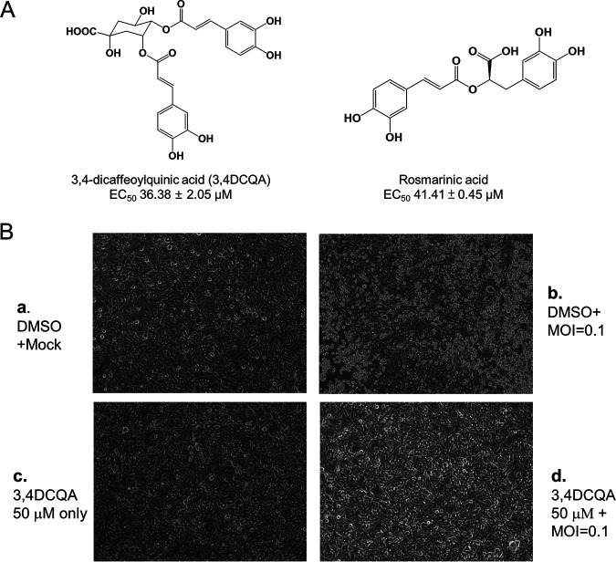
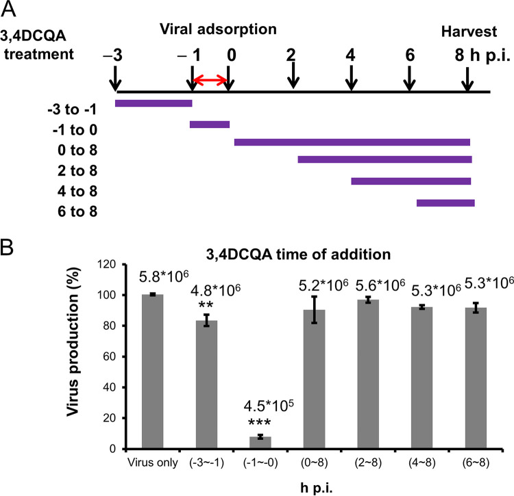
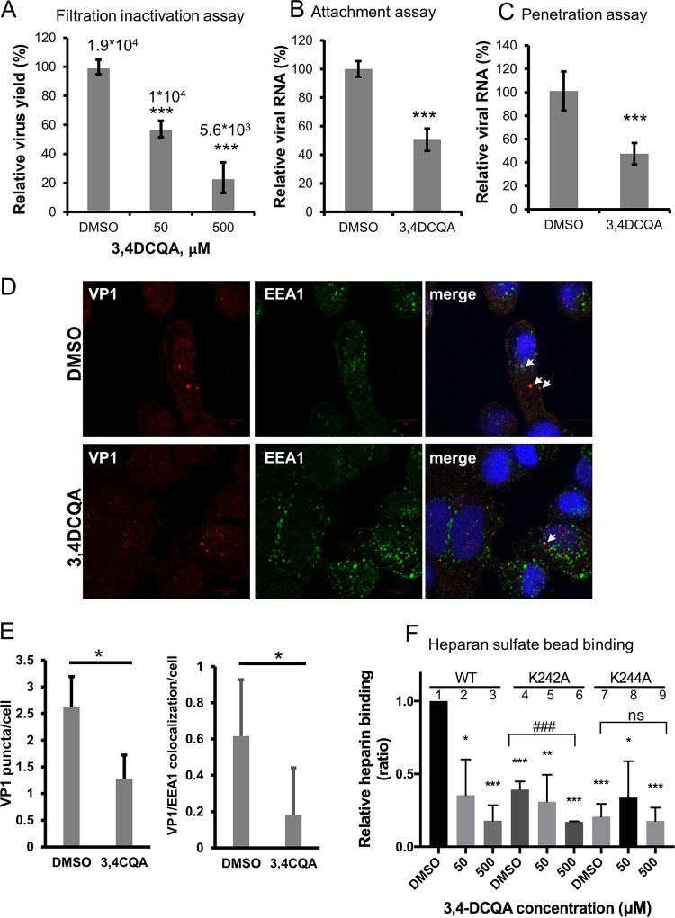
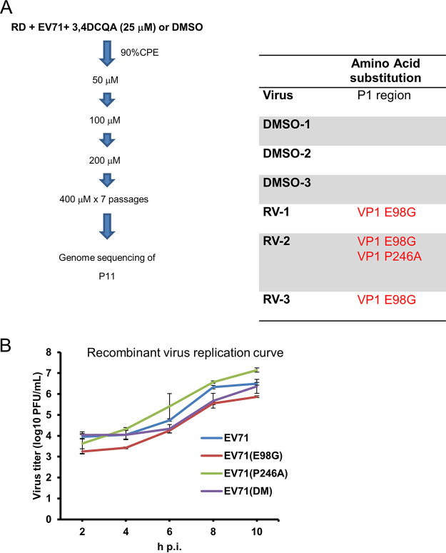
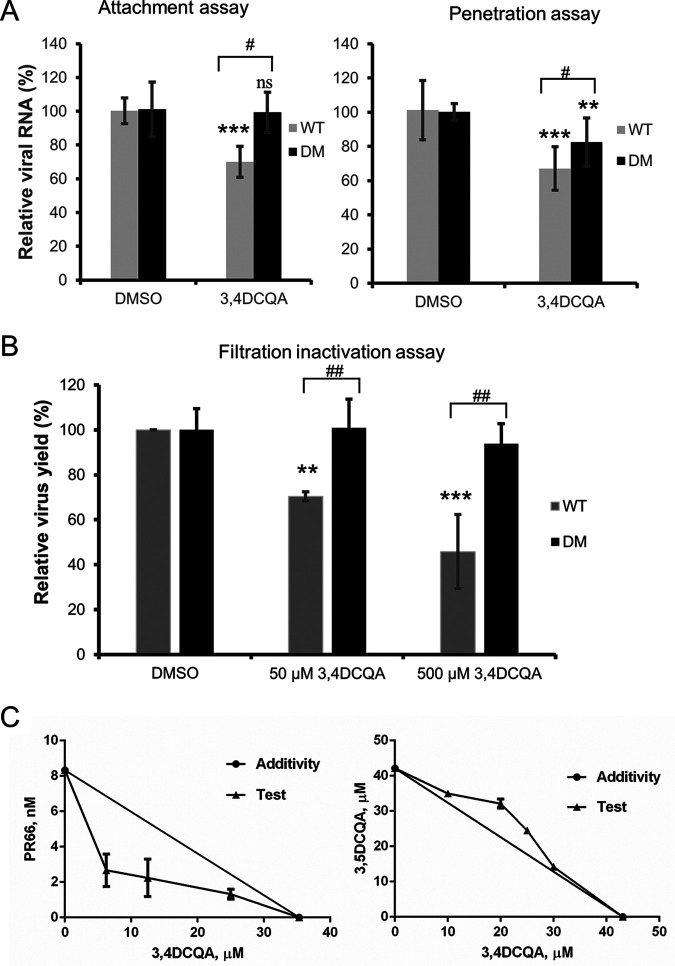
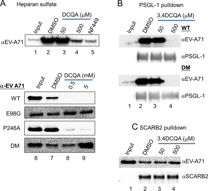
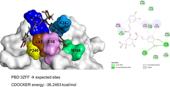
While infections by enterovirus A71 (EV-A71) are generally self-limiting, they can occasionally lead to serious neurological complications and death. No licensed therapies against EV-A71 currently exist. Using anti-virus-induced cytopathic effect assays, 3,4-dicaffeoylquinic acid (3,4-DCQA) from Ilex kaushue extracts was found to exert significant anti-EV-A71 activity, with a broad inhibitory spectrum against different EV-A71 genotypes. Time-of-drug-addition assays revealed that 3,4-DCQA affects the initial phase (entry step) of EV-A71 infection by directly targeting viral particles and disrupting viral attachment to host cells. Using resistant virus selection experiments, we found that 3,4-DCQA targets the glutamic acid residue at position 98 (E98) and the proline residue at position 246 (P246) in the 5-fold axis located within the VP1 structural protein. Recombinant viruses harboring the two mutations were resistant to 3,4-DCQA-elicited inhibition of virus attachment and penetration into human rhabdomyosarcoma (RD) cells. Finally, we showed that 3,4-DCQA specifically inhibited the attachment of EV-A71 to the host receptor heparan sulfate (HS), but not to the scavenger receptor class B member 2 (SCARB2) and P-selectin glycoprotein ligand-1 (PSGL1). Molecular docking analysis confirmed that 3,4-DCQA targets the 5-fold axis to form a stable structure with the E98 and P246 residues through noncovalent and van der Waals interactions. The targeting of E98 and P246 by 3,4-DCQA was found to be specific; accordingly, HS binding of viruses carrying the K242A or K244A mutations in the 5-fold axis was successfully inhibited by 3,4-DCQA.The clinical utility of 3,4-DCQA in the prevention or treatment of EV-A71 infections warrants further scrutiny. IMPORTANCE The canyon region and the 5-fold axis of the EV-A71 viral particle located within the VP1 protein mediate the interaction of the virus with host surface receptors. The three most extensively investigated cellular receptors for EV-A71 include SCARB2, PSGL1, and cell surface heparan sulfate. In the current study, a RD cell-based anti-cytopathic effect assay was used to investigate the potential broad spectrum inhibitory activity of 3,4-DCQA against different EV-A71 strains. Mechanistically, we demonstrate that 3,4-DCQA disrupts the interaction between the 5-fold axis of EV-A71 and its heparan sulfate receptor; however, no effect was seen on the SCARB2 or PSGL1 receptors. Taken together, our findings show that this natural product may pave the way to novel anti-EV-A71 therapeutic strategies.

Keywords: 3; 4-dicaffeoylquinic acid; 5-fold axis; enterovirus-A71; heparan sulphate.


Conflict of interest statement

The authors declare no conflict of interest.

We have no conflicts of interest to declare.

Figures

FIG 1
FIG 1
3,4-DCQA suppresses EV-A71-induced cytopathic effects in RD cells. (A) Chemical structures of 3,4-DCQA and rosmarinic acid. (B) Microscopy findings revealed that 3,4-DCQA prevented EV-A71-induced cytopathic effects in RD cells. (a) Mock: mock infection with DMSO; (b) Virus: infection with EV-A71 (multiplicity of infection [MOI] = 0.1) in DMSO; (c) 3,4-DCQA: mock infection and concomitant exposure to 3,4-DCQA (50 μM); (d) infection with EV-A71 (MOI = 0.1) in DMSO and concomitant exposure to 3,4-DCQA (50 μM).
FIG 2
FIG 2
Anti-EV-A71 activity of 3,4-DCQA assessed by time-of-addition assays in RD cells. (A) Schematic representation of 3,4-DCQA exposure at different time points during the viral life cycle. Purple lines denote the duration of incubation with 3,4-DCQA, whereas the red double arrow indicates the duration of viral infection (between −1 and 0 hours postinfection [hpi]). (B) Bar chart showing the extent of reduction in EV-A71 titers related to different time points of 3,4-DCQA exposure. All data were normalized to control conditions (virus-only group arbitrarily set to 100%). Data are presented as means ± standard deviations of four replicates from two independent experiments; comparisons were carried out using the Student’s t test (**, P < 0.01 and ***, P < 0.001). We also showed the results in mean log viral titer (PFU/mL) to ensure a significant viral titer load reduction.
FIG 3
FIG 3
(A) Centrifugal filtration inactivation assay. Data are presented as means ± standard deviations from three independent experiments, and two replicates for each assay were used. Viral titers were also expressed as PFU/mL. (B) Attachment inhibition assay and (C to E) penetration inhibition assay. (B to C) RNA was extracted and analyzed with RT-qPCR to detect viral attachment and viral penetration into cells. Data are presented as means ± standard deviations from three independent experiments. (D to E) Inhibition of penetration by 3,4-DCQA by confocal immunofluorescence microscopic analysis. Bar: 10 μm. Images of endocytosed VP1 inhibited by 3,4-DCQA were analyzed with IN Cell Investigator high-content analysis software. Viral penetration into the cells was reflected by cell-associated VP1 puncta (panel D and left part of panel E). Additionally, colocalization of VP1 and EEA1 (arrows) and their relative proportions were quantified (panel D and right part of panel E). (F) Heparan sulfate pulldown assay. The wild-type 5865/sin/000009 strain as well as mutants carrying the VP1-K242A and VP1-K244A variants were pretreated with DMSO or 3,4-DCQA before binding to heparan sulfate beads. Virus attachment was assessed with RT-qPCR. RNA copy numbers for each experiment were normalized to the wild-type dimethyl sulfoxide (DMSO) control (arbitrarily set to 1). Results are from three independent experiments. *, P < 0.05, **, P < 0.01, and ***, P < 0.001. ***, P < 0.001 ns for comparisons between DMSO and 3,4-DCQA (500 μM) treatment.
FIG 4
FIG 4
Selection and identification of 3,4-DCQA-resistant viruses. (A) Experimental flow chart for the selection and identification of 3,4-DCQA-resistant viruses. 3,4-DCQA- or DMSO-treated viruses were plaque-purified and Sanger-sequenced (three independent clones each). (B) Growth kinetics of wild-type and mutant EV-A71 TW/2231/1998 recombinant viruses. RD cells were infected (MOI = 1) with different recombinant viruses (wild-type, E98G mutant, P246A mutant, and double-mutant) for 1 h. Samples were harvested at the reported time points and virus titers were determined using plaque assays. Data are presented as means ± standard deviations from two independent experiments; duplicates were included in each experiment.
FIG 5
FIG 5
Attachment, penetration, and filtration inactivation assays of wild-type and E98G/P246A mutant viruses. (A) Attachment (left graph) and penetration (right graph) assays. Subsequently, RNA was extracted and analyzed with RT-qPCR. Data are presented as means ± standard deviations from three independent experiments. **, P < 0.01 and ***, P < 0.001 compared with control (DMSO). ##, P < 0.01 compared with the DM group. (B) Filtration inactivation assays. Data are presented as means ± standard deviations from three independent experiments. (C) 3,4-DCQA was used in combination with either PR66 (left) or 3,5-DCQA (right).
FIG 6
FIG 6
Receptor pulldown assays. Heparan sulfate receptor pulldown of double- or single-mutant viruses (A). 3,4-DCQA, NF449 (50 μM), or DMSO were initially mixed on ice for 1 h with either the wild-type EV-A71 recombinant virus or viruses carrying E98G and/or P246A mutations; subsequently, heparan sulfate bead pulldown was performed. 3,4-DCQA-induced inhibition was monitored with Western blotting using anti-EV71 antibodies. (B to C) PSGL-1 and SCARB2 receptor pulldown assays. Results are from three independent and reproducible experiments (control: 10% of input).
FIG 7
FIG 7
Structural docking of 3,4-DCQA and the EV-A71 VP1 protein. The expected binding site of 3,4-DCQA in the 5-fold axis of VP1 (PBD ID: 3ZFF) was predicted using Discovery Studio software. Amino acid residues which play crucial roles in the binding process are highlighted with different colors.

Similar articles

Cited by

References

    1. Brown BA, Pallansch MA. 1995. Complete nucleotide sequence of enterovirus 71 is distinct from poliovirus. Virus Res 39:195–205. 10.1016/0168-1702(95)00087-9. - DOI - PubMed
    1. Racaniello VR. 2007. Picornaviridae: the viruses and their replication, p 795–838. In Knipe DavidM, Martin MalcolmA., Martin DianeE, Griffin RobertA, Lamb Bernard, Roizman, Straus Stephen E (ed), Fields virology, 5th ed, vol 1. Lippincott Williams & Wilkins, Philadelphia.
    1. Plevka P, Perera R, Cardosa J, Kuhn RJ, Rossmann MG. 2012. Crystal structure of human enterovirus 71. Science 336:1274. 10.1126/science.1218713. - DOI - PMC - PubMed
    1. Yamayoshi S, Fujii K, Koike S. 2014. Receptors for enterovirus 71. Emerg Microbes Infect 3:e53. 10.1038/emi.2014.49. - DOI - PMC - PubMed
    1. Baggen J, Thibaut HJ, Strating J, van Kuppeveld FJM. 2018. The life cycle of non-polio enteroviruses and how to target it. Nat Rev Microbiol 16:368–381. 10.1038/s41579-018-0005-4. - DOI - PubMed

MeSH terms

LinkOut - more resources