. 2022 Apr 26;7(2):e0146621.
doi: 10.1128/msystems.01466-21. Epub 2022 Mar 23.

High-Content Screening and Computational Prediction Reveal Viral Genes That Suppress the Innate Immune Response

Affiliations

High-Content Screening and Computational Prediction Reveal Viral Genes That Suppress the Innate Immune Response

Tai L Ng et al. mSystems. .

Abstract

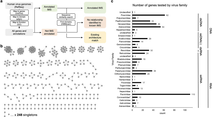
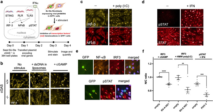
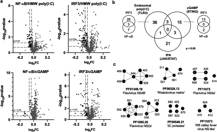
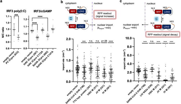
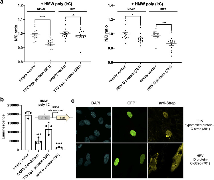
Suppression of the host innate immune response is a critical aspect of viral replication. Upon infection, viruses may introduce one or more proteins that inhibit key immune pathways, such as the type I interferon pathway. However, the ability to predict and evaluate viral protein bioactivity on targeted pathways remains challenging and is typically done on a single-virus or -gene basis. Here, we present a medium-throughput high-content cell-based assay to reveal the immunosuppressive effects of viral proteins. To test the predictive power of our approach, we developed a library of 800 genes encoding known, predicted, and uncharacterized human virus genes. We found that previously known immune suppressors from numerous viral families such as Picornaviridae and Flaviviridae recorded positive responses. These include a number of viral proteases for which we further confirmed that innate immune suppression depends on protease activity. A class of predicted inhibitors encoded by Rhabdoviridae viruses was demonstrated to block nuclear transport, and several previously uncharacterized proteins from uncultivated viruses were shown to inhibit nuclear transport of the transcription factors NF-κB and interferon regulatory factor 3 (IRF3). We propose that this pathway-based assay, together with early sequencing, gene synthesis, and viral infection studies, could partly serve as the basis for rapid in vitro characterization of novel viral proteins. IMPORTANCE Infectious diseases caused by viral pathogens exacerbate health care and economic burdens. Numerous viral biomolecules suppress the human innate immune system, enabling viruses to evade an immune response from the host. Despite our current understanding of viral replications and immune evasion, new viral proteins, including those encoded by uncultivated viruses or emerging viruses, are being unearthed at a rapid pace from large-scale sequencing and surveillance projects. The use of medium- and high-throughput functional assays to characterize immunosuppressive functions of viral proteins can advance our understanding of viral replication and possibly treatment of infections. In this study, we assembled a large viral-gene library from diverse viral families and developed a high-content assay to test for inhibition of innate immunity pathways. Our work expands the tools that can rapidly link sequence and protein function, representing a practical step toward early-stage evaluation of emerging and understudied viruses.

Keywords: expression systems; virus-host interactions.


Conflict of interest statement

The authors declare no conflict of interest.

Figures

FIG 1
FIG 1
Assembly of a viral gene library to test in immune suppression screens. (a) Overview of the bioinformatic workflow to generate a list of viral proteins for testing. We designated viral genes as (i) immunosuppressive (IMS) by gene ontology (GO) or Pfam search, (ii) predicted IMS based on sequence similarity or permissive hmmscan, or (iii) uncharacterized viral proteins. (b) Sequence similarity network of 605 viral proteins to test clusters of sequence-related proteins and singletons. (c) Distribution of 605 genes by viral family. One hundred ninety-five additional genes from Coronaviridae were tested during the COVID-19 pandemic and are not included in this figure.
FIG 2
FIG 2
Activation and suppression of antiviral innate immune pathways assayed in fibroblasts via medium-throughput fluorescence microscopy. (a) Innate immune system signaling pathways that respond to viral infection. (Top left) Transcription factors IRF3 and NF-κB are activated by the presence of nucleic acids in an inappropriate cellular compartment, signifying viral infection. Together, IRF3 and NF-κB activate expression of IFN-α and IFN-β, which are secreted and locally stimulate the JAK/STAT pathway. IRF3, NF-κB, and pSTAT1 are all nuclear translocated proteins during signaling. Viruses often encode proteins that disrupt these pathways, by directly or indirectly inhibiting nuclear translocation, or by causing degradation of the transcription factor. (Top right) The pathways can be initiated in vitro by addition of cGAMP (a second messenger), extracellular double-strand RNA (dsRNA), or IFN-α. (Bottom) Experimental workflow for testing virus genes for modulation of the IRF3, NF-κB, and pSTAT1 signaling pathways. BJ-5ta ΔcGAS cells are transiently transfected with a viral gene expression vector, treated with various stimuli, , stained with antibodies against IRF3, NF-κB, and/or pTyr701-STAT1, incubated with secondary antibodies, and then imaged. (b) Knockout of cGAS prevents transfection-mediated stimulation of IRF3 translocation while allowing downstream activation of IRF3 via cGAMP. Double-stranded DNA (dsDNA) in the cytoplasm activates cGAS to create the cyclic dinucleotide cGAMP, which then acts on STING to activate IRF3. Parental cGAS+ BJ-5ta cells show nuclear IRF3 translocation in response to transfected DNA and exogenous cGAMP, while BJ-5ta cells with a CRISPR cGAS knockout show an IRF3 response only after treatment with cGAMP. This allowed us to assay elements of the STING/IRF3 pathway without interference by transfected DNA. (c) BJ-5ta ΔcGAS cells treated with poly(I·C) show translocation of IRF3 and NF-κB into the nucleus. (d) IFN-α treatment caused translocation of cytoplasmic nuclear STAT1 and pSTAT1 into the nucleus. For this study, we chose to stain only for pSTAT1. (e) Images of BJ-5ta ΔcGAS cells that were cotransfected with plasmids encoding GFP and rotavirus A (RV-A) nonstructural protein 1 (NS1) and stained for NF-κB and IRF3. Cells were also cotransfected with plasmids encoding GFP and parainfluenza virus 5 (PIV5) V protein and stained for pSTAT1. Only transfected cells (arrows) show inhibition of nuclear localization or formation of phospho-STAT1. (f) Quantitative results for nuclear-to-cytoplasmic (N/C) ratio for IRF3 and pSTAT1 for BJ-5ta ΔcGAS cells expressing no viral protein, rotavirus A NS1, or PIV5 V protein. Some cells expressing these proteins were treated with cGAMP, HMW poly(I·C), or IFN-α to activate the immune signaling axes of interest. Viral inhibition of transcription factor translocation resulted in lower N/C ratio in the presence of stimuli. Data are means and standard deviations (SD) for >5 replicates. Statistical significance values were calculated with an unpaired t test. ****, P < 0.0001; ***, P < 0.001; **, P < 0.01.
FIG 3
FIG 3
(a) Volcano plots highlight hits with stringent (P < 0.05) and permissive (P < 0.1) cutoffs. Eight hundred genes (including the 195 coronavirus genes) are plotted. Cutoff values for log2 fold change (log2FC) and P values were determined by comparing the data to the corresponding no-treatment controls, which do not result in robust nuclear translocation of transcription factors. Results for no treatment controls and IFN-α-treated cells are depicted in Fig. S2 to S4. Raw source data are provided in Data Set S2. (b) Venn diagram depicting 79 stringent hits (P < 0.05) among 800 viral genes across the four assays. For the endosomal HMW poly(I·C)- and STING (cGAMP)-stimulated pathways, the additional Venn diagrams report the number of viral genes that inhibited IRF3 and/or NF-κB. (c) Examples of sequence-related positive innate immune inhibitors, grouped by Pfam domains, found in our screen and/or among inhibitors reported in the literature. The full list of permissive hits can be found in Data Set S3. Plots depicting log2FC and statistical significance for selected hits are compiled in Fig. S5.
FIG 4
FIG 4
(a) Consolidated imaging results for nuclear translocation of IRF3 in cGAMP-stimulated BJ-5ta ΔcGAS cells transfected with wild-type and variant viral genes. Cells expressing parechovirus (PV) 3Cpro exhibited lowered nuclear IRF3 intensity when stimulated with poly(I·C). Cells transfected with hepatovirus A (HAV) 3Cpro and salivirus A (SalVA) 3Cpro exhibited lower nuclear IRF3 intensity, while their corresponding active site mutants did not exhibit these effects. (b and c) Matrix proteins from viruses in the family Rhabdoviridae inhibit nuclear import and export of RFP probe in U2OS cells. In the presence of 447-nm light, a fusion protein with a LOV2 domain undergoes a conformational change that reveals either a nuclear localization signal (NLS) (b) or nuclear export signal (NES) (c) that increases or decreases nuclear RFP localization. Import and export rates were measured in single cells with a confocal microscope. These results demonstrated that M from vesicular stomatitis virus (VSIV), Isfahan virus (IV), and Jurona vesiculovirus (JV), which scored positive in our assay, inhibited nuclear import and export of proteins as expected. Torque teno virus 10 (TTV) hypothetical protein (381) and louping ill virus (LIV) NS2a (536) were also tested in the assay as additional negative controls. Data are means and SD for >5 replicates or cells. Statistical significance values were calculated with an unpaired t test. ****, P < 0.0001; ***, P < 0.001.
FIG 5
FIG 5
(a) High-content imaging results for nuclear translocation of IRF3 when BJ-5ta ΔcGAS cells express TTV hypothetical (hyp.) protein or HRV3 D protein. Cells transfected with these genes exhibited lower nuclear IRF3 intensity. Data are means and SD for >5 replicates. Statistical significance values were calculated with an unpaired t test. ****, P < 0.0001; ***, P < 0.001; *, P < 0.05. (b) Reduced luminescence readout was observed for the TTV hypothetical protein and HRV D protein in A549-Dual (InvivoGen) cells when HMW poly(I·C) was used to stimulate the cells. ISRE, interferon stimulated response element; luci, gene encoding luciferase. Data are means and SD for 3 replicates. Statistical significance values were calculated with an unpaired t test. ****, P < 0.0001; ***, P < 0.001; *, P < 0.05. (c) Cellular localization of the hypothetical TTV and HRV D proteins seen by immunofluorescence staining of C-terminally streptavidin-tagged proteins of interest expressed in BJ-5ta ΔcGAS cells. Results of SDS-PAGE of the overexpressed and purified viral proteins are shown in Fig. S7.

Similar articles

Cited by

References

    1. Crowl JT, Gray EE, Pestal K, Volkman HE, Stetson DB. 2017. Intracellular nucleic acid detection in autoimmunity. Annu Rev Immunol 35:313–336. doi:10.1146/annurev-immunol-051116-052331. - DOI - PMC - PubMed
    1. Lee H-C, Chathuranga K, Lee J-S. 2019. Intracellular sensing of viral genomes and viral evasion. Exp Mol Med 51:1–13. doi:10.1038/s12276-019-0299-y. - DOI - PMC - PubMed
    1. Nelemans T, Kikkert M. 2019. Viral innate immune evasion and the pathogenesis of emerging RNA virus infections. Viruses 11:961. doi:10.3390/v11100961. - DOI - PMC - PubMed
    1. Katze MG, He Y, Gale M. 2002. Viruses and interferon: a fight for supremacy. Nat Rev Immunol 2:675–687. doi:10.1038/nri888. - DOI - PubMed
    1. Versteeg GA, García-Sastre A. 2010. Viral tricks to grid-lock the type I interferon system. Curr Opin Microbiol 13:508–516. doi:10.1016/j.mib.2010.05.009. - DOI - PMC - PubMed

Publication types

LinkOut - more resources