. 2022 Apr;36(4):e22262.
doi: 10.1096/fj.202101275R.

NR1D1 downregulation in astrocytes induces a phenotype that is detrimental to cocultured motor neurons

Affiliations

NR1D1 downregulation in astrocytes induces a phenotype that is detrimental to cocultured motor neurons

Kelby M Killoy et al. FASEB J. 2022 Apr.

Abstract

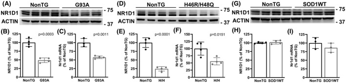
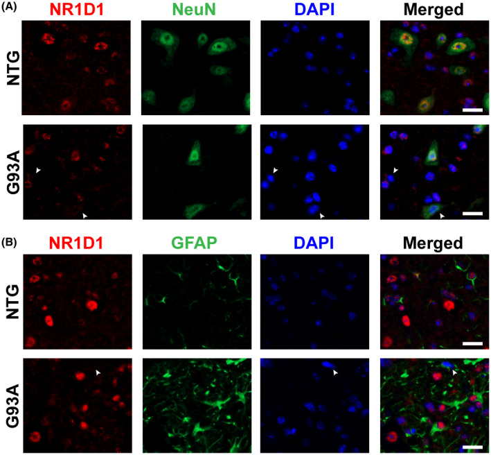
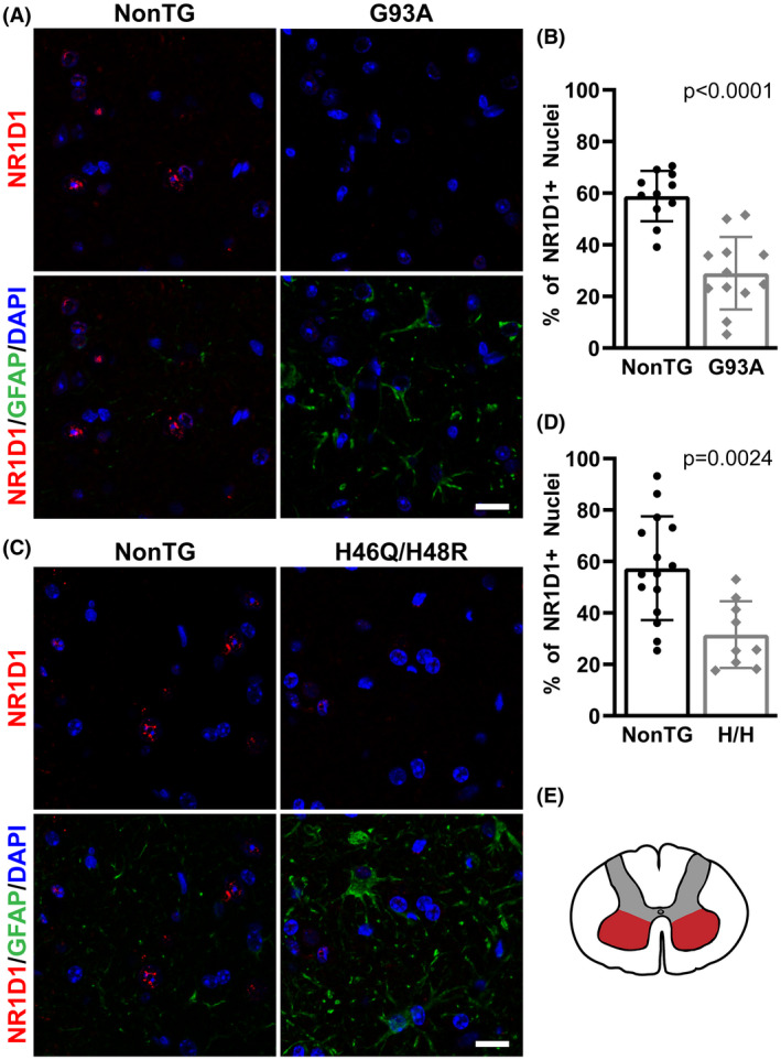
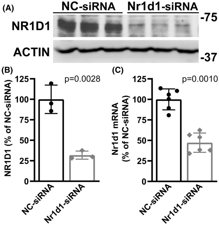
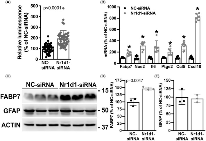
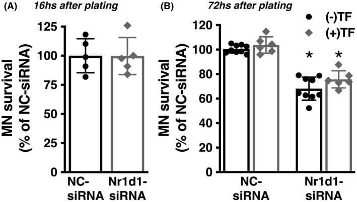
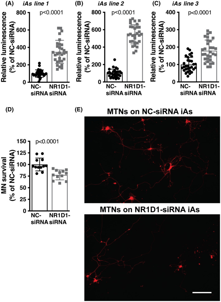
Nuclear receptor subfamily 1 group D member 1 (NR1D1, also known as Rev-erbα) is a nuclear transcription factor that is part of the molecular clock encoding circadian rhythms and may link daily rhythms with metabolism and inflammation. NR1D1, unlike most nuclear receptors, lacks a ligand-dependent activation function domain 2 and is a constitutive transcriptional repressor. Amyotrophic lateral sclerosis (ALS) is the most common adult-onset motor neuron disease, caused by the progressive degeneration of motor neurons in the spinal cord, brain stem, and motor cortex. Approximately 10%-20% of familial ALS is caused by a toxic gain-of-function induced by mutations of the Cu/Zn superoxide dismutase (SOD1). Dysregulated clock and clock-controlled gene expression occur in multiple tissues from mutant hSOD1-linked ALS mouse models. Here we explore NR1D1 dysregulation in the spinal cord of ALS mouse models and its consequences on astrocyte-motor neuron interaction. NR1D1 protein and mRNA expression are significantly downregulated in the spinal cord of symptomatic mice expressing mutant hSOD1, while no changes were observed in age-matched animals overexpressing wild-type hSOD1. In addition, NR1D1 downregulation in primary astrocyte cultures induces a pro-inflammatory phenotype and decreases the survival of cocultured motor neurons. NR1D1 orchestrates the cross talk between physiological pathways identified to be disrupted in ALS (e.g., metabolism, inflammation, redox homeostasis, and circadian rhythms) and we observed that downregulation of NR1D1 alters astrocyte-motor neuron interaction. Our results suggest that NR1D1 could be a potential therapeutic target to prevent astrocyte-mediated motor neuron toxicity in ALS.

Keywords: NR1D1; Rev-erbα; amyotrophic lateral sclerosis; astrocytes; inflammation; motor neurons.


Figures

FIGURE 1
FIGURE 1
Decreased NR1D1 expression in the spinal cord of mutant hSOD1‐linked mouse models. (A, D, and G) Western blot analysis of NR1D1 expression in the lumbar spinal cord of age‐matched nontransgenic (NonTG) and symptomatic hSOD1G93A (G93A, about 140 days old) mice (A) or symptomatic hSOD1H46R/H48Q (H46R/H48Q, about 210 days old) mice (D) or age‐matched control hSOD1WT mice (about 210 days old) (G). (B, E, and H) Quantification of NR1D1 protein levels shown in A, D, and G, respectively. NR1D1 expression was quantified, normalized by ACTIN levels and expressed as a percentage of age‐matched NonTG mice (each lane corresponds to a different animal, mean ± SD). Both bands shown in the NR1D1 western blot were used for intensity quantification. (C, F, and I) Nr1d1 mRNA levels in the lumbar spinal cord of age‐matched NonTG and symptomatic G93A (C), or symptomatic H46R/H48Q (F), or age‐matched hSOD1WT (I) mice. Nr1d1 mRNA levels were determined by real‐time PCR and corrected by Actin mRNA levels (n = 3 or 4 mice, mean ± SD)
FIGURE 2
FIGURE 2
Decreased NR1D1 expression in the spinal cord of symptomatic hSOD1G93A mice. (A) Representative images showing NR1D1 (red) and NeuN (green) immunofluorescence in the ventral horn of the lumbar spinal cord from symptomatic hSOD1G93A (G93A) and age‐matched nontransgenic (NTG) mice. Nuclei were counterstained with DAPI (blue). The arrowheads point to NR1D1‐negative nuclei from NeuN‐negative and NeuN‐positive cells. Scale bar: 20 μm. (B) Representative images showing NR1D1 (red) and GFAP (green) immunofluorescence in the ventral horn of the lumbar spinal cord from symptomatic G93A and age‐matched NonTG mice. Nuclei were counterstained with DAPI (blue). The arrowhead points to a NR1D1‐negative, GFAP‐positive nucleus. Scale bar: 20 μm
FIGURE 3
FIGURE 3
Decreased NR1D1 immunostaining in the spinal cord of mutant hSOD1‐linked mouse models. (A and C) Representative images showing GFAP (green) and NR1D1 (red) immunofluorescence in the ventral horn of the lumbar spinal cord from symptomatic hSOD1G93A (G93A), hSOD1H46R/H48Q (H46R/H48Q), and age‐matched nontransgenic (NonTG) mice. Nuclei were counterstained with DAPI (blue). Scale bar: 20 μm. (B, D) Quantification of NR1D1‐positive nuclei in the ventral horn of the lumbar spinal cord from mice in A and C. Data are expressed as the percentage of NR1D1‐positive nuclei from the total number of nuclei analyzed for each genotype (n = 4 or 5 mice per group and two to four images per animal; mean ± SD). (E) Schematic representation of the spinal cord indicating the area (red) in which the analysis was performed
FIGURE 4
FIGURE 4
NR1D1 silencing in primary nontransgenic spinal cord astrocytes. Confluent primary astrocyte cultures from nontransgenic mice were treated with a negative control siRNA (NC‐siRNA) or an Nr1d1‐siRNA for 48 h. (A) Western bolt analysis of NR1D1 protein levels in astrocytes following siRNA treatment. (B) Quantification of NR1D1 protein levels shown in A. NR1D1 expression was quantified, normalized by ACTIN levels, and expressed as a percentage of NC‐siRNA‐treated cultures (mean ± SD). (C) Nr1d1 mRNA levels in Nr1d1‐siRNA‐treated astrocytes. Nr1d1 mRNA levels were determined by real‐time PCR and corrected by Actin mRNA levels (mean ± SD)
FIGURE 5
FIGURE 5
NR1D1 downregulation induces a pro‐inflammatory phenotype in nontransgenic astrocytes. (A) Relative luminescence produced by a firefly luciferase expressed under an NF‐κB‐driven promoter 48 h after NC‐siRNA or Nr1d1‐siRNA treatment of nontransgenic astrocytes. Relative firefly luciferase luminescence was corrected by the amount of Renilla luciferase activity controlled by a constitutive promoter and expressed as a percentage of NC‐siRNA‐treated control cells. (B) Fabp7, Nos2, Il6, Ptgs2, Ccl5, and Cxcl10 mRNA levels in astrocytes 48 h after NC‐siRNA or Nr1d1‐siRNA treatment. mRNA levels were determined by real‐time PCR and corrected by Actin mRNA levels. Data are expressed as a percentage of NC‐siRNA‐treated control cells. (C) Western blot analysis of FABP7 and GFAP expression in astrocytes treated as in A. (D, E) Quantification of the images shown in (C). FABP7 and GFAP protein levels were quantified, corrected by ACTIN levels and expressed as a percentage of NC‐siRNA‐treated control cells. For all graph panels, data are expressed as mean ± SD (*< .05, significantly different from NC‐siRNA‐treated cells)
FIGURE 6
FIGURE 6
The phenotype induced by NR1D1 downregulation in primary nontransgenic astrocytes is detrimental to cocultured motor neurons. (A) Spinal cord nontransgenic astrocytes were treated with NC‐siRNA or Nr1d1‐siRNA. Forty‐eight hours later embryonic motor neurons were plated on top. Motor neuron (MN) survival was determined 16 h later (data obtained from two independent coculture experiments, two or three replicas per coculture). (B) The same coculture setup as in A but motor neuron survival was determined 72 h later. The addition of trophic factors (TF: GDNF 10 ng/ml and BDNF 0.1 ng/ml) to the coculture does not prevent the motor neuron loss induced by Nr1d1‐siRNA treated astrocytes. Cocultures were treated at the time of motor neuron plating with (+TF) or without (−TF) trophic factors, and motor neuron survival was determined 72 h later. Data were obtained from at least three independent coculture experiments, two or three replicas per coculture. For all panels, data are expressed as percentage of NC‐siRNA control mean ± SD (*< .05, significantly different from NC‐siRNA‐treated cells)
FIGURE 7
FIGURE 7
NR1D1 downregulation in transdifferentiated human astrocytes is detrimental to cocultured motor neurons. (A, B, C) Relative luminescence produced by a firefly luciferase expressed under an NF‐κB‐driven promoter 48 h after NC‐siRNA or Nr1d1‐siRNA treatment of three different transdifferentiated human astrocytes (iAs) lines derived from control (nondisease) healthy subjects. Relative firefly luciferase luminescence was corrected by the amount of Renilla luciferase activity controlled by a constitutive promoter and expressed as a percentage of NC‐siRNA‐treated cells. (D) Control iAs (line 1) were treated with NC‐siRNA or Nr1d1‐siRNA. Forty‐eight hours later embryonic motor neurons were plated on top. Motor neuron (MN) survival was determined 72 h later. Data were obtained from four independent coculture experiments, three replicas per coculture. For all panels, data are expressed as percentage of NC‐siRNA control (mean ± SD). (E) Representative images showing motor neurons cultured on the top of NC‐siRNA or Nr1d1‐siRNA‐treated iAs. Scale bar: 20 μm

Similar articles

Cited by

References

    1. Raghuram S, Stayrook KR, Huang P, et al. Identification of heme as the ligand for the orphan nuclear receptors REV‐ERBalpha and REV‐ERBbeta. Nat Struct Mol Biol. 2007;14:1207‐1213. - PMC - PubMed
    1. Yin L, Wu N, Curtin JC, et al. Rev‐erbalpha, a heme sensor that coordinates metabolic and circadian pathways. Science. 2007;318:1786‐1789. - PubMed
    1. Renaud JP, Harris JM, Downes M, Burke LJ, Muscat GE. Structure‐function analysis of the Rev‐erbA and RVR ligand‐binding domains reveals a large hydrophobic surface that mediates corepressor binding and a ligand cavity occupied by side chains. Mol Endocrinol. 2000;14:700‐717. - PubMed
    1. Harding HP, Lazar MA. The orphan receptor Rev‐ErbA alpha activates transcription via a novel response element. Mol Cell Biol. 1993;13:3113‐3121. - PMC - PubMed
    1. Harding HP, Lazar MA. The monomer‐binding orphan receptor Rev‐Erb represses transcription as a dimer on a novel direct repeat. Mol Cell Biol. 1995;15:4791‐4802. - PMC - PubMed

Publication types

MeSH terms

Substances