. 2022 May;9(15):e2105408.
doi: 10.1002/advs.202105408. Epub 2022 Mar 23.

An Injectable Dual-Function Hydrogel Protects Against Myocardial Ischemia/Reperfusion Injury by Modulating ROS/NO Disequilibrium

Affiliations

An Injectable Dual-Function Hydrogel Protects Against Myocardial Ischemia/Reperfusion Injury by Modulating ROS/NO Disequilibrium

Tian Hao et al. Adv Sci (Weinh). 2022 May.

Abstract

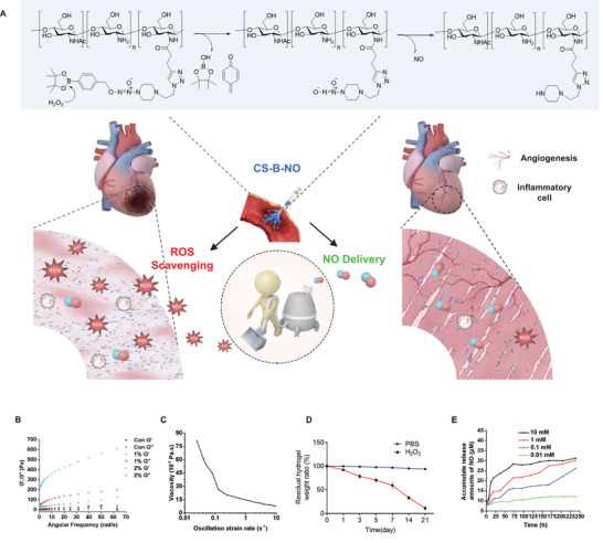
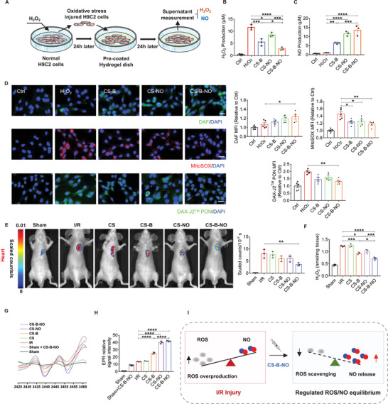
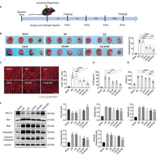
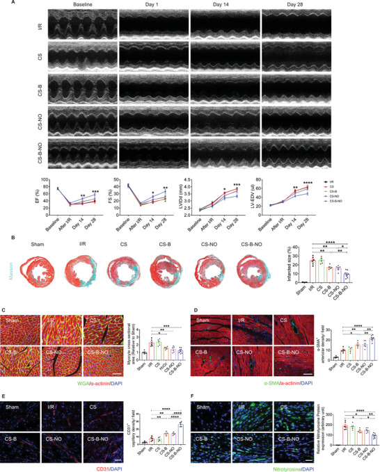
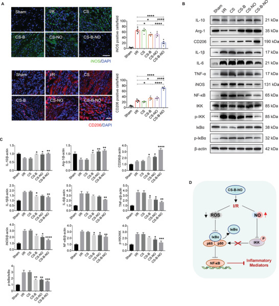
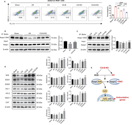
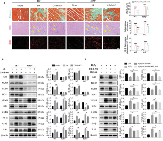
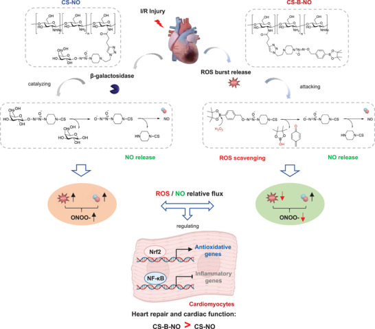
Acute myocardial infarction (MI) is the leading cause of death worldwide. Exogenous delivery of nitric oxide (NO) to the infarcted myocardium has proven to be an effective strategy for treating MI due to the multiple physiological functions of NO. However, reperfusion of blood flow to the ischemic tissues is accompanied by the overproduction of toxic reactive oxygen species (ROS), which can further exacerbate tissue damage and compromise the therapeutic efficacy. Here, an injectable hydrogel is synthesized from the chitosan modified by boronate-protected diazeniumdiolate (CS-B-NO) that can release NO in response to ROS stimulation and thereby modulate ROS/NO disequilibrium after ischemia/reperfusion (I/R) injury. Furthermore, administration of CS-B-NO efficiently attenuated cardiac damage and adverse cardiac remodeling, promoted repair of the heart, and ameliorated cardiac function, unlike a hydrogel that only released NO, in a mouse model of myocardial I/R injury. Mechanistically, regulation of the ROS/NO balance activated the antioxidant defense system and protected against oxidative stress induced by I/R injury via adaptive regulation of the Nrf2-Keap1 pathway. Inflammation is then reduced by inhibition of the activation of NF-κB signaling. Collectively, these results show that this dual-function hydrogel may be a promising candidate for the protection of tissues and organs after I/R injury.

Keywords: inflammation; ischemia/reperfusion injury; nitric oxide; oxidative stress; reactive oxygen species/nitric oxide equilibrium.


Conflict of interest statement

The authors declare no conflict of interest.

Figures

Figure 1
Figure 1
Preparation and characterization of the injectable chitosan hydrogel with ROS‐responsive NO‐releasing function (CS‐B‐NO). A) Schematic illustration of the treatment of I/R heart injury by CS‐B‐NO. B) Rheology properties of CS‐B‐NO was evaluated by frequency sweep experiments at a constant strain of 1%. C) Oscillatory strain sweeps of CS‐B‐NO. D) In vitro degradation of CS‐B‐NO hydrogel in the presence or absence of H2O2 (100 µm), and the residual weight of dried hydrogel was measured at different intervals. Data were presented as mean ± SEM (n = 3 individual experiments). E) In vitro generation of NO from the CS‐B‐NO hydrogel (5mg) in 5 mL of PBS buffer (pH 7.4) with hydrogen peroxide of various concentrations. The cumulative releasing amount of NO was calculated. Data were presented as mean ± SEM (n = 3 individual experiments).
Figure 2
Figure 2
CS‐B‐NO hydrogel modulated the myocardial ROS/NO disequilibrium after I/R injury by simultaneously releasing NO and scavenging ROS. A) Schematic illustration of H9C2 cells oxidative stress model of H2O2‐induced oxidative. B,C) Quantitative analysis of H2O2 and NO release with different treatments one day after oxidative stress stimuli (n = 3 individual experiments). D) After 24 h of H2O2 stimulation followed with different hydrogel treatments, H9C2 cells were stained with DAF‐FM DA, MitoSOX Red, and DAX‐J2 PON Green 99 to detect the production of NO, superoxide, and peroxynitrite (ONOO), respectively. The mean fluorescence intensity was quantified respectively. (scale bar = 20 µm, n = 5 individual experiments). E) In vivo imaging of H2O2 and quantitative analysis of fluorescence intensity. F) Quantitative analysis on the H2O2 level of heart homogenate after different treatments at one‐day post‐surgery (n = 3). G) Representative EPR spectra reflecting NO generation in the presence of (DETC)2Fe. H) NO levels were determined by quantitation of (DETC)2Fe‐NO complex using 2,2,5,5‐tetramethyl piperidine 1‐oxyl (TEMPO), n = 3 animals for each group. I) Schematic illustration demonstrating that CS‐B‐NO hydrogel treatment restored the local balance of ROS/NO in infarcted myocardium after I/R injury. Data are expressed as mean ± SEM. Significant differences were detected by one‐way ANOVA with Tukey's multiple comparisons test, *p<0.05, **p<0.01, ***p<0.001, ****p<0.0001.
Figure 3
Figure 3
CS‐B‐NO hydrogel ameliorated myocardial I/R injury in mice at early stage. A) Experimental schedule for the treatment of I/R injury via intramyocardial injection of CS‐B‐NO hydrogel. B) TTC staining (scale bar = 2 mm), was performed to evaluate the ischemia injured myocardium via quantifying the infarct area (n = 6 animals per group). C) Representative images of TUNEL staining at 1 day after surgery to detect apototic nucleus (scale bar = 100 µm), and positive staining nucleus was quantified (n = 5–6 animals per group). D,E) Serum cTnT, LDH levels were analyzed, respectively (n = 3–6 animals per group). F) Representative Western blot images and quantitative data showing the expression of BCL2, Bax, Bad, Caspase3, cleaved Caspase3 in the hearts at 3 days after different treatments (n = 5 animals per group). Data are expressed as mean ± SEM. Significant differences were detected by one‐way ANOVA with Tukey's multiple comparisons test, *p<0.05, **p<0.01, ***p<0.001, and ****p<0.0001.
Figure 4
Figure 4
CS‐B‐NO hydrogel improved heart function, stimulated angiogenesis, and reduced adverse cardiac remodeling in mice after myocardial I/R injury. A) Cardiac echo measurement was performed at different time‐points post‐surgery, and cardiac function indicators of left ventricular‐ejection fraction (LV‐EF), left ventricular‐fractional shortening (LV‐FS), left ventricular internal diameter at end diastole (LVIDd), and left ventricular end‐diastolic volume (LV‐EDV) were evaluated accordingly. Data are expressed as mean ± SEM, n = 5–8 animals per group. Significant differences were detected by two‐way ANOVA with Tukey's multiple comparisons test, *p<0.05, **p<0.01, ***p<0.001, and ****p<0.0001. The hearts were collected for histological analyses at 28 days post‐surgery. B) Masson's trichrome (scale bar = 400 µm) was performed, and infarcted size was quantified accordingly. C) Representative images of WGA immunofluorescence staining and the cross‐section area of cardiomyocytes were measured (scale bar = 50 µm). D) Representative images of α‐SMA immunofluorescence staining and number of the α‐SMA positive arterioles were counted (scale bar = 100 µm). E) Representative images of CD31 immunofluorescence staining and number of the CD31 positive capillaries were quantified (scale bar = 50 µm). F) Representative images of nitrotyrosine immunostaining and quantification of the relative nitrotyrosine protein expression (scale bar = 25 µm). Data are expressed as mean ± SEM, n = 5–8 animals for each group. Significant differences were detected by one‐way ANOVA with Tukey's multiple comparisons test, *p<0.05, **p<0.01, ***p<0.001, and ****p<0.0001.
Figure 5
Figure 5
CS‐B‐NO hydrogel inhibited NF‐κB inflammatory signaling pathway by regulating local ROS/NO balance after myocardial I/R injury. A) Macrophage polarization was detected by immunofluorescence staining targeting iNOS and CD206, the markers of M1 and M2 macrophage phenotype, respectively (scale bar = 50 µm). B,C) Representative Western blot images and quantitative data showing the expression of IL‐10, Arg‐1, CD206, IL‐1β, IL‐6, TNF‐α, iNOS, NF‐κB, IKK, p‐IKK, IκBα, and p‐IκBα, in the hearts after I/R injury with different treatments. Data are expressed as mean ± SEM, n = 5 animals for each group, Significant differences were detected by one‐way ANOVA with Tukey's multiple comparisons tests, *p<0.05, **p<0.01, ***p<0.001, and ****p<0.0001. D) Schematic illustration summarizing the mechanism of CS‐B‐NO hydrogel on inhibiting the NF‐κB signaling pathway after I/R injury.
Figure 6
Figure 6
CS‐B‐NO hydrogel ameliorated oxidative stress induced by ischemic/reperfusion injury via adaptive regulation of the Nrf2‐Keap1 signaling pathway. A) Flow cytometric analysis was performed to quantify the number of ROS positive cells in myocardial tissue at 1‐day post‐surgery (n = 3 animals per group). B) Mice after I/R injury were treated by myocardial injection of CS‐B‐NO hydrogel, and 7 days post‐surgery Keap1 S‐nitrosylation of the heart tissues was detected by biotin switch (## p<0.01 vs Sham, *p<0.05 vs I/R, n = 5 animals per group). C) H9C2 cells were stimulated with H2O2 for 24 h and followed by various treatments. Keap1 S‐nitrosylation was further detected by biotin switch assay. (#### p <0.0001 vs Ctrl, *p<0.05, ***p<0.001 vs H2O2 stimulated group, n = 5 individual experiments) D) Representative Western blot images and quantitative data showing the expression of Nrf2, SOD1, SOD2, HO‐1, HO‐2, NQO‐1, and CAT in mice hearts in mice hearts after I/R injury with different treatments (n = 5 animals per group). Data are expressed as mean ± SEM, Significant differences were detected by one‐way ANOVA with Tukey's multiple comparisons test, *p<0.05, **p<0.01, ***p<0.001, and ****p<0.0001. E) Schematic illustration summarizing the mechanism of CS‐B‐NO hydrogel on activation of the Nrf2 pathway against oxidative stress via enhancing Keap1 S‐nitrosylation.
Figure 7
Figure 7
Nrf2 deficiency abrogates the cardioprotection of CS‐B‐NO hydrogel in mice after ischemic/reperfusion injury. A) H&E (scale bar = 100 µm) and DHE staining (scale bar = 50 µm) were performed in wild‐type C57BL/6 mice (WT) and Nrf2 deficient mice (Nrf2−/−) at 3 days post‐surgery. Myocardium fibrosis was indicated by yellow arrows. Masson's trichrome was performed at 28 days post‐surgery (scale bar = 100 µm). The infarction area, inflammatory cell infiltration, and DHE positive staining were quantified, respectively. (#### p<0.0001 vs Sham, *p<0.05, **p<0.01, ***p<0.001, ****p<0.0001 vs I/R, n = 5 animals per group). B) WT and Nrf2−/− mice after I/R surgery were treated by CS‐B‐NO hydrogel. At 7 days post‐surgery, western blotting analysis was performed to quantify the expression level of Nrf2, NQO1, SOD1, HO‐1, NF‐κB, IκBα, p‐IκBα, TNFα, IL‐1β, and IL‐6 in hearts (n = 5 animals per group). C) H9C2 cells were treated with 5 µm ML385 to inhibit Nrf2 expression in vitro. 200 µm H2O2 was introduced to stimulate H9C2 cells with or without Nrf2 inhibition for 24 h, followed by administration of CS‐B‐NO hydrogel for 48 h. Protein expression level of Nrf2, NQO1, SOD1, HO‐1, NF‐κB, IκBα, p‐IκBα, TNFα, IL‐1β, and IL‐6 in H9C2 cells was evaluated by Western blotting analysis (n = 5 individual experiments). Data are expressed as mean ± SEM. Significant differences were detected by one‐way ANOVA with Tukey's multiple comparisons test, *p<0.05, **p<0.01, ***p<0.001, and ****p<0.0001.
Figure 8
Figure 8
Mechanistic summary of CS‐B‐NO hydrogel for myocardial repair after ischemic/reperfusion injury by a head‐to‐head comparison with CS‐NO.

Similar articles

Cited by

References

    1. a) Schwalm J. D., McKee M., Huffman M. D., Yusuf S., Circulation 2016, 133, 742; - PMC - PubMed
    2. b) Li Y., Chen B., Yang X., Zhang C., Jiao Y., Li P., Liu Y., Li Z., Qiao B., Bond Lau W., Ma X. L., Du J., Circulation 2019, 140, 751. - PubMed
    1. a) Finn A. V., Nakano M., Narula J., Kolodgie F. D., Virmani R., Arterioscler., Thromb., Vasc. Biol. 2010, 30, 1282; - PubMed
    2. b) Rentrop K. P., Feit F., Am. Heart J. 2015, 170, 971. - PubMed
    1. Farah C., Michel L. Y. M., Balligand J. L., Nat. Rev. Cardiol. 2018, 15, 292. - PubMed
    1. Liu X., Huang Y., Pokreisz P., Vermeersch P., Marsboom G., Swinnen M., Verbeken E., Santos J., Pellens M., Gillijns H., Van de Werf F., Bloch K. D., Janssens S., J. Am. Coll. Cardiol. 2007, 50, 808. - PubMed
    1. a) Cohen M. V., Yang X. M., Downey J. M., Cardiovasc. Res. 2006, 70, 231; - PubMed
    2. b) Weerateerangkul P., Chattipakorn S., Chattipakorn N., Med. Sci. Monit. 2011, 17, RA44. - PMC - PubMed

Publication types

MeSH terms

LinkOut - more resources