. 2022 May;9(15):e2200063.
doi: 10.1002/advs.202200063. Epub 2022 Mar 23.

Development of Neutralization Breadth against Diverse HIV-1 by Increasing Ab-Ag Interface on V2

Affiliations

Development of Neutralization Breadth against Diverse HIV-1 by Increasing Ab-Ag Interface on V2

Nan Gao et al. Adv Sci (Weinh). 2022 May.

Abstract

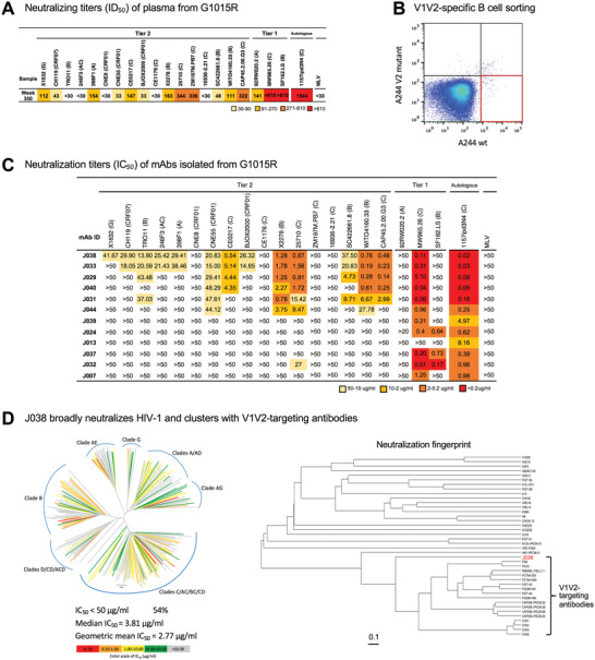
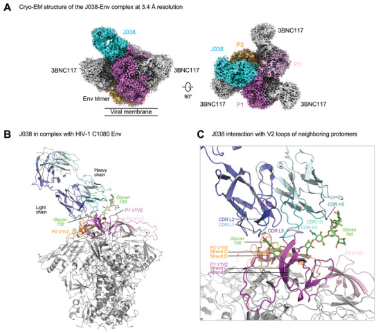
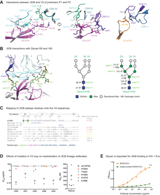
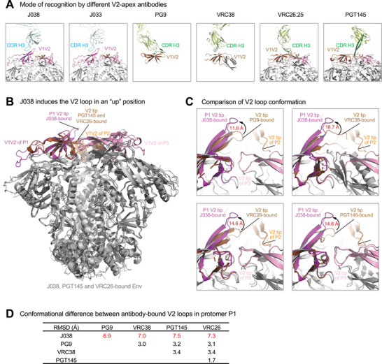
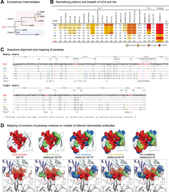
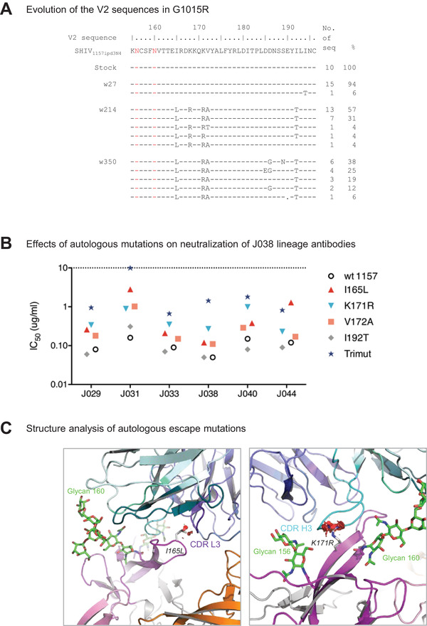
Understanding maturation pathways of broadly neutralizing antibodies (bnAbs) against HIV-1 can be highly informative for HIV-1 vaccine development. A lineage of J038 bnAbs is now obtained from a long-term SHIV-infected macaque. J038 neutralizes 54% of global circulating HIV-1 strains. Its binding induces a unique "up" conformation for one of the V2 loops in the trimeric envelope glycoprotein and is heavily dependent on glycan, which provides nearly half of the binding surface. Their unmutated common ancestor neutralizes the autologous virus. Continuous maturation enhances neutralization potency and breadth of J038 lineage antibodies via expanding antibody-Env contact areas surrounding the core region contacted by germline-encoded residues. Developmental details and recognition features of J038 lineage antibodies revealed here provide a new pathway for elicitation and maturation of V2-targeting bnAbs.

Keywords: antibody evolution; broadly neutralizing antibodies; humoral immunity; nonhuman primates; simian/human immunodeficiency virus; virus variation.


Conflict of interest statement

The authors declare no conflict of interest.

Figures

Figure 1
Figure 1
Isolation of broadly neutralizing antibodies from G1015R. A) Neutralization breadth of week 350 plasma post infection from G1015R infected with SHIV1157ipd3N4. Data are shown as ID50 and neutralization potency is color coded. B) Fluorescence‐activated cell sorting (FACS) plots of sorted V1V2‐specific single B cells from week 350 peripheral blood mononuclear cells (PBMCs) from G1015R that bind d11_A244_gp120_SavBV421 but not glycan knockout mutant N156_160K_d11_A244_gp120_SavAF647 (red box). C) Neutralization profiles of 12 mAbs were determined with 17 tier 2 viruses, three tier 1 viruses and the autologous virus. Murine leukemia virus (MLV) was used as a nonspecific control. Neutralization titers are represented as IC50 (µg mL−1). X2 test was used to compare neutralization breadth of bnAbs using 17 tier 2 viruses. D) J038 neutralizes 112 strains of a 208‐virus panel (54%). Stains in HIV‐1 phylogenetic tree are colored according to their respective J038 potency based on IC50. Median and geometric mean titers are calculated only for samples with IC50<50 µg mL−1. Neutralization profile of J038 shows that its clusters with V1V2‐targeting antibodies.
Figure 2
Figure 2
Antibody J038 targets the V2‐apex of the HIV‐1 Env. A) Overall cryo‐EM structure of the J038 Fab‐Env complex, with EM reconstruction density shown in gray. The CD4bs antibody 3BNC117 was used to aid the resolution. Protomers of the trimer are labeled as P1, P2, and P3. B) Antibody J038 recognition of the HIV‐1 Env. Heavy and light chains of J038 are colored in cyan and blue, respectively. The V1V2 loops of the Env are colored magenta, pink and orange for the three protomers, respectively. Glycans N156 and N160 on P1 interacting with J038 are colored green. The CD4bs antibody 3BNC117 was omitted for clarify. C) J038 interaction with V2‐loops of the neighboring protomers P1 and P2. The complementarity determining regions (CDR) of J038 are shown in shades of cyan and blue. The strands of V2 that interacted with J038 are labeled.
Figure 3
Figure 3
J038 interaction with V2‐apex of HIV‐1 Env. A) J038 interaction of V2 of protomers P1 (left and middle) and P2 (right). Key interacting residues are shown in sticks and numbered with hydrogen bonds shown in dashed lines. B) Interactions between J038 and glycans on Asn156 and Asn160. Key residues that interact with glycans are shown in sticks and numbered with hydrogen bonds shown in dashed lines. A schematic for each glycan is shown next to the cryo‐EM structure with GlcNAc as blue squares and mannose residues as green circles. Buried surface areas (Å2) and hydrogen bonds with J038 for each glycan moieties are labeled. C) Alignment of V2 sequences from HIV‐1 strains, shown relative to their neutralization sensitivities. J038 contacts are marked as g (glycan contacts), s (side chain contacts) and b (both side chain and main chain contacts). D) Effects of mutations in V2 on neutralization potency of the J038 lineage antibodies. Mutations removing the Asn160 glycosylation site on V2 make the virus resistant to J038 lineage antibodies. (E) Deglycosylation reduces binding of HIV‐1 BG505 Env to J038. Paired t‐test was used for statistical analysis of 8‐seiral dilution ELISA binding data. Data were presented with mean ± SD. ** = p <0.01.
Figure 4
Figure 4
J038 uses a different mode of gp120 recognition. A) Comparison of heavy chain CDR H3 interactions with the V2‐apex of HIV‐1 Env. CDR H3s of antibodies (J008, J033, PG9, VRC38, VRC26, and PGT145) are shown in cyan or dark green. The V1V2 loops are colored as in Figure 1 with that of protomer 1 in the PG9‐, VRC38‐, VRC26‐, and PGT145‐bound shown in brown for comparison. B) Binding of J038 induces the strands B and C in P1 V2‐loop in an “up” conformation. The structures of J038, VRC26 and PGT145 in complex with HIV‐1 Env are superposed and the V1V2 loops are colored the same as in (A). C) Comparison of conformation of the P1 V2‐loops in J038, PG9‐, VRC38‐, VRC26‐, and PGT145‐bound structures, distance shown are between Cα atoms of Glu165 at the tip of loop between strands B and C. D) Cα‐RMSD between antibody‐bound V2 loop strands B and C (residues 154–177) in P1.
Figure 5
Figure 5
Development of neutralization breadth of the J033 lineage Abs. A) The maximum likelihood phylogenetic tree of the J038 lineage sequences. UCA and IAs at evolutionary nodes were computationally inferred. Two different clades are shaded with different colors. B) Neutralization profiles of UCA and IAs were determined with 17 tier 2 viruses, three tier 1 viruses and the autologous virus. Murine leukemia virus (MLV) was used as a nonspecific control. Neutralization titers are represented as IC50 (µg mL−1). X2 test was used to compare neutralization breadth of bnAbs using 17 tier 2 viruses. C) Sequence alignment of the UCA, intermediates and mature J038 Ab sequences. Heavy chain shown in the upper set and light chain shown in the lower set. Env contacts are shown as g (glycan contacts), s (side chain contacts), and b (both side chain and main chain contacts). D) Mapping of the development of J038 paratope residues on models of inferred intermediate antibodies. Paratope residues directly from UCA were colored red, those from IA5, IA3, and IA2 were colored green, light blue and olive, respectively. Paratope surface areas estimated from UCA and increase from intermediate antibodies are indicated. Top panels: on antibody. Bottom panels: interface of antibody and V2‐apex.
Figure 6
Figure 6
Limited mutations accumulated at the J038‐Env contact site during infection. A) Identification of neutralization escape mutations in the env gene from longitudinal samples in G1015R. Amino acid sequences of V2 from the viral stock and weeks 27, 214, and 350 post infection were compared to the SHIV1157ipd3N4 reference sequence. The amino acid positions are based on the HIV‐1 HXB2 sequence. B) Effects of the mutations in V2 on neutralization potency of the J038 lineage antibodies. I165L, K171R and V172A render the virus resistant to J038 lineage antibodies at various levels. C) Structural basis for neutralization resistance by the I165L and K171R mutations. Leu165 and Arg171 are shown in stick representation. Potential clashes with CDR l3 and CDR H3 caused by these mutations are indicated as red discs.

Similar articles

Cited by

References

    1. UNAIDS , UNAIDS Fact‐sheet 2020. https://www.unaids.org/en/resources/fact-sheet (accessed: September, 2021).
    1. Rerks‐Ngarm S., Pitisuttithum P., Nitayaphan S., Kaewkungwal J., Chiu J., Paris R., Premsri N., Namwat C., de Souza M., Adams E., Benenson M., Gurunathan S., Tartaglia J., McNeil J. G., Francis D. P., Stablein D., Birx D. L., Chunsuttiwat S., Khamboonruang C., Thongcharoen P., Robb M. L., Michael N. L., Kunasol P., Kim J. H., Investigators M.‐T., N. Engl. J. Med. 2009, 361, 2209. - PubMed
    1. Gray G. E., Bekker L. G., Laher F., Malahleha M., Allen M., Moodie Z., Grunenberg N., Huang Y., Grove D., Prigmore B., Kee J. J., Benkeser D., Hural J., Innes C., Lazarus E., Meintjes G., Naicker N., Kalonji D., Nchabeleng M., Sebe M., Singh N., Kotze P., Kassim S., Dubula T., Naicker V., Brumskine W., Ncayiya C. N., Ward A. M., Garrett N., Kistnasami G., et al., N. Engl. J. Med. 2021, 384, 1089. - PubMed
    1. Malim M. H., Emerman M., Cell 2001, 104, 469. - PubMed
    1. a) Burton D. R., Mascola J. R., Nat. Immunol. 2015, 16, 571; - PMC - PubMed
    2. b) Kwong P. D., Mascola J. R., Immunity 2018, 48, 855; - PubMed
    3. c) Haynes B. F., Burton D. R., Mascola J. R., Sci. Transl. Med. 2019, 11, aaz2686. - PMC - PubMed

Publication types

Substances

LinkOut - more resources