. 2022 Mar 25;8(12):eabk1410.
doi: 10.1126/sciadv.abk1410. Epub 2022 Mar 23.

Somatostatin venom analogs evolved by fish-hunting cone snails: From prey capture behavior to identifying drug leads

Affiliations

Somatostatin venom analogs evolved by fish-hunting cone snails: From prey capture behavior to identifying drug leads

Iris Bea L Ramiro et al. Sci Adv. .

Abstract

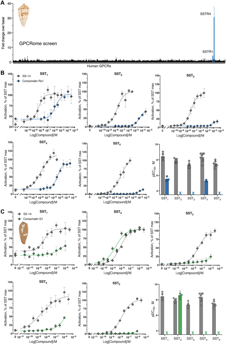
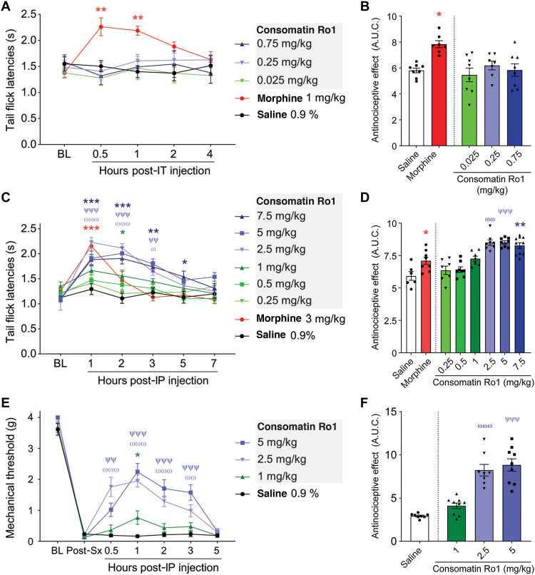
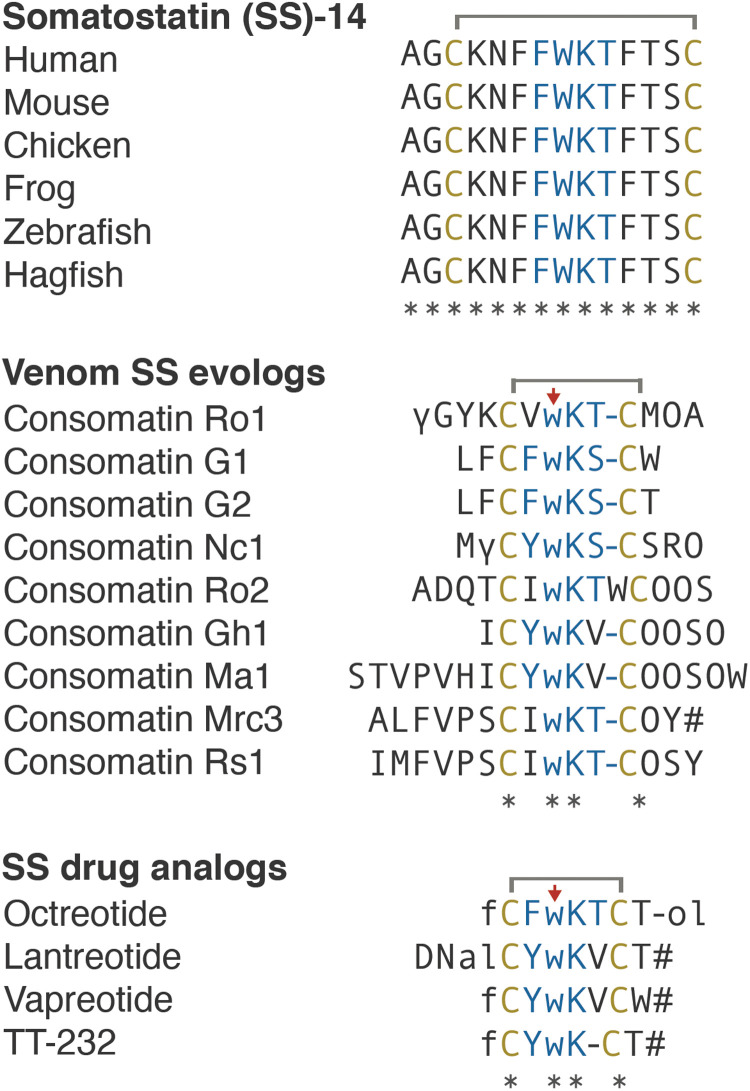
Somatostatin (SS) is a peptide hormone with diverse physiological roles. By investigating a deep-water clade of fish-hunting cone snails, we show that predator-prey evolution has generated a diverse set of SS analogs, each optimized to elicit specific systemic physiological effects in prey. The increased metabolic stability, distinct SS receptor activation profiles, and chemical diversity of the venom analogs make them suitable leads for therapeutic application, including pain, cancer, and endocrine disorders. Our findings not only establish the existence of SS-like peptides in animal venoms but also serve as a model for the synergy gained from combining molecular phylogenetics and behavioral observations to optimize the discovery of natural products with biomedical potential.


Figures

Fig. 1.
Fig. 1.. Evolution of distinct predation strategies in different clades of fish-hunting cone snails.
(A) Phylogenetic tree of the eight recognized clades of fish hunters. Bootstrap values and shell images of neotypes are shown (1: Conus radiatus, 2: Conus sulcatus, 3: Conus pergrandis, 4: Conus geographus, 5: Conus kinoshitai, 6: Conus magus, 7: Conus ermineus, 8: Conus bullatus). Arrows show the species used in this work, with color corresponding to the proposed hunting strategy shown in (B). (B) Still images of three distinct predation strategies filmed for three different species of fish hunters [from top to bottom: C. neocostatus (Asprella clade, gray arrow marks proboscis loaded with radular tooth), C. geographus (Gastridium clade), and C. bullatus (Textilia clade)]. Images were extracted from supporting movie files (movies S1 to S3). Video/photo credit: Dylan Taylor and Baldomero M. Olivera, University of Utah.
Fig. 2.
Fig. 2.. Identification of Consomatin Ro1.
(A) Reversed-phase chromatography of crude venom of the Asprella snail C. rolani and (B) subfractionation and (C) purification of Consomatin Ro1 from subfraction #16-12 (shown in red). (D) ETD MS/MS spectrum of the quadruple-charged ion of the venom peptide after reduction and alkylation with 2-methylaziridine acquired on the Orbitrap Lumos Tribrid with 30,000 resolution (at 400 m/z) and three microscans. N-terminal fragment ions (c-type ions) are indicated by ⌉, and C-terminal fragment ions (z,y-type ions) are indicated by ⌊. Doubly charged ions are indicated with ++, and z ions resulting from cleavage at cysteine and loss of the cysteine side chain are indicated with *. [M+4H]2+•• and [M+4H]1+••• indicate charge-reduced species. Because of space limitations, not all different charge states of already labeled peptide bond cleavages are indicated in the figure artwork. The mass accuracy for all fragment ions is better than 10 ppm. γ, γ-carboxyglutamate; O, hydroxyproline; w, d-tryptophan. (E) Organization of the prepropeptide of Consomatin Ro1 identified by transcriptome sequencing depicting posttranslational intermediates and processing events. AU, absorbance units.
Fig. 3.
Fig. 3.. Sequences and schematic representation of human SS, Consomatin Ro1, and the SS drug analog octreotide.
Cysteines and amino acids of the core SS receptor binding motif are shown in yellow and blue, respectively. Modifications: γ, γ-carboxyglutamate; w, d-tryptophan (marked by red arrow); O, hydroxyproline; f, d-phenylalanine; #, amidation; -ol, alcohol.
Fig. 4.
Fig. 4.. Consomatin Ro1 and G1 are structural mimetics of the SS drug analog octreotide.
(A) X-ray structure of Consomatin Ro1 at 1.95-Å resolution (PDB ID: 7SMU). (B) Nuclear magnetic resonance (NMR) solution structure of human SS-14 obtained during heparin-induced fibril formation (PDB ID: 2MI1). (C) Alignment of the structure of Consomatin Ro1 (orange) with that of SS-14 (gray). (D) NMR solution structure of octreotide (PDB ID: 1SOC). (E) Alignment of the structure of Consomatin Ro1 (orange) with that of octreotide (purple) showing nearly identical backbone conformation and orientation of d-Trp7, Lys8, Thr9, and the disulfide bond, but differences in the amino acid composition and side-chain arrangements of Val/Phe6 and of residues outside the cyclic core. (F) Alignment of a homology model of Consomatin G1 (green), based on the structure of Ro1, with that of octreotide (purple), suggesting that the molecules share strong structural similarities. Numbering of residues according to that of Consomatin Ro1.
Fig. 5.
Fig. 5.. Consomatin Ro1 and G1 selectively activate the human SS receptors.
(A) PRESTO-Tango screen of Consomatin Ro1 (10 μM) at 318 human GPCRs (x axis) identified SST4 and SST1 as the molecular target. Plotted values represent means ± SD of four technical replicates. (B and C) Representative concentration-response curves at the five human SS receptor subtypes comparing (B) Consomatin Ro1 or (C) Consomatin G1 with human SS-14 using the β-arrestin recruitment assay (error bars represent SD of three technical replicates), as well as bar graphs showing the respective pEC50 values (error bars represent CI95 of three to seven independent repeats). RLU, relative luminescence units. Illustration credit: Paula Flórez Salcedo, University of Utah.
Fig. 6.
Fig. 6.. Consomatin Ro1 provides analgesia in two mouse models of acute pain.
(A and B) Sensitivity to thermal noxious stimuli was evaluated by dipping mouse tails into hot water (52°C) and recording tail flick latencies (TFLs). (A) Mouse TFLs after a single intrathecal injection of saline, morphine, or multiple doses of Consomatin Ro1. TFLs were captured over a 4-hour period [n = 7 to 8 per group, two-way analysis of variance (ANOVA) with Dunnett’s post hoc test]. (B) Area under the curve between baseline (BL) and 4-hour condition, presenting global TFLs when animals received intrathecal (IT) injections (Kruskal-Wallis followed by Dunn’s post hoc test). (C) Mouse TFLs after a single intraperitoneal (IP) injection of saline, morphine, or multiple doses of Consomatin Ro1. TFLs were captured over a 7-hour period (n = 6 to 10 per group, two-way ANOVA with Dunnett’s post hoc test). (D) Area under the curve (A.U.C.) between baseline and 7-hour condition, presenting global TFLs when animals received intraperitoneal injections (Kruskal-Wallis followed by Dunn’s post hoc test). (E) Analgesic effect of Consomatin Ro1 on acute postsurgical pain. Plantar incision surgery intraperitoneal injections. Mechanical sensitivity was assessed using von Frey filaments (n = 8 to 9 per group, two-way ANOVA with Dunnett’s post hoc test). (F) Area under the curve between baseline and 5-hour condition, presenting mechanical withdrawal thresholds when animals received intraperitoneal injections (Kruskal-Wallis followed by Dunn’s post hoc test). Results correspond to means ± SEM. */ω/ψP < 0.05, **/ωω/ψψP < 0.01, and ***/ωωω/ψψψP < 0.001. Red asterisks illustrate significant difference between morphine and saline condition, blue asterisks: 7.5 mg versus saline, ψ: 5 mg versus saline, ω: 2.5 mg versus saline, green asterisks: 1 mg/kg versus saline.
Fig. 7.
Fig. 7.. Comparative sequence alignments of vertebrate SS, selected Consomatin evologs, and SS drug analogs.
Identical amino acids are denoted by an asterisk (*). Mature toxins are shown with posttranslational modifications based on processing and modification of Consomatin Ro1 (modifications and color codes as shown in Fig. 3). See fig. S3 for precursor sequences of all consomatin sequences reported in this study.

Similar articles

Cited by

References

    1. Olivera B. M., Seger J., Horvath M. P., Fedosov A. E., Prey-capture strategies of fish-hunting cone snails: Behavior, neurobiology and evolution. Brain Behav. Evol. 86, 58–74 (2015). - PMC - PubMed
    1. King G. F., Venoms as a platform for human drugs: Translating toxins into therapeutics. Expert. Opin. Biol. Ther. 11, 1469–1484 (2011). - PubMed
    1. Safavi-Hemami H., Brogan S. E., Olivera B. M., Pain therapeutics from cone snail venoms: From ziconotide to novel non-opioid pathways. J. Proteome 190, 12–20 (2019). - PMC - PubMed
    1. Yarotskyy V., Elmslie K. S., Omega-conotoxin GVIA alters gating charge movement of N-type (CaV2.2) calcium channels. J. Neurophysiol. 101, 332–340 (2009). - PubMed
    1. Miljanich G. P., Ziconotide: Neuronal calcium channel blocker for treating severe chronic pain. Curr. Med. Chem. 11, 3029–3040 (2004). - PubMed