. 2022 Mar 25;8(12):eabj9019.
doi: 10.1126/sciadv.abj9019. Epub 2022 Mar 23.

Opposite effects of stress on effortful motivation in high and low anxiety are mediated by CRHR1 in the VTA

Affiliations

Opposite effects of stress on effortful motivation in high and low anxiety are mediated by CRHR1 in the VTA

Ioannis Zalachoras et al. Sci Adv. .

Abstract

Individuals frequently differ in their behavioral and cognitive responses to stress. However, whether motivation is differently affected by acute stress in different individuals remains to be established. By exploiting natural variation in trait anxiety in outbred Wistar rats, we show that acute stress facilitates effort-related motivation in low anxious animals, while dampening effort in high anxious ones. This model allowed us to address the mechanisms underlying acute stress-induced differences in motivated behavior. We show that CRHR1 expression levels in dopamine neurons of the ventral tegmental area (VTA)-a neuronal type implicated in the regulation of motivation-depend on animals' anxiety, and these differences in CRHR1 expression levels explain the divergent effects of stress on both effortful behavior and the functioning of mesolimbic DA neurons. These findings highlight CRHR1 in VTA DA neurons-whose levels vary with individuals' anxiety-as a switching mechanism determining whether acute stress facilitates or dampens motivation.


Figures

Fig. 1.
Fig. 1.. Effects of acute exposure to stress on PR test performance.
(A) All rats were phenotyped for trait anxiety in the EPM before the beginning of experiments. Bar graphs represent the distribution of the time spent in the open arms (%OA) in a representative batch of 130 rats. (B) Scheme describing the experiment probing the effects of acute stress exposure on PR test performance of LA and HA rats. (C) Stress exposure resulted in a higher breakpoint in LA rats compared to HA rats. Two-way ANOVA revealed a significant stress × anxiety interaction (F1,36 = 15.46, P = 0.0004), but no significant effects of anxiety or stress (F1,36 = 3.736, P = 0.061 and F1,30 = 0.1244, P = 0.726, respectively, n = 9 to 11 per group). (D) LA rats exposed to stress performed significantly more nosepokes than HA rats exposed to stress. Two-way ANOVA revealed a significant stress × anxiety interaction (F1,36 = 12.35, P = 0.001), but no significant effects of anxiety or stress (F1,36 = 3.010, P = 0.091 and F1,36 = 0.141, P = 0.71, respectively, n = 9 to 11 per group). (E) LA rats exposed to stress acquired more rewards than HA rats exposed to stress. Two-way ANOVA revealed a significant stress × anxiety interaction (F1,36 = 12.42, P = 0.001), but no significant effects of anxiety or stress (F1,36 = 3.304, P = 0.077 and F1,36 = 0.0001, P = 0.99, respectively, n = 9 to 11 per group). Asterisks denote significant differences in the respective post hoc tests (**P < 0.01, *P < 0.05, and #P < 0.1).
Fig. 2.
Fig. 2.. Trait anxiety is associated with a Crhr1 common variant and with different CRHR1 expression in VTA DA neurons.
(A to C) All rats (n = 26 HA, n = 32 IA, and n = 35 LA) were genotyped for SNP on rs106600307 located on chr10:92191940. (A) Allele pair distribution is associated with time spent in the open arms in the EPM (Kruskal-Wallis χ2 = 6.2139, df = 2, P = 0.04474). (B) Heterozygous rats spent significantly more time in the open arms of the EPM compared to homozygous rats (Wilcoxon rank sum test, W = 1397.5, P = 0.01309). Bars represent the percentage of time spent in the open arms (%OA) for the different allele pairs. (C) A significantly higher proportion of LA rats than HA rats are heterozygous for the Crhr1 SNP (χ2 test6.146,1, P = 0.0132). (D to F) CRHR1 expression in DA cells in the VTA was probed with RNAscope. Expression of CRHR1 mRNA (red) in TH+ cells (green), along with nuclear staining with DAPI (blue) and merged image in the VTA of LA (D) and HA (E) rats. (F) Left: Higher magnification images of the marked area of panels in (D) and (E) in LA and HA rats. Right: LA rats had higher CRHR1 mRNA expression in DA neurons in the VTA than HA rats (Mann-Whitney test, U = 2.000, P = 0.032). (G) Left: CRHR1 expression in GABAergic cells in the VTA. Merged images of expression of CRHR1 mRNA (red) in GAD1+ cells (magenta), along with nuclear staining with DAPI (blue) in LA and HA rats. Right: CRHR1 mRNA expression was not different between LA and HA rats in GABAergic neurons (Mann-Whitney test, U = 11.00, P = 0.816) and when quantified in all nuclei (Mann-Whitney test, U = 8.500, P = 0.46). *P < 0.05. Scale bars, 100 μm.
Fig. 3.
Fig. 3.. Effects of intra-VTA CRH administration on PR test performance.
(A) Scheme describing the experiment probing the effects of intra-VTA CRH administration on PR test performance of LA and HA rats. (B) Vehicle-treated HA rats had a higher breakpoint compared to vehicle-treated LA rats. Intra-VTA CRH treatment in LA rats resulted in a higher breakpoint compared to CRH-treated HA rats. Two-way ANOVA revealed a significant treatment × anxiety interaction (F1,26 = 17.01, P = 0.0003), but no significant effects of anxiety or treatment (F1,26 = 0.0005, P = 0.98 and F1,26 = 1.560, P = 0.223, respectively, n = 7 to 8 per group). (C) Intra-VTA CRH administration in LA rats resulted in higher number of correct nosepokes performed compared to CRH-treated HA rats. Two-way ANOVA revealed a significant treatment × anxiety interaction (F1,26 = 17.93, P = 0.0003), but no significant effects of anxiety or treatment (F1,26 = 0.204, P = 0.65 and F1,26 = 2.170, P = 0.153, respectively, n = 7 to 8 per group). (D) Vehicle-treated HA rats acquired a higher number of rewards compared to vehicle-treated LA rats. However, intra-VTA CRH treatment in LA rats resulted in a higher number of acquired rewards compared to CRH-treated HA rats. Two-way ANOVA revealed a significant treatment × anxiety interaction (F1,26 = 21.50, P < 0.0001), but no significant effects of anxiety or treatment (F1,26 = 0.468, P = 0.50 and F1,26 = 2.086, P = 0.161, respectively, n = 7 to 8 per group). Asterisks denote significant differences between anxiety groups at respective time points (**P < 0.01, *P < 0.05 and #P < 0.1).
Fig. 4.
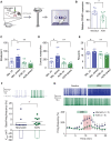
Fig. 4.. Effects of CRH on VTA DA neuron firing.
(A) Bath application of CRH (500 nM) induced a reversible increase in DA VTA cell firing, more pronounced in LA rats, and abrogated in LA rats by CRHR1 antagonist CP-154526 (3 μM) (Kruskal-Wallis: 14.16, P = 0.0008, Dunn’s post hoc: LA versus HA, P = 0.04; LA versus LA + CP, P = 0.0002; n = 10-11-5). Color-coded representative traces recorded before and during CRH application on the left. (B) Dose-response curves for submillimolar concentrations of CRH on DA VTA cell firing in LA and HA rats (fit comparison: F4,49 = 3.726, P = 0.010, n = 4, 4, 6, 9, and 5 for LA and n = 3, 4, 4, 11, and 7 for HA for increasing concentration values). (C) Spontaneous firing frequency recorded in DA VTA neurons with no current injection appeared reduced in LA rats (Mann-Whitney: U = 66.5, P = 0.0163, n = 15 to 17). Representative voltage traces at the top. (D) Data presented in (A) are here represented as absolute firing values. (E) The anxiety-related differential effect of CRH on DA VTA cells persisted in the presence of the GABAAR blocker picrotoxin (100 μM) and the GABABR blocker CGP55845 (2 μM) (Mann-Whitney test comparing mean values in the shaded area, U = 2.0, P = 0.0317, n = 5). (F) Quantification of CRHR1 expression levels in VTA tissue from WT and DAT-CRHR1 KO mice (two-tailed t test, t7 = 2.551, P = 0.0381). (G) Bath application of CRH (500 nM) induced a reversible increase in cell firing in WT mice, which was absent in DAT-CRHR1 KO mice (Mann-Whitney test comparing mean values in the shaded area, U = 2, P = 0.0317, n = 5 to 6). Color-coded representative traces recorded before and during CRH application on the left. *P < 0.05.
Fig. 5.
Fig. 5.. Antisense-mediated CRHR1 down-regulation in the VTA blocks stress effects on PR performance in LA rats.
(A) Schematic representation of AON treatment and PR test. (B) Treatment with AONs decreased CRHR1 expression in the VTA (one-tailed t test, t11 = 2.199, P = 0.025). (C) Significant AON treatment × stress interaction, significant treatment effect, and a nonsignificant effect of stress on breakpoint (two-way ANOVA: F1,24 = 6.789, P = 0.016, F1,24 = 10.27, P = 0.004, and F1,24 = 1.872, P = 0.184, respectively, n = 7). (D) Significant AON treatment effect, significant interaction, and nonsignificant effect of stress on the number of correct nosepokes (two-way ANOVA: F1,24 = 9.566, P = 0.005, F1,24 = 6.346, P = 0.019, and F1,24 = 2.146, P = 0.156, respectively, n = 7). (E) Two-way ANOVA revealed a significant AON treatment × stress interaction, a significant effect of AON treatment, and a nonsignificant effect of stress on the number of rewards (F1,24 = 5.526, P = 0.027, F1,24 = 9.566, P = 0.005, and F1,24 = 0.75, P = 0.395, respectively, n = 7). (F) Ex vivo basal firing rate of DA neurons in the VTA, following AON-mediated CRHR1 down-regulation. Treatment with AONs down-regulating CRHR1 resulted in a higher spontaneous firing rate in DA neurons compared to the mismatch treated group (Mann-Whitney test, U = 17.5, P = 0.0222, n = 9 to 10). (G) Bath application of CRH (500 nM) induced a reversible increase in cell firing in LA rats, which was partially blocked by AON treatment (Mann-Whitney test comparing mean values in the shaded area, U = 6, P = 0.0451, n = 5 to 8). Representative voltage traces are shown at the top. Asterisks denote significant differences in the respective post hoc tests (**P < 0.01, *P < 0.05, and #P < 0.1).
Fig. 6.
Fig. 6.. Genetic manipulation of CRHR1 expression affects motivated behavior after stress.
(A) Schematic representation of the viral strategy for CRHR1 OE or GFP expression in NAc-projecting VTA neurons in HA rats, generating CRHR1 OE rats and control GFP rats, which were tested for PR performance following stress exposure. (B to D) CRHR1 OE rats reached a higher breakpoint (F1,19 = 12.43, P = 0.0027), performed more nosepokes (F1,19 = 13.23, P = 0.0018), and obtained more rewards (F1,19 = 12.06, P = 0.0025) both under basal conditions and after stress. (E) PR performance after stress was tested in DAT-CRHR1 mice in which CRHR1 was selectively down-regulated in DA neurons. (F to H) Stress in DAT-CRHR1 mice led to impaired performance. (F) Stress in DAT-CRHR1 mice led to a lower breakpoint. Two-way repeated-measures ANOVA revealed a significant stress × genotype interaction (F1,20 = 8.028, P = 0.01), a marginally nonsignificant genotype effect, and a nonsignificant stress effect (F1,20 = 4.016, P = 0.059 and F1,20 = 0.004, P = 0.95, n = 7 to 15). (G) Stress reduced the number of correct nosepokes in DAT-CRHR1 mice. Two-way repeated-measures ANOVA revealed a significant stress × genotype interaction (F1,20 = 5.836, P = 0.025), a marginally nonsignificant genotype effect, and a nonsignificant stress effect (F1,20 = 4.013, P = 0.059 and F1,20 = 0.056, P = 0.82, n = 7 to 15). (H) Stress in DAT-CRHR1 mice reduced reward acquisition. Two-way repeated-measures ANOVA revealed a significant stress × genotype interaction (F1,20 = 10.14, P = 0.005), a significant effect of genotype, and a nonsignificant effect of stress (F1,20 = 7.150, P = 0.014 and F1,20 = 0.416, P = 0.53, n = 7 to 15 per group). Asterisks denote significant differences in the t test or post hoc test (**P < 0.01, *P < 0.05, and #P < 0.1).

Similar articles

Cited by

References

    1. Korte S. M., Koolhaas J. M., Wingfield J. C., McEwen B. S., The Darwinian concept of stress: Benefits of allostasis and costs of allostatic load and the trade-offs in health and disease. Neurosci. Biobehav. Rev. 29, 3–38 (2005). - PubMed
    1. McEwen B. S., Akil H., Revisiting the stress concept: Implications for affective disorders. J. Neurosci. 40, 12–21 (2020). - PMC - PubMed
    1. Crocker L. D., Heller W., Warren S. L., O’Hare A. J., Infantolino Z. P., Miller G. A., Relationships among cognition, emotion, and motivation: Implications for intervention and neuroplasticity in psychopathology. Front. Hum. Neurosci. 7, 261 (2013). - PMC - PubMed
    1. Treadway M. T., Bossaller N. A., Shelton R. C., Zald D. H., Effort-based decision-making in major depressive disorder: A translational model of motivational anhedonia. J. Abnorm. Psychol. 121, 553–558 (2012). - PMC - PubMed
    1. Pulcu E., Trotter P. D., Thomas E. J., McFarquhar M., Juhasz G., Sahakian B. J., Deakin J. F. W., Zahn R., Anderson I. M., Elliott R., Temporal discounting in major depressive disorder. Psychol. Med. 44, 1825–1834 (2014). - PMC - PubMed